



Key takeaways
- 1Ankle arthroscopy lets the surgeon look inside the joint with a small camera and treat problems through cuts of about 5 mm, rather than opening the ankle widely.
- 2The operation usually takes about 60 minutes, is done under general or spinal anaesthesia, and is normally a day case with 0 hospital nights.
- 3It works best for specific, mechanical problems such as bone spurs that pinch, loose fragments, scar tissue, a small cartilage defect or an unstable ligament, not for widespread arthritis.
- 4Many people walk in a boot or protective shoe within days, return to desk work in about 1 to 2 weeks and need around 8 weeks before the ankle feels close to normal.
- 5Ankle arthroscopy risks include stiffness, numbness over the top of the foot, swelling that lingers and, rarely, infection or a blood clot, and most are reduced by careful portal placement.
- 6Planned ankle arthroscopy in turkey can suit stable patients with scans in hand who can stay about 5 to 7 days for the first wound check before flying home.
Overview
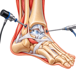
Ankle arthroscopy is a keyhole operation in which a surgeon looks inside the ankle joint through a thin camera and treats problems with miniature instruments. It uses cuts of about 5 mm instead of a long incision, takes about 60 minutes, and is normally done as a day case. The recovery time is around 8 weeks, depending on what is treated.
What is ankle arthroscopy?
The word comes from Greek: "arthro" for joint and "skopein" for to look. A narrow metal telescope, the arthroscope, is placed into the joint between the tibia (shin bone) and the talus (the bone below it). A screen shows the lining, cartilage and ligaments in magnified detail. Through a second small cut, the surgeon uses a shaver, grasper, burr or knife to repair what is found.
The procedure is sometimes called an ankle scope, or arthroscopic ankle surgery. Because the ankle is a tight joint with little spare room, surgeons often use a small strap or a pin to gently pull the bones apart, which opens the space and gives a better view.
What problems does ankle arthroscopy treat?
Ankle arthroscopy is used when a mechanical or inflammatory problem inside the joint explains symptoms. Common targets are anterior impingement, where bony spurs or thick tissue at the front of the ankle pinch during dorsiflexion, osteochondral lesions of the talus (a damaged patch of cartilage and underlying bone), loose bodies, scarred synovium and tendon-related problems around the joint.
It is also used for chronic ankle instability, where repeated sprains have stretched the lateral ligaments, which is described on our chronic ankle instability page. In selected cases the surgeon repairs the ligament with the scope, and the joint inspection catches damage that scans missed.
Does ankle arthroscopy help arthritis?
Only in a limited way. Cleaning the joint can ease catching or pinching in early ankle osteoarthritis, but it does not regrow cartilage. Studies suggest benefit is shorter-lived when the joint space is already narrow. For advanced wear, surgeons discuss ankle fusion or total ankle replacement instead.
Where does this fit in foot and ankle care?
Ankle arthroscopy is one tool within the wider foot and ankle field. It sits between non-surgical care and larger operations, so it often serves as a first operative step. Surgeons also use it to confirm a diagnosis before choosing a bigger procedure, which can spare you an unnecessary reconstruction.
Why use keyhole surgery for the ankle?
The ankle has little soft tissue cover, so open cuts can heal slowly and scar. Keyhole surgery leaves a few small marks, causes less swelling in many cases, and usually allows an earlier return to walking. It also shows structures that open surgery might not reach, such as the back of the talus, without cutting through ligaments.
How is the operation different from open ankle surgery?
Open surgery gives direct access and is preferred for fractures, deformity correction and joint replacement. Ankle arthroscopy gives a magnified internal view with less trauma to the tissue around the joint. It is a diagnostic and treatment tool, rather than a way to reshape or rebuild the whole ankle. Many surgeons combine both methods, starting with the scope and adding a small open step if needed.
Is ankle arthroscopy major surgery?
It is considered minor to moderate. Blood loss is very small, and there is rarely any need for an overnight stay. Even so, it is a real operation under anaesthetic, with swelling and a period of protection afterwards. The effect depends on what is done inside: a simple spur trim recovers quickly, while a cartilage repair needs weeks of protected weight-bearing.
| Feature | Ankle arthroscopy | Open ankle surgery |
|---|---|---|
| Skin cuts | 2 to 3 small portals, about 5 mm | One longer incision, often 8 to 15 cm |
| Typical stay | Day case | Day case to 2 nights |
| Swelling and scarring | Usually less | Usually more |
| View of the joint | Magnified, internal | Direct, wider |
| Best for | Pinching, loose bodies, small cartilage lesions | Fractures, deformity, implants |
Who should think twice?
Be cautious if the main complaint is a diffuse ache from advanced arthritis, if the ankle is badly misaligned, or if circulation or skin is poor. Surgery is also unwise during an active infection. A clear mechanical problem, supported by symptoms and imaging, is the best predictor of a good result, so your surgeon should explain exactly what the scope is expected to fix.
Conditions treated
Who it's for
- Anterior ankle impingement, with bony spurs or thickened soft tissue pinching at the front when the foot flexes up, and pain that persists after rest, activity change and injections.
- Posterior ankle impingement, often from a prominent bone at the back of the talus (os trigonum), common in dancers and footballers.
- An osteochondral lesion of the talus, a localised cartilage and bone injury that is debrided or treated with microfracture or other cartilage techniques.
- Loose bodies or fragments of cartilage that cause catching, locking or sudden giving way.
- Chronic synovitis or scarred lining that causes swelling and pain and has not responded to medicines.
- Chronic lateral ankle instability, in selected people where the ligament repair can be done arthroscopically.
- Early ankle osteoarthritis with mechanical symptoms such as catching, in people who are not ready for fusion or replacement.
- Persistent pain after an ankle sprain or fracture where scans do not explain the problem, and diagnostic arthroscopy may show the cause.
- Removal of infected or inflamed material from the joint, or retrieval of a broken instrument or foreign body.
Good candidates
The best candidates for ankle arthroscopy are people with a clearly localised mechanical problem, symptoms that match the scan, and months of failed non-operative care. Younger and middle-aged adults and athletes are common candidates. The more specific the target, the more likely the keyhole procedure is to help, which is why the diagnosis should be firm before anyone books a date.
Who is a good candidate for arthroscopic ankle surgery?
A good candidate has pinching at the front or back of the ankle, catching or locking, a recognised cartilage lesion, or ligament laxity after repeated sprains. Symptoms have usually lasted at least 3 to 6 months despite physiotherapy, bracing, activity changes and, where useful, an injection. You should also be ready for a structured rehabilitation plan, since the scope itself is only the first step.
Who is less likely to benefit?
People whose pain comes mainly from widespread arthritis, a deformed or tilted ankle, or nerve and tendon problems outside the joint often gain little from a scope. The same applies if the pain is vague and the scans are normal. In those cases, a clearer diagnosis, a different operation, or continued non-surgical care is usually the wiser path.
How do surgeons decide between options?
The decision rests on examination, weight-bearing X-rays and usually an MRI or CT scan, which show spurs, cartilage defects and ligament status. Surgeons also weigh age, sport, job, general health and the failure of earlier treatment. If you are not sure, a second opinion from a foot and ankle specialist is reasonable before committing to surgery.
| Situation | Arthroscopy likely appropriate | Non-surgical first | Another operation or not advised |
|---|---|---|---|
| Front ankle spur pinching for over 6 months | Yes | Rest, physiotherapy, heel raise | |
| Small talus cartilage defect with catching | Yes | Protected loading first | |
| Repeated sprains with a loose ankle | Often, after bracing and rehab | Strength and balance training | Open reconstruction if tissue is poor |
| Advanced arthritis with bone-on-bone pain | Injections, bracing | Fusion or replacement | |
| Fresh ankle fracture | Urgent fracture care instead | ||
| Normal scans, vague pain | Rarely, diagnostic only | Yes |
A candid conversation helps. Ask what finding the surgeon expects, what happens if the inside of the joint looks different, and how likely it is that more than one step is needed. Clear expectations make recovery easier to accept.
Before surgery
Preparing for ankle arthroscopy means confirming exactly what is wrong, making sure you are well enough for anaesthesia and arranging a few days of rest. Imaging, a medicines review and a plan for crutches, work and transport are the main steps. The earlier you start, the smoother the day of surgery will feel.
What tests and scans are needed?
Standing X-rays show alignment, bone spurs and joint space. MRI reveals cartilage damage, loose fragments, ligament injury and bone swelling, while CT is useful for subtle bone detail. Stress views can show instability. A heart tracing and blood tests are added for older patients or those with other conditions. Bring all old images, because comparing them can help.
How should I prepare my health?
Stop smoking for several weeks if possible, since nicotine slows healing. Keep diabetes well controlled and treat any skin infection near the foot. Your doctor will advise on blood thinners, anti-inflammatories and herbal products. Do not stop prescribed treatment without advice. Tell the team about previous clots, allergies, and bad reactions to anaesthesia.
What non-surgical steps come first?
Before surgery is booked, most surgeons expect a trial of physiotherapy focused on calf flexibility, balance and peroneal strength, supportive footwear, bracing for instability, activity changes, and sometimes an injection. If those steps have been tried and symptoms remain, arthroscopy becomes a more reasonable choice. A record of what you have tried helps your surgeon.
What should I send for a remote review?
A remote opinion needs recent MRI or CT reports, ideally the images, standing X-rays, a short history of sprains or injuries, treatments tried, and a medicines list. Photos of the ankle, a video of how you walk, and notes on which movements hurt are useful. Our medical record review guide explains how specialists use these files.
How do I plan home and work?
- Arrange a lift home and someone for the first 24 hours.
- Set up a resting place where the foot can be raised above the heart.
- Collect crutches, a boot or shoe, ice packs and simple painkillers.
- Plan for 1 to 2 weeks off desk work, or 4 to 6 weeks for physical jobs.
- Check whether you can drive; a right-sided procedure usually needs a pause of 1 to 3 weeks.
What questions should I ask the surgeon?
Useful questions include what the scans show, what the surgeon expects to find, which steps may be added during the procedure, what the weight-bearing rules will be and how many such operations they perform each year. Also ask who you contact after hours and how your results will be reviewed. Clear answers build trust and prevent surprises.
What happens on the day before and morning of surgery?
You will be told when to stop eating and drinking, usually about 6 hours for food and 2 hours for clear water. Shower with the supplied wash, remove nail varnish and jewellery, and take only the medicines you were told to take. The surgeon will mark the correct ankle, and you will sign a consent form after a final discussion.
If your problem is a sprain that has just happened, or an ankle that is swollen after a fall, urgent local assessment comes first. Treatment abroad is for stable patients with planned, non-urgent keyhole surgery.
How the operation is performed
During ankle arthroscopy the surgeon makes two or three small cuts around the joint, inserts a camera and instruments, examines every part of the joint in a set order, and then trims, repairs or smooths whatever is causing the problem. The operation usually takes about 60 minutes, though simple spur trims are shorter and cartilage or ligament repairs are longer.
What anaesthesia is used?
General or spinal anaesthesia is most common, sometimes with a sciatic or popliteal nerve block that numbs the foot for several hours after surgery. Light sedation with a nerve block can be enough in some cases. Your anaesthetist chooses the plan according to your health, the length of the operation and whether you want to be awake.
How are you positioned and how is the joint opened up?
You lie on your back with the leg supported, and a thigh tourniquet may limit bleeding. Some surgeons add non-invasive distraction, a soft strap around the ankle and foot, that gently pulls the joint apart by a few millimetres. Others prefer to keep the leg free and flex the ankle, avoiding traction pressure on the skin.
What are the key steps of the procedure?
- The surgeon marks landmarks, including the front tibial tendon, the peroneal nerve branches and the vessels, to keep the portals safe.
- An anteromedial portal is made, followed by a small anterolateral portal, and fluid gently fills the joint to give a clear view.
- The arthroscope is inserted, and a systematic inspection follows: cartilage of the tibia and talus, ligaments, gutters and the lining.
- Instruments remove inflamed tissue, loose bodies and bone spurs with a shaver or burr.
- If a cartilage lesion is found, the surgeon debrides the unstable edge and treats the base, for example by microfracture.
- For ligament repair, anchors and sutures are placed through the scope.
- The portals are closed with a stitch or a strip, and a soft dressing and bandage are applied.
What is a posterior ankle arthroscopy?
For problems at the back of the ankle, surgeons use a two-portal technique with you lying on your front or side. The portals sit beside the Achilles tendon. This route gives access to the os trigonum, the flexor hallucis longus tendon and posterior spurs, and avoids a larger cut. It requires special training because nerves and vessels are close.
How are cartilage lesions treated?
Surgeons clear unstable cartilage, then stimulate healing in the bone beneath. Microfracture makes tiny holes so that blood and marrow cells form repair tissue. For larger or failed lesions, options include bone marrow concentrate, grafts or other cartilage techniques, which may need an open step. The choice depends on lesion size, depth and the patient's activity goals.
How is ankle instability treated through the scope?
The surgeon assesses the cartilage and any impinging tissue first, then repairs the stretched lateral ligaments, usually the anterior talofibular and calcaneofibular, by tightening them with anchors and sutures. This is the arthroscopic version of the Broström repair. Where tissue is poor or the ankle is very loose, an open repair or graft reconstruction is the better choice.
Which variants exist and how do surgeons choose?
| Variant | Typical use | Strength | Limit |
|---|---|---|---|
| Anterior arthroscopy | Front spurs, loose bodies, synovitis | Straightforward, quick | Limited view of the back |
| Posterior arthroscopy | Os trigonum, back impingement | Excellent rear access | Needs specialised training |
| Arthroscopic cartilage surgery | Talus lesions | Treats and inspects in one step | Large lesions may need open work |
| Arthroscopic ligament repair | Chronic lateral instability | Treats the joint and ligament together | Less suited to poor tissue |
| Diagnostic scope | Unexplained pain after injury | Direct look inside | Only worthwhile if it will change care |
What is done at the end, and how is the joint protected?
Before closing, the surgeon washes out the joint to clear debris, checks that no instrument tip or fragment remains, and tests movement. Local anaesthetic may be placed around the portals to ease the first hours. The skin is closed with fine nylon or adhesive strips, and a padded dressing with a posterior splint or boot supports the ankle. Photographs of the findings may be taken for your notes.
Surgeons also record exactly what was treated, because this determines the weight-bearing instructions. A clean-out, a microfracture and a ligament repair each have different rules, so ask for these in writing before you leave the unit.
How long does ankle arthroscopy take?
Most operations take about 30 to 90 minutes, and the usual figure is around 60 minutes for a typical procedure. A simple clean-out is at the shorter end, while a combined ligament repair and cartilage treatment is at the longer end. Add time for anaesthesia and waking up, so allow 2 to 3 hours from entering theatre to returning to the recovery area.
Hospital stay
Nearly all patients go home the same day after ankle arthroscopy, so the usual plan is 0 hospital nights. You wake up in recovery, have a drink and a light snack, learn to use crutches, and leave with a bandage, painkillers and instructions. An overnight stay is reserved for people with other illnesses, poor support at home, or a longer combined procedure.
What happens on the day?
After the operation, a nurse checks your pulse, pain, nausea and the colour and warmth of your toes. A physiotherapist teaches safe walking, with crutches and a boot or surgical shoe as advised. Most surgeons allow some weight-bearing if the operation was a simple clean-out, but protect the ankle fully after a cartilage or ligament repair.
How is pain managed?
The block can leave the foot numb and heavy for 8 to 24 hours. Start regular paracetamol and an anti-inflammatory if allowed before it wears off, with a short supply of stronger medicine for the first nights. Ice through a cloth and elevation above heart level reduce swelling more than most people expect. Most pain settles within a week.
What are the discharge criteria?
You can usually leave when you are awake and comfortable, can pass urine, are not heavily nauseous, can walk with crutches, and have a responsible adult with you. You are given written wound care, dressing and medicine advice, plus the contact number for the surgical team and the date of your first review at about 10 to 14 days.
How do I look after the wound?
Leave the dressing in place for the first 2 days if advised, keep it dry, and then change it as directed. The small portals can leak pink fluid at first, which is normal. Stitches or strips are removed at around 10 to 14 days. Avoid soaking in a bath or pool until the skin is sealed and the surgeon agrees.
What should international patients know?
Planned ankle arthroscopy in turkey is practical because the procedure is short and the stay is small. Plan 5 to 7 days locally for the first check, bring your imaging, and ask about flight timing and clot precautions. Our guides on travel and accommodation, surgery day and flying after surgery help with preparation.
A companion is very helpful for the first 48 hours because crutches, bags and stairs are tiring when the foot is swollen. See the companion guide for what to organise, and agree how your own physiotherapist will receive the operation report.
Recovery
Recovery after ankle arthroscopy is usually quicker than after open ankle surgery. Expect pain to ease over about a week, swelling for several weeks and a return to normal shoes and everyday walking at around 2 to 4 weeks. The ankle arthroscopy recovery time of about 8 weeks is a typical figure for simple procedures, while cartilage or ligament repairs take longer.
What do the first 2 weeks feel like?
The ankle is swollen, bruised and tender, and often feels tight in the evening. Most of the day should be spent sitting with the foot raised, with short walks to the bathroom and kitchen. Move the toes and flex the ankle gently as soon as the surgeon allows, because stiffness builds quickly when a joint is left still.
How much weight can I put on the foot?
That depends on what was done. After a spur trim or loose-body removal, most people walk with a boot and crutches for comfort and drop the crutches within 1 to 2 weeks. After microfracture, partial or no weight-bearing is often needed for 4 to 6 weeks. After ligament repair, a boot or brace for about 4 to 6 weeks is typical.
When does physiotherapy begin?
Early movement starts within days to protect range. Between weeks 2 and 6, a physiotherapist adds calf stretching, ankle circles, balance on one leg and strengthening of the peroneal muscles, which stabilise the outer ankle. By 6 to 8 weeks, cycling, swimming and then jogging in straight lines are usually possible. Sport-specific work builds up gradually thereafter.
When can I drive, work and fly?
- Driving: after a left-sided procedure in an automatic car, often within 1 week; after a right-sided one, usually 2 to 4 weeks, when you can brake firmly.
- Desk work: about 1 to 2 weeks with the foot raised.
- On-your-feet work: 4 to 8 weeks, with graded duties.
- Flying: commonly after the first check, with clot precautions.
What does normal healing look like?
| Time | Typical status | What you may notice |
|---|---|---|
| 2 weeks | Portals healed, stitches out | Swollen at night, walking short distances |
| 6 weeks | Normal shoes, strength rebuilding | Walking normally, a little stiffness on stairs |
| 12 weeks | Most activities resumed | Occasional ache after long days |
| 6 months | Sport and heavy work | Swelling mostly gone, confidence restored |
How long does swelling last?
Mild swelling around the ankle can persist for 3 to 6 months, especially after a long day, heat or exercise. Raise the foot, wear a light compression sock if allowed and keep walking in moderation. Increasing pain, redness or warmth are different, and need review.
Which symptoms are red flags?
Call your surgeon if you have a fever, spreading redness, pus or a foul smell, persistent bleeding, a hot tender calf, or chest pain or breathlessness, which need urgent care. New numbness that does not improve, a foot that turns cold or blue, or fast-growing swelling should also be checked quickly. Early advice almost always prevents bigger problems.
How do I protect the ankle during everyday life?
Wear a firm, supportive shoe even indoors once out of the boot, avoid uneven ground for the first weeks, and use the stairs one step at a time. A lace-up brace or tape can help on holidays, crowded streets and in the gym. Sleep with the foot slightly raised, and avoid sitting with the leg dangling for long periods, because the ankle will swell.
What can slow progress?
Doing too much too soon, skipping physiotherapy, smoking, uncontrolled diabetes and a large cartilage lesion can slow recovery. People who return to running at week 4 often develop a flare of swelling that costs them extra weeks. A steady, planned build-up is faster in the long run. Our rehabilitation guide gives general advice, and the follow-up guide explains how to continue care at home.
Recovery timeline
- Rest and elevate1Rest and elevate
Days 0–3
Stay at home with the ankle raised above heart level, ice through a cloth and take pain tablets on schedule. Use crutches and a boot as instructed. Wiggle the toes often. Keep the dressing dry. Watch for fever, spreading redness or calf pain, and call the team if any appear.
- Settle the swelling2Settle the swelling
Days 4–14
Walk short, frequent distances with support. Begin gentle ankle motion if cleared, and keep elevating through the day. Stitches or strips are checked or removed at about 10 to 14 days. Desk work may restart, and sleeping becomes easier as the pain falls.
- Wean the boot3Wean the boot
Weeks 2–4
After a simple clean-out, move to a normal supportive shoe and drop the crutches. After cartilage or ligament work, keep the boot and the weight-bearing limit set by your surgeon. Calf stretches and balance work begin with a physiotherapist.
- Restore motion4Restore motion
Weeks 4–6
Range of movement approaches normal. Stationary cycling and pool walking are added. Standing work builds up. The ankle may still swell after long days, which is expected. Protected weight-bearing after microfracture usually ends by about 6 weeks.
- Back to normal walking5Back to normal walking
Weeks 6–8
Most people walk without a limp, climb stairs and drive. Peroneal and calf strengthening progresses, with single-leg balance and gentle resisted exercises. This is the point where a simple arthroscopy is considered recovered for most daily activities.
- Build strength6Build strength
Weeks 8–12
Add brisk walking, swimming, cross-trainer and low-impact gym work. A run-walk programme starts if the ankle is calm. Stiffness in the morning and a little puffiness at night are normal. Cartilage repairs still avoid impact.
- Return to running7Return to running
Months 3–4
Jogging and agility drills begin on flat ground, followed by change-of-direction work. Sport-specific training progresses if swelling stays low and strength is symmetrical. Bracing or taping may be advised for sport after ligament repair.
- Full activity8Full activity
Months 4–6
Most people return to full sport, with a final check of strength, balance and range. After cartilage procedures, the ankle can continue to improve for up to a year. Swelling and stiffness usually fade over this period.
Outcomes and success rates
Most people feel clearly better after ankle arthroscopy when the operation targets a specific mechanical problem, with less pinching, catching and swelling. Success means a quieter ankle that supports your normal activity, not a perfect joint. Results are less predictable in established arthritis, and they depend heavily on what the surgeon finds inside the joint.
What is the ankle arthroscopy success rate?
The ankle arthroscopy success rate varies with the reason for surgery. Studies suggest that most patients treated for impingement or loose bodies, often roughly 7 or 8 in 10, report meaningful improvement. Results after cartilage treatment depend on lesion size, and results in arthritis are less reliable. Exact figures differ between studies, so use them as a rough guide only.
How does the result depend on the diagnosis?
- Anterior impingement: often among the better results, particularly in athletes with a clear spur.
- Small talus cartilage lesions: good outcomes in many people, although repaired tissue is not identical to normal cartilage.
- Instability: good stability in most when tissue quality and technique are suitable.
- Arthritis: pain relief is less predictable and may fade over time.
What factors improve the result?
A firm diagnosis, a problem that can be fixed through the scope, correct alignment, sensible weight, not smoking and committed physiotherapy all help. A surgeon who regularly performs ankle arthroscopy is also important, since portal placement and a systematic joint check are technical skills. Being clear about goals beforehand improves satisfaction as well.
What factors worsen it?
Widespread arthritis, a deformed or tilted ankle, large cartilage defects, ongoing ligament laxity, high body weight, nerve pain and smoking tend to give weaker results. Returning to impact too soon also lets swelling and pain return. If stiffness persists, an early review can sometimes prevent it from becoming fixed.
How long do the benefits last?
Many people enjoy years of relief after a well-chosen operation. A cartilage lesion, if healed, may remain stable for a long time, and a repaired ligament can protect against sprains. However, if the underlying joint wear continues, symptoms can return, and a later operation such as fusion or replacement may become relevant.
When is another operation needed?
A repeat procedure is uncommon but possible if spurs regrow, a cartilage repair fails, or instability returns. It may also be needed when the original scope revealed more damage than expected. Not every return of symptoms means failure, and many can be managed with physiotherapy, bracing or an injection before considering revision.
What should I do to keep the benefit?
Keep calf and peroneal strength up with regular exercise, warm up before sport, and wear supportive shoes on uneven ground. Maintain a healthy weight, because each extra kilogram adds load through the ankle. If pinching, swelling or giving way returns, seek review early rather than waiting, since small problems are easier to treat than established ones.
How satisfied are patients?
Satisfaction is generally good when the indication was clear and expectations were realistic. Complaints usually relate to ongoing swelling, a slow recovery or an arthritic ankle that still aches. Discussing what the scope can and cannot achieve beforehand is one of the best ways to be happy with the final result.
Implants and technology
Ankle arthroscopy depends on a small high-definition camera, a fluid pump, miniature cutting and burring tools, and in some cases suture anchors or cartilage-repair products. These tools allow surgeons to see and treat tight corners of the joint with minimal damage to surrounding tissue. Where a plate or implant is not used, the technology is about vision and precision rather than hardware.
What cameras and scopes are used?
Ankle scopes are usually 2.7 mm to 4 mm across, with a 30-degree angled lens, because the joint is tight. A camera head and light cable feed a high-definition monitor, and some systems offer 4K images. Smaller scopes are used for narrow gutters and for posterior work. Good image quality helps find subtle cartilage flaps and early lining changes.
What do the fluid pump and tourniquet do?
Sterile fluid expands the joint, flushes debris and controls minor bleeding, and a pump keeps the pressure steady and low. A thigh tourniquet can give a bloodless view for part of the operation. Careful control of fluid pressure matters, as too much can leak into the leg and cause swelling afterwards.
Which instruments are used inside the joint?
Motorised shavers remove inflamed tissue, burrs shave spurs and smooth bone, and small graspers pull out loose bodies. Radiofrequency probes seal bleeding vessels and smooth edges. Curettes and picks prepare cartilage lesions. For microfracture, angled awls make fine holes in the bone to trigger repair. These tools are single-use or sterilised between patients.
Are anchors and cartilage products used?
For ligament repair, small anchors loaded with sutures fix the stretched tissue to the bone. Cartilage procedures may use scaffolds, bone marrow concentrate or grafts, where evidence and local availability allow. Evidence for some biologic products is still developing, so a surgeon should explain what is proven and what is more experimental before using them.
Is there a role for navigation, robots or invisible tools?
Fluoroscopy, or live X-ray, helps confirm positions during anchor placement and bone work. Navigation and robotics are not standard in ankle arthroscopy. Intraoperative traction devices, distraction frames and specialised retractors improve access instead. The most valuable technology remains an experienced surgeon, careful planning and a good set of images.
What happens to the images and records?
Many surgeons save still images or video of the findings, which help explain what was done and guide rehabilitation. Ask for a copy of the operation note and key images, particularly if you return home for follow-up care or may need another opinion.
Risks and how they are managed
All surgery carries risk. Partner hospitals follow enhanced-recovery and infection-prevention protocols, and your surgeon will discuss the risks specific to your case before consent.
- Stiffness and swelling: scarring or lingering fluid can limit motion for weeks, and early movement, elevation and physiotherapy help most people regain a good range.
- Nerve irritation: the superficial peroneal nerve runs close to the front portals and may be bruised, causing numbness or tingling on the top of the foot, which often settles over months and is reduced by careful portal marking.
- Infection: a wound or joint infection is uncommon, and sterile technique, antibiotics before surgery and good wound care reduce it; fever or leaking should be reviewed promptly.
- Persistent pain: symptoms may continue if the main cause was arthritis or lay outside the joint, which is why a firm diagnosis and realistic expectations matter.
- Portal leakage or sinus: a small hole can leak fluid for days, and it usually closes with a dressing, though rarely needs a stitch.
- Blood clot: a deep vein thrombosis is uncommon after short keyhole surgery, but boot use and reduced walking raise the chance, so movement, hydration and sometimes blood thinners are used.
- Cartilage or tendon injury: instruments or traction can damage structures, though it is rare, and gentle technique and training minimise it.
- Complex regional pain syndrome: rarely, an exaggerated, long-lasting pain and swelling reaction develops, and early physiotherapy and pain specialist care are used.
- Failure of repair: a ligament or cartilage repair may not hold or heal, so protected weight-bearing and rehabilitation are followed carefully.
- Anaesthetic risks: nausea, drowsiness and rare more serious events occur with any anaesthesia, and pre-operative screening limits them.
Alternatives
- Physiotherapy and activity change: calf stretching, balance and strengthening exercises, chosen first for most impingement and instability problems.
- Bracing and supportive footwear: an ankle brace or heel raise that reduces pinching and sprains, useful during sport or while waiting for surgery.
- Injections: corticosteroid or hyaluronic acid injections into the joint to settle inflammation, used selectively for flare-ups, with benefit that is often short-lived.
- Open ankle surgery: a larger incision for ligament reconstruction, large cartilage lesions or deformity that cannot be handled through a scope.
- Ankle fusion: a fusion operation for severe arthritis, described on our <a href='/procedures/ankle-fusion'>ankle fusion</a> page.
- Total ankle replacement: a joint replacement for end-stage arthritis in suitable patients, covered under <a href='/procedures/total-ankle-replacement'>total ankle replacement</a>.
- Watchful waiting: monitoring when symptoms are mild and function is acceptable, which carries little risk.
What ankle arthroscopy costs
The contracted Turkey partner package next to approved self-pay benchmarks. Benchmarks are 20th–80th percentile ranges of approved records, normalised to USD.
Turkey package
$3,000 – $5,500
United States self-pay
$10,500 – $26,200
United Kingdom self-pay
$4,050 – $12,200
Germany self-pay
$3,450 – $10,500
Typical self-pay range by country
Surgeons who perform ankle arthroscopy
All surgeonsHospitals offering this procedure
Antalya Yaşam Hospital
Private hospital in Muratpaşa, Antalya, with an orthopedics and traumatology department
BHT Clinic Istanbul Tema Hospital
Large private hospital in Atakent, Küçükçekmece, with an orthopedics and traumatology unit
Biruni University Hospital
University hospital in Küçükçekmece, Istanbul, with a published orthopedics and traumatology department
Çankaya Hospital for Orthopedic Care
Çankaya Hospital for Orthopedic Care is an orthopedic hospital in central Ankara with robotic-arm assisted surgery
Sources and references
Peer-reviewed guidance and institutional sources used to write and review this page.
- 01Ankle Arthroscopy
American Academy of Orthopaedic Surgeons (OrthoInfo), 2023
https://orthoinfo.aaos.org/en/treatment/ankle-arthroscopy/
- 02Ankle Arthroscopy
FootCareMD, American Orthopaedic Foot and Ankle Society, 2023
https://www.footcaremd.org/conditions-treatments/ankle/ankle-arthroscopy
- 03Anterior Ankle Impingement
StatPearls, National Library of Medicine, 2023
https://www.ncbi.nlm.nih.gov/books/NBK538234/
- 04
- 05
- 06
- 07