




Key takeaways
- 1Ankle osteoarthritis is far less common than hip or knee arthritis, and in most patients it follows an old fracture, sprains or ligament injury rather than simple ageing.
- 2Pain deep in the front of the ankle, stiffness and swelling after activity are the usual early signs, and walking on uneven ground often hurts most.
- 3Weight-bearing X-rays show the narrowing of the joint space and are the key test, with CT or MRI used to plan surgery.
- 4Activity changes, supportive footwear, bracing, physiotherapy and carefully chosen injections come first and help many people for years.
- 5Three operations are common: ankle arthroscopy for early problems, ankle fusion for a durable, strong result and total ankle replacement to keep movement.
- 6The best surgery depends on age, activity, bone quality, alignment and the state of the neighbouring joints, so the choice is individual.
- 7Planned surgery for ankle osteoarthritis in turkey can suit people who have tried non-surgical care and whose scans and records have been reviewed by the surgical team.
Overview
What is ankle osteoarthritis?
Ankle osteoarthritis is a long-term wearing of the cartilage inside the main ankle joint, and it usually appears years after an injury. It causes pain, stiffness and a limp, yet many people manage well with simple measures. This page explains how it develops, how doctors stage it, which treatments work, and how planned treatment in turkey is organised.
What is ankle osteoarthritis?
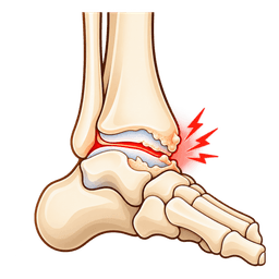
The ankle joint is lined with a thin layer of cartilage, a rubbery, slippery tissue that lets the bones glide. In ankle osteoarthritis that layer thins and roughens. The bone beneath it hardens, small spurs (osteophytes) grow at the edges and the lining of the joint can swell.
The result is a joint that is painful and tight. Because the ankle is small and carries your whole body weight on a surface about the size of a matchbox, even a modest loss of cartilage can be felt clearly.
Who is affected?
Most cases are post-traumatic. Studies suggest that a large majority of ankle arthritis follows a fracture, severe sprains or repeated instability, often 10 to 20 years before the symptoms start. Primary arthritis with no injury is less common than in the knee. Inflammatory disease such as rheumatoid arthritis, past infection and bleeding disorders account for the rest.
People tend to be younger than those who have knee or hip arthritis, which affects how surgery is chosen.
How serious is it?
The condition is not life-threatening, but it can limit walking, work and sport. It tends to progress slowly, though the pace varies between people. Many reach a point where shoes, a stick and medicines no longer give enough relief, and that is when surgery enters the conversation.
How this page is organised
We explain the anatomy, the stages and the causes first, then the tests and look-alike conditions. After that we cover non-surgical care, self-help and the three main operations. We close with treatment abroad, risks, prevention and outlook. For wider reading, visit the foot and ankle hub.
Anatomy
What happens in the body with ankle osteoarthritis
The ankle joint is a tight hinge between three bones, and it handles forces of several times your body weight with every step. Ankle osteoarthritis appears when the cartilage covering those bones wears out. A short tour of the anatomy shows why the joint behaves as it does and why treatment choices differ from the knee.
How is the healthy ankle built?
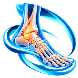
The talus, the dome-shaped ankle bone, sits inside a socket formed by the lower tibia (shin bone) and the fibula (outer leg bone). This socket is called the mortise. Cartilage about 1 to 2 mm thick covers the top of the talus and the underside of the tibia. That is thinner than in the knee, but it is stiffer and better suited to the high load.
Ligaments on both sides keep the talus centred, and tendons from the calf and the shin cross the joint to move the foot.
Which joints sit next to it?
Just below the talus is the subtalar joint, which allows the foot to tilt side to side. Beyond it lie the midfoot joints. The ankle moves mainly up and down, and the nearby joints supply the sideways motion. If the ankle is fused, these neighbours take over some of the work, which is why their health matters in surgical planning.
What changes with osteoarthritis?
Cartilage thins and cracks, and the joint space on X-ray narrows. Spurs form on the front of the tibia and talus, which block upward bending of the foot. The lining thickens and makes extra fluid. The joint may drift into varus (inward tilt) or valgus (outward tilt), because the bone surface wears unevenly.
Why does the ankle behave differently from the knee?
Ankle cartilage is better at resisting wear, so primary arthritis is uncommon. When injury changes the shape of the joint, though, the uneven loading speeds up the damage. That explains the strong link with old fractures and the gradual onset after a long symptom-free period.
Symptoms & causes
Ankle osteoarthritis symptoms and causes
Common symptoms
- Deep, aching pain at the front of the ankle that builds with walking, stairs or standing for long periods and eases with rest.
- Stiffness after sitting or first thing in the morning, usually settling within about 30 minutes as the joint warms up.
- Swelling around the joint that increases through the day and may feel puffy or tight over the front of the ankle.
- Reduced ability to pull the foot upward, so that squatting, climbing steps or walking up slopes feels blocked.
- Pain on uneven ground, grass or cobbles, where the ankle has to adapt quickly to changes in surface.
- A grinding, clicking or catching feeling when the ankle moves, which comes from rough cartilage and bone spurs.
- A limp, often with a shorter stride or a stiff, flat-footed gait that avoids bending the ankle.
- Visible bumps at the front of the joint, caused by bone spurs, which can rub on the shoe tongue.
- Night aching in more advanced disease, which may disturb sleep and respond poorly to simple painkillers.
- Tilting of the ankle inward or outward in late stages, with the hindfoot visibly out of line.
Causes and risk factors
- Previous ankle fracture, especially if the joint surface healed out of line or cartilage was damaged at the moment of injury.
- Repeated ankle sprains or chronic ankle instability, which let the talus rock and scuff the cartilage over many years.
- Ligament injuries and syndesmosis tears that left the bones slightly out of position, so load fell unevenly.
- Cartilage and bone injuries inside the joint, such as osteochondral lesions of the talus, that never healed completely.
- Inflammatory arthritis, including rheumatoid arthritis, gout and psoriatic arthritis, which attack the joint lining and cartilage.
- Previous infection of the joint, which can destroy cartilage quickly and leave scarring.
- Avascular necrosis of the talus, where poor blood supply causes the bone to collapse, often after a fracture.
- Hindfoot or foot alignment problems, including a high arch or a flatfoot, which tilt the joint and focus pressure on one side.
- Bleeding disorders such as haemophilia, which cause repeated bleeds into the joint and damage the cartilage.
- Primary osteoarthritis linked with age, body weight and genetics, which is less common in the ankle than elsewhere.
Types
Types and stages of ankle osteoarthritis
Doctors describe ankle osteoarthritis by its cause, its stage on X-ray and the alignment of the joint. These labels guide the choice between joint-preserving operations, fusion and replacement. The groups below are the ones surgeons use most.
Primary and secondary arthritis
- Post-traumatic: follows a fracture, ligament injury or instability. It is by far the commonest form.
- Inflammatory: part of a body-wide disease such as rheumatoid arthritis.
- Primary: no clear cause, linked with age and genetics.
- Infective or haemophilic: after joint infection or repeated bleeding.
How are ankle osteoarthritis stages described?
A simple way to understand ankle osteoarthritis stages is to look at the joint space, the spurs and the alignment on standing X-rays. Several grading scales exist, and the one below is a plain-language summary rather than a single official system.
| Stage | What X-rays show | Typical treatment direction |
|---|---|---|
| Early | Small spurs, joint space preserved | Exercise, footwear, injections, sometimes arthroscopy |
| Moderate | Partial space narrowing, more spurs | Bracing, injections, joint-preserving options |
| Advanced | Space largely gone, bone-on-bone contact | Fusion or replacement discussed |
| End-stage with deformity | Joint tilted, bone loss or cysts | Fusion, or replacement with correction |
Why does alignment matter?
When the joint tilts, one side carries most of the load. Surgeons may need to correct alignment with a bone cut above or below the ankle, or with ligament work, before a replacement can last. Marked tilt is one reason a fusion is chosen over a replacement.
Do the neighbouring joints matter?
Yes. Arthritis in the subtalar or midfoot joints influences which operation is safest, and it can explain pain that persists after surgery. CT scanning can show these joints in detail.
Diagnosis
How is ankle osteoarthritis diagnosed?
Ankle osteoarthritis is diagnosed by combining your history and examination with weight-bearing X-rays, and more advanced scans help when surgery is considered. The goal is to confirm that the joint is the source of pain, grade it and find any neighbouring problems. This is how the process normally goes.
What does the doctor ask?
The doctor will ask when the pain began, whether you ever broke or sprained the ankle, what makes it worse and how far you can walk. They want to know about night pain, swelling, medicines, smoking, diabetes and any inflammatory disease. A past injury 10 or 20 years ago is often the missing piece.
What does the examination include?
The doctor watches you walk and stand, looking at the heel alignment and the arch. They press along the joint line to find tenderness, check swelling, and move the ankle up and down, comparing it with the other side. They also test the subtalar joint and the stability of the ligaments, and they check pulses and skin.
Which imaging answers which question?
Standing X-rays from the front, side and a view of the hindfoot show joint space, spurs and tilt. They are the foundation. CT scans show bone cysts, spurs and neighbouring joint damage in detail. MRI shows cartilage lesions and tendon problems when the X-ray looks mild but pain is high. SPECT-CT or injection tests sometimes pinpoint the painful joint.
What should you bring to a remote review?
- Standing X-ray images, and any CT or MRI, on disc or by secure link.
- A timeline of injuries, operations and treatments, including injections and bracing.
- Photos of your standing feet from behind and the side.
- A list of medicines, allergies, smoking status and conditions such as diabetes.
- A short note on your daily activity, job and walking distance.
Tests you may have
- Weight-bearing X-rays of the ankle and foot, which show joint space narrowing, spurs, tilt and the general alignment of the leg.
- Hindfoot alignment view, a special X-ray that shows whether the heel sits in line with the leg or tilts to one side.
- CT scan, which shows bone cysts, spurs and the state of the joints below the ankle and is vital in surgical planning.
- MRI scan, which shows cartilage defects, bone bruising and tendon problems when X-rays do not explain the pain.
- Diagnostic injection of local anaesthetic into the joint, which helps confirm that the ankle is the main source of the pain.
- Blood tests for inflammation and rheumatoid markers, ordered when an inflammatory disease or infection is suspected.
- Gait and range-of-movement assessment, measuring how far the foot bends up and down compared with the other ankle.
Look-alikes
Conditions that can feel like ankle osteoarthritis
Pain around the ankle has many causes, and ankle osteoarthritis can look like several of them. Doctors rely on the history, the exam and imaging to tell them apart, because treatments differ widely. The table lists the most common look-alikes.
| Look-alike condition | How it differs | How doctors tell |
|---|---|---|
| Anterior ankle impingement | Pinching at the front when bending up, spurs but good joint space | X-ray shows spurs with preserved space; arthroscopy treats it |
| Subtalar arthritis | Pain below the ankle, worse on uneven ground | CT shows the subtalar joint; injection test |
| Chronic ankle instability | Repeated giving way; joint space normal | Drawer and tilt tests; MRI |
| Osteochondral lesion of the talus | Deep pain and catching from a localised cartilage defect | MRI shows a focal lesion |
| Gout or inflammatory arthritis | Hot, swollen joint, often several joints, flares and remissions | Blood tests, joint fluid analysis |
| Peroneal or tibialis tendon problems | Pain along tendons rather than in the joint | Ultrasound or MRI |
| Ankle fracture or stress fracture | Sudden onset or injury-related, bony tenderness | X-ray, CT or MRI |
Why does the correct label matter?
An impingement problem with good cartilage may need only keyhole surgery, whereas advanced arthritis needs a bigger decision. A person with gout needs medicines, not a fusion. Getting the label right can save you from the wrong operation.
Can two conditions coexist?
Often. A worn ankle can also have a tendon tear, and arthritis in the ankle may sit beside arthritis in the hindfoot. A careful review of your scans is worth the time before any procedure.
Non-surgical
Non-surgical treatment for ankle osteoarthritis
Non-surgical treatment is the first step for ankle osteoarthritis and gives real, lasting relief to many people. It will not regrow cartilage, but it can reduce pain, protect the joint and delay or avoid surgery. Here is the usual order doctors follow.
How do activity and weight changes help?
Every extra kilogram adds several kilograms of force across the ankle with each step. Even a modest loss of 5% to 10% of body weight can ease pain. Switching from running and jumping to cycling, swimming or rowing keeps you fit without pounding the joint. Pace your day with short rests.
Which footwear and braces work?
- Rocker-soled shoes: a curved sole lets you roll through the step with less ankle bend.
- Cushioned insoles and heel wedges: these soften impact and may shift load.
- Lace-up ankle brace: it limits painful motion and gives a feeling of support.
- Custom ankle-foot orthosis (AFO): a more rigid brace for severe pain or tilt.
- Walking stick: used in the opposite hand, it takes load off the sore ankle.
What does physiotherapy add?
A physiotherapist works on calf and shin strength, balance and gait. They may use gentle stretching to keep what movement you have, and they teach pacing. Programmes of 6 to 12 weeks are common, followed by a home routine.
Which medicines are used?
Paracetamol and topical anti-inflammatory gels are the usual first choices. Oral anti-inflammatory tablets can help flares if your doctor judges them safe for your stomach, kidneys and heart. Stronger painkillers have limited roles and carry risks, so they are used cautiously.
What about injections?
A corticosteroid injection can calm a flare for a few weeks to months, and it can also help confirm that the joint is the pain source. Repeated injections are limited because of possible cartilage and tendon effects. Hyaluronic acid and platelet-rich plasma injections have been studied, but evidence in the ankle is limited, so ask your clinician what is realistic.
How long should you try?
Most clinicians suggest at least 3 to 6 months of well-organised non-surgical care before surgery is advised, unless deformity is severe. Review your progress at regular intervals and keep a pain and walking diary.
Self-care
Exercises and self-care for ankle osteoarthritis
Self-care for ankle osteoarthritis means moving the joint as freely as it allows, protecting it from overload and pacing your day. Always check with your doctor or physiotherapist before starting or changing exercises, particularly if your pain is rising or you have had surgery.
Which gentle exercises can you do?
- Ankle pumps: point and flex the foot 15 to 20 times, 3 times a day.
- Alphabet tracing with the toes, to keep the joint moving in all directions.
- Seated calf stretch with a towel around the ball of the foot, holding for 30 seconds.
- Heel raises holding a counter, building from 10 to 20 repetitions.
- Stationary cycling or pool walking for 15 to 30 minutes.
How can you pace the day?
Break long walks into shorter stretches, sit during tasks that can be done sitting, and alternate heavy and light days. Plan rest before the pain peaks rather than after. Many people find that a 10-minute break every hour prevents the evening flare.
Which home measures ease a flare?
Rest, raise the foot above hip level and use a cool pack through a cloth for 15 minutes. Warm soaks may suit morning stiffness. Compression sleeves can reduce puffiness. Simple painkillers taken as advised help you keep moving.
What habits support the joint?
- Wear supportive shoes indoors as well as outdoors.
- Keep your weight in a healthy range.
- Stop smoking, which affects bone and wound healing before any surgery.
- Use a stick on bad days without feeling ashamed of it.
What should you avoid?
Avoid repeated jumping, long runs on hard surfaces and shoes with thin, flat soles. Do not push through sharp pain, and do not stop moving altogether, because stiffness makes the problem worse.
Check with your doctor or physiotherapist before starting new exercises, and stop any movement that causes sharp pain.
Treatment
Ankle osteoarthritis treatment options
Surgery for ankle osteoarthritis falls into three groups: keyhole cleaning of the joint, fusion and replacement. Each has a clear place. The right option depends on the stage, your age, activity and the state of nearby joints. Each linked procedure has its own detailed page.
When does ankle arthroscopy help?
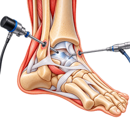
In ankle arthroscopy, the surgeon uses a camera and small tools through cuts of about 5 mm. It can remove spurs, scar tissue and loose fragments, and treat small cartilage defects. It works best when the joint space is still reasonable and pain comes from pinching rather than complete wear. It does not rebuild cartilage, so it suits early disease.
What is ankle fusion?
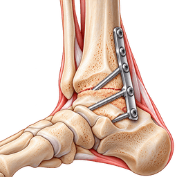
Ankle fusion (arthrodesis) removes the damaged cartilage and joins the tibia and talus with screws or a plate until they heal into one bone. It removes pain from the joint and is very durable. You lose up-and-down motion at the ankle, but the nearby joints compensate and most people walk well. It can be done open or arthroscopically.
What is total ankle replacement?
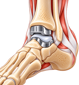
Total ankle replacement removes the worn surfaces and fits metal components with a plastic bearing between them. The aim is to keep movement and reduce pain. It suits selected patients with good bone, reasonable alignment, a stable joint and moderate activity demands. Revision or conversion to fusion may be needed in the long term.
Which joint-preserving options exist?
- Supramalleolar osteotomy: a bone cut above the ankle to shift load to healthier cartilage in tilted joints.
- Cartilage procedures: microfracture or grafts for localised defects in earlier stages.
- Distraction arthroplasty: a frame temporarily opens the joint in selected younger patients.
How do the main options compare?
| Option | Best suited to | Main trade-off |
|---|---|---|
| Ankle arthroscopy | Early disease, impingement, loose fragments | Does not repair cartilage; benefit may fade |
| Ankle fusion | Advanced disease, deformity, younger or heavy patients | Loss of ankle motion; neighbouring joints take strain |
| Total ankle replacement | Advanced disease with good bone and alignment | Implant wear; possible later revision |
| Osteotomy | Tilted joint with preserved cartilage | Longer bone healing |
What recovery follows?
After arthroscopy you often walk within days. After fusion or replacement you spend about 6 weeks in a splint and boot, with limited weight-bearing, and then rehabilitate over several months. See the rehabilitation guide.
When surgery is considered
Consider surgery for ankle osteoarthritis when pain, stiffness or deformity keep limiting daily life after a fair trial of non-surgical care. There is no fixed X-ray threshold. The person, not the picture, decides, and good timing balances relief against the limits of implants and fusions.
Which signs suggest the time has come?
- Pain that wakes you at night or limits walking to a short distance despite shoes, braces and medicines.
- Injections that now relieve pain for only a few weeks or not at all.
- Progressive tilt of the ankle or hindfoot.
- Loss of independence at work or at home, or falls caused by a stiff, painful ankle.
How do you choose between fusion and replacement?
Replacement tends to suit older, lighter, less active patients with good bone and alignment who value movement. Fusion tends to suit younger, heavier, more active patients, people with poor bone or marked deformity, and those with prior infection. Neither is a universal winner, and a good surgeon explains why one fits you.
Is early surgery ever better?
Waiting too long can let deformity and bone loss build up, which narrows your options. In younger patients with limited disease, joint-preserving surgery may be wise before cartilage is lost completely.
What should you ask your surgeon?
- Which option do you advise and what would you do if it were your own ankle?
- How many of these operations do you do each year?
- What happens if the result fails, and what is the plan B?
- How long will I be off my feet, and when can I drive and fly?
A free case review can give you a second opinion before you decide.
Procedures
Procedures that may treat ankle osteoarthritis
Costs
Ankle osteoarthritis treatment cost in Turkey
| Procedure | Turkey package | US self-pay | Saving |
|---|---|---|---|
| Total Ankle Replacement | $10,000 – $15,000 | — | — |
| Ankle Arthroscopy | $3,000 – $5,500 | $18,350 | ~77% |
| Ankle Fusion | $6,000 – $9,500 | $36,625 | ~79% |
Packages are contracted partner prices with validity dates; benchmarks are source-backed estimates. Open a cost guide for the full country comparison.
In Turkey
Treating ankle osteoarthritis in Turkey
Planned surgery for ankle osteoarthritis in turkey can be a sound option because the condition develops slowly and the decision can be made calmly. After you have tried non-surgical care and reviewed your scans with a surgical team, you can plan travel with a clear programme. Any acute injury still needs local care first.
When does treatment in turkey suit ankle osteoarthritis?
It can suit people who have long waits for surgery at home, want a second opinion on fusion versus replacement, or need revision of an earlier operation. It is less suitable if you have an active infection, poor skin over the ankle, uncontrolled diabetes or a medical condition that makes a long journey risky. Your own doctors should agree that travel is sensible.
What does the pathway look like?
- Remote review: you send X-rays, scans, history and photos through medical record review.
- Plan: the surgeon proposes arthroscopy, fusion or replacement, explains the reasoning and the alternatives.
- Admission: you arrive a day or two before surgery for examination, fresh imaging and anaesthetic checks.
- Surgery: operations usually take 1 to 3 hours, followed by a hospital stay of a few days.
- Follow-up: a written protocol and remote check-ins support rehabilitation at home.
What do people ask about ankle arthritis surgery in turkey?
The most common questions concern whether fusion or replacement is better, how many days to stay and how rehabilitation works after returning home. Ankle arthritis surgery in turkey follows the same principles as elsewhere: the right implant for the right patient, careful alignment and a structured recovery. The answers depend on your scans, so a review comes before any promise.
What should you send for total ankle replacement in turkey?
For total ankle replacement in turkey, the team needs standing X-rays, a CT scan if you have one, details of previous injuries or operations, your height and weight, and a medicine list. They also need photos of your standing alignment. Replacement is more sensitive to bone quality and alignment than other options, so complete records matter.
What about ankle fusion in turkey?
Ankle fusion in turkey is planned in a similar way. Surgeons consider bone quality, smoking, diabetes and the neighbouring joints, and they explain the 6 to 12 weeks of protected weight-bearing that fusion needs. Stopping smoking before surgery improves the chance of the bones joining.
How can you judge a hospital?
- Choose an accredited hospital with a foot and ankle service.
- Ask how many replacements, fusions and arthroscopies the lead surgeon performs.
- Ask which implant system is used and how long it has been in use.
- Confirm how complications, revisions and returns to the operating theatre are handled.
What about timing and practical travel?
Plan roughly 7 to 14 days in turkey after surgery, depending on the operation and wound healing. Read the flying after surgery guide, the companion guide and the follow-up page. Antalya and other cities offer foot and ankle services.
Where can you read more?
Visit total ankle replacement in turkey, ankle fusion in turkey and ankle arthroscopy in turkey. The cost guides for replacement, fusion and arthroscopy explain what influences price, and the orthopedics in turkey hub gives context.
Complications
Complications of ankle osteoarthritis
Ankle osteoarthritis tends to worsen slowly, and every treatment carries some risk. Knowing both sides helps you decide when to act. Most people do well, but you should understand what can go wrong and how teams reduce the odds.
What happens if it is left untreated?
- Pain and stiffness often increase, and walking distance shortens.
- The joint may tilt, which strains the knee, hip and back through an altered gait.
- Bone loss and cysts can develop, which makes later surgery harder.
- Neighbouring joints in the hindfoot can wear faster.
- Activity falls, with weight gain and loss of fitness as knock-on effects.
What are the risks of arthroscopy?
Risks include nerve irritation at the front of the ankle, wound problems and stiffness. Relief may fade if the arthritis continues to progress.
What are the risks of fusion?
- Nonunion, meaning the bones fail to join, which is more likely in smokers and people with diabetes.
- Arthritis in the neighbouring joints after many years.
- Wound problems, infection and irritation from hardware.
- Reduced motion and a stiffer gait, particularly on stairs and slopes.
What are the risks of replacement?
- Loosening or wear of the implant over time, which can mean revision surgery.
- Wound breakdown or infection, which can be serious.
- Fracture around the ankle during or after the operation.
- Persistent pain or stiffness, and a need for conversion to fusion in some cases.
What risks do all operations share?
Blood clots in the leg, nerve injury, anaesthetic complications and persistent swelling can occur. Your team will assess your personal risk and take preventive steps.
Urgent care
When to seek urgent care for ankle osteoarthritis
- Sudden severe pain with fever, redness and a hot, swollen ankle: seek urgent medical assessment the same day, as this could be infection or an acute inflammatory flare.
- A swollen, tender calf or chest pain and breathlessness after surgery or a flight: call emergency services, since a blood clot is possible.
- Numb, cold, pale or blue toes at any time: get emergency help, because blood supply may be reduced.
- After an operation, fever, wound leakage, spreading redness or an opening wound: contact your surgeon the same day.
- A sudden fall or injury with inability to bear weight: go to emergency, as a fracture can occur on top of arthritis.
- Rapidly increasing tilt of the foot or a new loud crack and pain after replacement: arrange urgent review, as an implant problem may be present.
Prevention
How to lower your risk of ankle osteoarthritis
Ankle osteoarthritis cannot always be prevented, but you can lower the risk and slow its progress. Because most cases follow injury, the best strategy is to treat ankle injuries thoroughly and to keep the joint strong, aligned and not overloaded.
How can you protect the joint after injury?
Have a significant ankle injury assessed, especially if you cannot walk 4 steps. Complete rehabilitation after sprains and fractures, including balance training, and treat chronic ankle instability early so repeated rolling does not scuff the cartilage. Realign displaced fractures accurately.
What lifestyle steps help?
- Keep your weight in a healthy range to reduce load through the joint.
- Stay active with low-impact exercise such as cycling and swimming.
- Wear supportive, well-fitting shoes and avoid high heels for long periods.
- Do not smoke, which harms bone and joint health.
Why does early action matter?
Worn ankle cartilage cannot be rebuilt, so the aim is to stop the damage before it starts. A fracture that heals within 1 to 2 mm of its normal position, and a sprain that gets full rehabilitation, both give the cartilage a much better chance of lasting a lifetime.
Can medicines prevent it?
No medicine reliably prevents osteoarthritis. People with inflammatory disease such as rheumatoid arthritis or gout benefit from disease-controlling treatment, which can protect the joint. Treat infection promptly.
What cannot be prevented?
You cannot change genetics, the shape of your foot or an injury that has already happened. Prevention lowers the chance and the pace of progression but never removes them. Honest expectations help you plan.
How does it connect to other injuries?
A heel cord problem such as an Achilles tendon rupture can alter gait and loading for months, so complete rehabilitation protects the ankle joint as well.
Outlook
Living with ankle osteoarthritis: outlook and recovery
Most people with ankle osteoarthritis keep walking and working for many years, and surgery gives good relief for those who need it. Expect a slowly changing condition, steady self-management and a clear decision point if symptoms outgrow simple measures.
What is the natural course?
Ankle osteoarthritis tends to progress over years rather than months, with good periods and flares. Some people stay stable for a decade. Early treatment, weight control and appropriate bracing help. Surgery is not inevitable.
What results can you expect from surgery?
Studies suggest that both fusion and replacement relieve pain substantially in most patients, with high satisfaction in selected groups. Fusion gives a durable result, while replacement keeps motion but carries a risk of later revision. Arthroscopy offers shorter, variable relief in early disease.
What is a typical recovery timeline after fusion or replacement?
| Stage | Typical timing | Main aims |
|---|---|---|
| Protection | 0 to 2 weeks | Splint, elevation, wound healing |
| Boot, limited weight-bearing | 2 to 6 weeks | Protect the fusion or implant |
| Progressive loading | 6 to 12 weeks | Walking in a boot, then shoes |
| Strengthening | 3 to 6 months | Calf strength, balance, stairs |
| Full recovery | 6 to 12 months | Swelling settles, maximum comfort |
When can you drive and return to work?
Desk work may resume after 2 to 6 weeks with the leg raised. Driving after a right-sided operation usually waits for about 6 to 8 weeks. Active jobs can take 3 to 6 months.
What activities suit you afterwards?
Walking, cycling, swimming and golf are usually fine. Running and jumping may be discouraged after replacement but are possible for some after fusion. Ask your surgeon for personal guidance and keep a long-term routine of calf strength and weight control.
Surgeons
Specialists who treat ankle osteoarthritis
FAQ
Ankle osteoarthritis: frequently asked questions
What are the first signs of ankle osteoarthritis?
What causes ankle osteoarthritis?
Can ankle osteoarthritis be reversed?
What are the best treatments for ankle arthritis pain?
Is walking good for ankle osteoarthritis?
How do doctors stage ankle osteoarthritis?
Which is better, ankle fusion or ankle replacement?
How long does recovery take after ankle replacement or fusion?
Can ankle arthroscopy cure arthritis?
Is ankle osteoarthritis treatment in turkey safe?
How long should I stay in turkey after ankle fusion or replacement?
Will I need another operation later?
Sources
Sources for this ankle osteoarthritis guide
Peer-reviewed guidance and institutional sources used to write and review this page.
- 01Ankle Arthritis
American Academy of Orthopaedic Surgeons (OrthoInfo), 2022
https://orthoinfo.aaos.org/en/diseases--conditions/arthritis-of-the-foot-and-ankle/
- 02
- 03Osteoarthritis
Mayo Clinic, 2023
https://www.mayoclinic.org/diseases-conditions/osteoarthritis/symptoms-causes/syc-20351925
- 04
- 05Osteoarthritis
National Institute of Arthritis and Musculoskeletal and Skin Diseases, 2023
https://www.niams.nih.gov/health-topics/osteoarthritis
- 06Foot and Ankle Conditions
American Orthopaedic Foot and Ankle Society (AOFAS), 2023
https://www.aofas.org/