








Key takeaways
- 1An ankle fracture is a break in the tibia, the fibula or the talus, and it is among the most common fractures treated by orthopedic teams.
- 2The key question is stability: if the ankle joint stays centred under the shin, a boot may be enough, but if it shifts, surgery is usually advised.
- 3Any suspected ankle fracture needs urgent local care, with the leg raised and an X-ray, before any thought of travel.
- 4Typical ankle fracture recovery time is about 6 weeks for bone healing, 3 months for walking comfortably and 6 to 12 months for full strength.
- 5Surgery uses plates, screws or a combination to restore the shape of the joint, and good alignment lowers the chance of later arthritis.
- 6Smoking, diabetes and poor skin condition raise the risk of wound problems and slow healing after an operation.
- 7Planned reconstruction or follow-up care in turkey is only considered once the swelling has settled, the patient is stable and the local team agrees that travel is safe.
Overview
What is ankle fracture?
An ankle fracture is a break in the bones that make up the ankle joint, and it is one of the most common reasons people attend an emergency department after a twist or a fall. If you think your ankle is broken, urgent local care comes first. This page explains how these breaks are classed, treated and rehabilitated, and where treatment in turkey fits.
What is an ankle fracture?
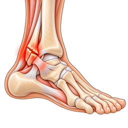
The ankle joint is formed by three bones. The tibia is the main shin bone, the fibula is the slim bone on the outside of the leg, and the talus is the small bone that sits between them and the heel. A fracture can involve one, two or all three, and the ligaments that hold the joint can tear at the same time.
Many people say "broken ankle" for any of these injuries. Doctors are more specific, because the exact pattern decides whether the joint is stable.
Who gets an ankle fracture?
Ankle fractures occur at all ages. Younger adults tend to break the ankle in sport or high-energy accidents, such as a fall from a ladder or a car crash. Older adults, especially women after the menopause, can fracture the ankle from a simple trip because their bones are thinner.
Children can also be affected, but their growth plates make the injury and its treatment different. This page focuses on adults.
How serious is this injury?
Most ankle fractures heal well when the joint is brought back into correct position and protected. The danger lies in a joint that heals out of line, because uneven loading can wear the cartilage and lead to arthritis years later. Open wounds, skin damage and blood vessel injury turn a fracture into an emergency.
How this page is organised
We start with anatomy, then explain the types and how doctors classify them. Next come diagnosis, look-alikes, treatment without and with surgery, recovery and the questions most patients ask. You can see the wider foot and ankle area for related topics.
Anatomy
What happens in the body with ankle fracture
The ankle is a hinge joint in which the talus fits snugly into a socket shaped by the tibia and fibula, so any break changes the fit of that socket. Seeing how the pieces lock together explains why even a small shift of the fibula can matter.
What is the normal structure of the ankle?
The bottom end of the tibia has two bony bumps called malleoli. The medial malleolus is the bump on the inside of the ankle, and the posterior malleolus is a rim at the back of the tibia. The lateral malleolus is the lower end of the fibula, on the outside. Together they form a mortise, like a wooden joint, around the top of the talus.
Strong ligaments tie the bones together. The deltoid ligament runs on the inside, the lateral ligaments run on the outside, and the syndesmosis is a set of ligaments that binds the tibia to the fibula just above the joint.
What changes when the ankle breaks?
When a bone breaks, the mortise can open up. If the fibula shortens or rotates, or if the deltoid or syndesmotic ligaments tear, the talus can slide sideways. A shift of only 1 to 2 mm has been shown to reduce the contact area in the joint substantially, which is why surgeons aim for a precise reduction.
Why do the soft tissues matter?
The skin over the inner and outer ankle is thin, with little padding. Swelling after a break can blister the skin and delay surgery. Nerves and tendons run close to the bones, so surgeons plan their incisions carefully.
Symptoms & causes
Ankle fracture symptoms and causes
Common symptoms
- Severe pain around the ankle immediately after a twist, roll or fall, usually worse when you try to move or put any weight on the foot.
- Rapid swelling that can extend from the ankle up the shin and into the foot within a few hours of injury.
- Bruising on the inside, outside or sole of the foot that develops over 24 to 48 hours and may spread towards the toes.
- Tenderness when pressing on the bony bumps on either side of the ankle or along the lower part of the fibula.
- A visibly crooked or displaced ankle, where the foot sits at an odd angle to the leg, which needs emergency help.
- Inability to put weight on the foot, or only able to take a few painful steps, which is a key sign doctors look for.
- A grinding or crunching feeling when the ankle moves, caused by bone ends rubbing together.
- Numbness, tingling or a cold, pale foot, which may suggest pressure on a nerve or blood vessel and needs urgent review.
- Blisters over the swollen skin, which can appear after 1 to 3 days and may affect when surgery is possible.
Causes and risk factors
- Rolling or twisting the ankle during sport, particularly football, basketball and skiing, where the foot is planted while the body keeps turning.
- Falls from standing height, including trips on stairs, kerbs or uneven ground, especially in older adults with thinner bones.
- High-energy trauma such as road traffic collisions or falls from a height, which can produce complex, multi-part breaks.
- Low bone density (osteoporosis), which makes fractures more likely after a minor stumble and slower to fix securely.
- Previous ankle injuries or weak ligaments, which can allow awkward movements that overload the bones.
- Smoking and diabetes, which do not usually cause the break but are strongly linked with delayed bone healing and wound problems.
- Wearing high heels or unstable shoes on rough surfaces, which increases the chance of a sudden inward or outward roll.
- Poor balance, vision or muscle strength, which raises the risk of falls at older ages.
- Stress from repetitive impact, such as long-distance running, which can sometimes cause a hairline stress fracture of the fibula or tibia.
Types
Types and stages of ankle fracture
Ankle fracture classification groups breaks by which bones are involved, where the fibula breaks and whether the ligaments are torn. These labels guide the choice between a boot and an operation, so it helps to know the main ones.
Which bones are broken?
- Lateral malleolus fracture: the most common single break, in the fibula on the outer ankle.
- Medial malleolus fracture: a break of the inner bump, often with a pulled deltoid ligament.
- Posterior malleolus fracture: a break of the back rim of the tibia, which can involve a large share of the joint surface.
- Bimalleolar fracture: two malleoli are broken, so the joint is usually unstable.
- Trimalleolar fracture: all three are broken, which is more complex and nearly always treated with surgery.
- Talus and pilon fractures: high-energy injuries to the ankle bone or the weight-bearing end of the tibia.
How does the Weber classification work?
The Weber system looks at the level of the fibula break relative to the syndesmosis. It is simple and widely used, and it gives a quick sense of how likely the ankle is to be unstable.
| Weber type | Level of fibula fracture | Usual stability |
|---|---|---|
| A | Below the syndesmosis | Often stable, may need only a boot |
| B | At the level of the syndesmosis | Stable or unstable, depends on the deltoid ligament |
| C | Above the syndesmosis | Syndesmosis torn, usually unstable, often needs surgery |
Open versus closed and displaced versus undisplaced
In a closed fracture the skin is intact, while an open fracture has a wound connecting to the bone and carries a high infection risk. An undisplaced fracture keeps its normal alignment, while a displaced one has moved. Displacement tends to push the decision toward surgery.
What is a stress or avulsion fracture?
A stress fracture is a hairline crack from repeated load, and an avulsion fracture is a small piece pulled off by a ligament. Both are usually minor but should still be checked.
Diagnosis
How is ankle fracture diagnosed?
Ankle fracture diagnosis rests on a careful history and examination followed by X-rays, and these quickly show whether a bone is broken and whether the joint is stable. Prompt assessment protects the skin and nerves and lets treatment start early. Here is how it usually unfolds.
What does the doctor ask and examine?
The doctor asks how you fell, which way the foot turned and whether you could stand afterwards. They look for deformity, open wounds, blisters and the colour and warmth of the foot. They feel for pulses and test sensation in the toes.
Tenderness over the bone, rather than only over the ligaments, raises concern for a fracture. The knee and the foot are also checked, since the fibula can break near the knee.
Which rules help decide if an X-ray is needed?
The Ottawa ankle rules are a widely used checklist. An X-ray is advised if there is pain near the ankle and either bony tenderness at the back or tip of the inner or outer ankle, or the person cannot take 4 steps both at the scene and in the clinic.
Which imaging is used?
Standard X-rays from three angles (front, side and a mortise view) are the first step. Stress views or weight-bearing X-rays can show whether the joint opens. A CT scan gives a detailed 3D picture for complex or posterior malleolus breaks, and MRI is used when ligament or cartilage injury is suspected.
What should you bring to a remote review?
- Your X-ray and CT images on disc or by secure link, not only the written report.
- The date of injury and the date of any surgery, with the operation note and implant sticker if available.
- Photos of the ankle showing swelling, skin condition and any wounds.
- Your medicines, allergies, smoking status and conditions such as diabetes or osteoporosis.
Tests you may have
- Plain X-rays in three views, which show the fracture lines, the position of the bone fragments and whether the ankle joint space is even.
- Weight-bearing or stress X-rays, which test whether the talus drifts sideways when load or pressure is applied to the ankle.
- Computed tomography (CT), which builds a 3D picture of the joint surface and is important for posterior malleolus and pilon fractures.
- MRI scan, used less often, to look for ligament tears, cartilage damage or a hidden stress fracture that X-rays miss.
- Ottawa ankle rules, a bedside checklist that helps decide who needs an X-ray after an ankle injury.
- Neurovascular examination, checking pulses, skin colour and toe sensation to exclude nerve or artery injury.
- Bone health assessment, for example blood tests or a bone density scan in older adults with a low-energy break.
Look-alikes
Conditions that can feel like ankle fracture
Not every painful, swollen ankle is a fracture, and not every fracture looks dramatic. Doctors separate a break from a sprain and other injuries using the exam and imaging, and the table below summarises the common look-alikes. If you cannot bear weight, assume the bone may be broken until proven otherwise.
| Look-alike condition | How it differs | How doctors tell |
|---|---|---|
| Ankle sprain | Ligament stretch or tear without bone injury; walking is often possible | No bony tenderness; normal X-ray |
| High (syndesmotic) ankle sprain | Pain above the joint, worse with twisting; no break visible | Squeeze and rotation tests; stress X-ray or MRI |
| Achilles tendon rupture | Snap at the heel cord, weak push-off, pain behind the ankle | Calf squeeze test; ultrasound |
| Talus or calcaneus fracture | Pain deeper in the hindfoot, often after a fall from height | CT scan shows the bone injury |
| Fifth metatarsal fracture | Pain on the outer edge of the midfoot rather than the ankle | Foot X-ray |
| Stress fracture | Gradual pain with activity, no single injury | MRI or repeat X-ray after 2 weeks |
| Chronic ankle instability | Repeated giving way over months, no acute break | History plus stress testing |
Why is a sprain sometimes missed as a fracture?
Some small avulsion fractures and fibula breaks produce modest swelling, and people walk on them. A persistent pain beyond 1 to 2 weeks after a "sprain" deserves a repeat assessment, particularly if the ankle feels unstable.
Can a break hide another injury?
Yes. A fibula break near the knee, called a Maisonneuve fracture, can come with a torn syndesmosis, and the ankle X-ray may look nearly normal. Doctors press along the full length of the fibula for this reason.
Non-surgical
Non-surgical treatment for ankle fracture
Non-surgical treatment works when the ankle joint is stable and the bones are lined up, and it avoids the risks of an operation. The principle is to protect the bone while it knits, then to restore movement and strength. This is how care normally runs.
What happens at the start?
The leg is placed in a splint with the ankle at 90 degrees, raised above heart level, and iced through a cloth. Pain relief usually combines paracetamol with stronger medicines if needed, according to your doctor. Swelling is monitored closely in the first 48 hours.
Who is suited to a cast or boot?
- Isolated, undisplaced fibula fractures below the syndesmosis (Weber A).
- Stable Weber B fractures where stress X-rays show the joint holds its position.
- Patients whose health or skin makes surgery risky, if the joint is still acceptably aligned.
How long do you stay protected?
A cast or walking boot is usually worn for about 6 weeks, with weight-bearing allowed earlier or later depending on stability. Repeat X-rays at 1 to 2 weeks check that nothing has moved. Healing is judged from the pain, the exam and the X-ray, not only from the calendar.
What else belongs in the plan?
- Blood-clot prevention, which may include injections or tablets while the leg is immobilised.
- Vitamin D and calcium review in older adults, with treatment of osteoporosis if present.
- Stopping smoking, which improves the chance of union.
- Early physiotherapy to maintain hip, knee and toe movement.
What does the evidence suggest?
Studies of carefully selected stable fractures show good results with a boot, similar to surgery for those cases. For unstable patterns, trials generally favour surgical fixation to hold the joint in place. The challenge is deciding which group a given fracture belongs to, which is why imaging and an expert opinion are valuable.
Self-care
Exercises and self-care for ankle fracture
Self-care after this injury means protecting the break, keeping swelling down and keeping everything else moving. Always check with your doctor or physiotherapist first, because limits on weight-bearing and movement differ widely between a stable boot-treated break and a surgically fixed ankle.
How do you control swelling?
Raise the foot above hip level for 20 to 30 minutes several times a day, especially in the first 2 weeks. Use ice through a towel, never directly on skin or inside a cast unless told. Notice whether a cast feels suddenly tight and report it.
How can you stay mobile on crutches?
- Practise with crutches, a knee scooter or a walker before you leave the clinic.
- Remove loose rugs and clear a path at home to prevent a second fall.
- Use a shower stool and a waterproof cover for the cast.
- Ask for help with carrying items, as both hands will often be needed for crutches.
Which exercises are safe early on?
While protected, you can usually do toe wiggles, knee extensions, straight leg raises and gentle hip exercises. These maintain blood flow and muscle bulk. Do not rotate the ankle or push the foot on the ground until the team says it is allowed.
How does movement resume?
Once the boot or cast is off, typical early work includes ankle pumps, alphabet tracing with the toes, towel stretches, and seated heel raises. Balance exercises on a stable surface follow, then calf raises and walking drills. The goal is to regain 90% or more of the other ankle's range over 8 to 12 weeks.
What should you avoid?
- Putting weight on the leg before you are cleared, which can displace the fracture.
- Smoking and vaping, which slow bone healing.
- Driving until you can brake safely and your doctor agrees.
- Pushing through sharp pain or increasing swelling.
Check with your doctor or physiotherapist before starting new exercises, and stop any movement that causes sharp pain.
Treatment
Ankle fracture treatment options
Surgery restores the shape of the joint using metal implants, and it is the usual route when the break is displaced or the ankle is unstable. The operation is described in more detail on the foot and ankle fracture surgery page.
What does fixation involve?
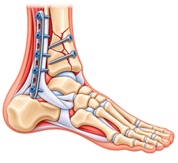
Under spinal or general anaesthesia and usually with a thigh tourniquet, the surgeon opens the fracture, lines the pieces up and holds them with plates and screws. The fibula is commonly fixed with a plate on its outer face, and the inner malleolus with screws or a plate. Operations often take 1 to 2 hours.
Which techniques are used for complex patterns?
- Syndesmosis fixation: a screw or suture-button device holds the tibia and fibula together while the ligament heals.
- Posterior malleolus fixation: a screw or plate restores the back rim when a large fragment is involved.
- External fixator: a frame outside the skin stabilises the leg when swelling or wounds delay definitive surgery.
- Primary fusion or replacement: rarely, for severely damaged joints in older adults.
What does a surgeon check before the operation?
Before any fixation the team reviews the skin, the pulses, your blood tests and the anaesthetic risk. They look at the CT to plan where each screw will sit. Many surgeons also ask about your home layout, because you will be non-weight-bearing for about 6 weeks and need to manage stairs, toilets and showers safely.
This planning step is also the best time to raise concerns. Ask what happens if the bone is softer than expected, and whether a different implant would then be used.
When is surgery delayed?
If the skin is blistered or swollen, the surgeon may wait 5 to 10 days, keeping the ankle splinted and elevated until the "wrinkle sign" returns. Operating too early on tight skin risks wound breakdown, so timing is a safety decision rather than a delay in care.
How do the options compare?
| Option | Best suited to | Main trade-off |
|---|---|---|
| Cast or boot | Stable, aligned fractures | Needs close X-ray follow-up in case of shift |
| Plate and screw fixation | Displaced or unstable fractures | Wound risk, possible hardware irritation |
| Syndesmosis fixation | Torn tibia-fibula ligaments | May need later screw removal |
| External fixator | Open or swollen injuries | Pin-site care, second operation |
What does ankle fracture surgery recovery look like?
After fixation, you usually spend about 2 weeks in a splint, then a boot, with protected weight-bearing from around 6 weeks depending on the fracture. Ankle fracture surgery recovery then builds with physiotherapy over several months. See the rehabilitation guide for a general outline.
When surgery is considered
You should consider surgery for an ankle fracture when the joint is displaced or unstable, because that is when a boot is least likely to give a good, even joint. The decision rests on imaging and examination, and it is best made by a foot and ankle or trauma surgeon.
Which findings point to surgery?
- The talus has shifted in the mortise, or the joint space is uneven on X-ray.
- Two or more malleoli are broken (bimalleolar or trimalleolar pattern).
- The syndesmosis is torn or the fibula is shortened or rotated.
- A large posterior malleolus fragment involves roughly a quarter or more of the joint surface.
- The fracture is open, or a cast fails to hold the position.
When is a boot reasonable?
When the fracture is isolated, aligned and proven stable, a boot often gives results as good as surgery. Older patients with fragile skin or poor circulation may also do better without an incision, accepting a small change in alignment.
What questions should you ask?
- Is my ankle stable, and how do you know?
- What implants will be used, and might they need removal?
- When can I put weight on the leg and when can I drive or fly?
- What are the main risks given my age, smoking status and health?
What if the fracture healed badly?
A malunion or nonunion can sometimes be corrected with a further operation. If the joint has already worn, options such as arthroscopy or fusion may come into play, as explained in the ankle osteoarthritis guide.
Procedures
Procedures that may treat ankle fracture
Costs
Ankle fracture treatment cost in Turkey
| Procedure | Turkey package | US self-pay | Saving |
|---|---|---|---|
| Foot & Ankle Fracture Surgery | $4,000 – $8,000 | $28,775 | ~79% |
Packages are contracted partner prices with validity dates; benchmarks are source-backed estimates. Open a cost guide for the full country comparison.
In Turkey
Treating ankle fracture in Turkey
Ankle fracture treatment in turkey can be considered for planned or follow-on care, but never in place of urgent local treatment. A fresh break needs an X-ray, a splint and often early surgery near where it happened. Travel is considered only once you are medically stable and your treating team agrees.
When can treatment in turkey suit an ankle fracture?
It can suit patients whose fracture has been stabilised locally and whose surgeon agrees that a delayed operation is acceptable, for example after swelling has settled. It can also suit people who need corrective surgery for a poorly healed break, or an implant revision. It does not suit anyone with an open wound, a suspected clot, uncontrolled infection or a leg that cannot be safely protected during a flight.
What does the pathway look like?
- Review: you send your imaging, operative notes and history using our medical record review service.
- Plan: a surgeon explains whether fixation, revision or conservative care fits, and how long you would stay.
- Admission: you arrive, are re-examined and have fresh imaging and fitness checks.
- Surgery: often 1 to 2 hours, followed by a short hospital stay.
- Follow-up: wound checks, a rehabilitation plan and remote review after you return home.
What should you send?
Prepare X-ray and CT images, the injury date, any treatment already given, photos of the skin, a medicine list and your smoking status. Clear records let the team judge urgency and plan implants properly.
How can you judge a hospital?
- Check recognised accreditation and an on-site trauma or foot and ankle service.
- Ask how many fixations of this kind the lead surgeon performs each year.
- Confirm the plan for wound problems, infection and revision surgery.
- Ask for a written discharge summary with implant details.
What about travel timing?
Most people plan to stay about 7 to 14 days after ankle fracture surgery in turkey so that wound checks and flight advice are completed. Read the flying after surgery guide and the companion guide before booking. Large centres in Ankara and other cities handle trauma regularly.
What do people often ask about broken ankle treatment in turkey?
Broken ankle treatment in turkey is usually discussed by people who were treated in a cast at home and later learned that the joint had shifted, or by those who want a second surgical opinion. The first question is always timing: bone that has already started to knit after 4 to 6 weeks is harder to realign. Send your scans early so the team can tell you honestly whether an operation is still worthwhile.
What are the limits of planned care?
Planned care cannot fix every problem. If the cartilage has already worn badly, realigning the bone may not relieve pain, and a different procedure could be better. A good team will say so plainly, even if that means advising you not to travel. Expect a candid discussion of benefits, limits and the realistic share of your recovery that will happen at home with local physiotherapy.
Where can you read more?
See the foot and ankle fracture surgery in turkey page, the cost guide and the orthopedics in turkey hub. A free case review will tell you whether travel is wise for your situation.
Complications
Complications of ankle fracture
Complications of this injury arise from the injury, from healing problems and from treatment. Most people recover well, but a minority face stiffness, arthritis or wound trouble, so it is helpful to know the possibilities and how they are reduced.
What can go wrong with the fracture itself?
- Malunion, where the bone heals out of line, which can unbalance the joint.
- Nonunion, where the bone fails to knit, more likely in smokers and people with diabetes.
- Post-traumatic arthritis, because cartilage injury at the time of the break may become visible years later.
- Persistent swelling and stiffness, which can last for 6 to 12 months.
What are the surgical risks?
- Wound infection or breakdown, particularly with smoking, diabetes or swollen skin.
- Irritation from plates or screws, sometimes needing removal after healing.
- Nerve irritation, giving numbness over the foot or ankle.
- Loss of fixation or a syndesmosis that loosens.
How likely is a blood clot?
Any leg that is immobilised has a higher risk of deep vein thrombosis, and a clot can travel to the lungs. Doctors assess your personal risk and may offer injections or tablets. Early movement of toes and knee, plus hydration, helps.
What is complex regional pain syndrome?
This rare condition causes pain out of proportion to the injury, swelling, colour change and sensitivity. Early mobilisation and good pain control reduce the chance, and specialist pain care helps if it develops.
How can you lower your risk?
Stop smoking, control blood sugar, follow weight-bearing orders, and attend every X-ray check. Report fever, wound leakage, severe pain or a toe that turns cold or pale.
Urgent care
When to seek urgent care for ankle fracture
- Severe, increasing pain, a tight cast or a foot that feels numb, cold, pale or blue: seek emergency care at once, as this can signal compartment syndrome or a blocked blood vessel.
- An open wound with bone visible or bleeding near the fracture: cover with a clean cloth and go to emergency now, because infection risk is high.
- Chest pain, breathlessness or coughing blood during recovery: call emergency services immediately for a possible pulmonary embolism.
- Fever, spreading redness, pus or wound opening after an operation: contact your surgeon the same day for an infection check.
- A swollen, warm, tender calf on the injured leg: arrange urgent assessment for a deep vein thrombosis.
- A visibly crooked ankle after injury: do not walk on it; call an ambulance or go to the emergency department.
Prevention
How to lower your risk of ankle fracture
You cannot prevent every break, but you can lower the odds by improving bone health, balance and footwear, and by treating ankle injuries properly. Prevention targets both the fall and the fragility that makes a fall break the bone.
How can you protect your bones?
Get enough calcium and vitamin D, do weight-bearing exercise such as walking, and avoid smoking and heavy alcohol use. Adults over about 50 years, especially women, should ask about a bone density scan if they have risk factors. Osteoporosis treatment can lower the chance of future fractures.
How can you improve balance?
- Practise single-leg standing for 30 seconds on each side, near a counter for safety.
- Try tai chi or a balance class, which are linked with fewer falls in older people.
- Strengthen the calves, hips and thighs with regular exercise.
- Have your eyesight and medicines reviewed if you feel unsteady.
What helps in sport?
Warm up before play, wear supportive shoes, and consider ankle braces or taping if you have a history of sprains. Rehabilitating a sprain fully, with balance training, lowers the chance of a repeat injury that could end in a fracture.
How can you make your home safer?
Fix loose carpets, add rails to stairs, improve lighting, and wear shoes with grip. Take care on wet or icy surfaces and avoid rushing down steps.
What cannot be prevented?
High-energy accidents and freak falls happen to healthy people. If you have had a break, good care of the injured ankle, including treatment of any chronic ankle instability, protects the joint long term.
Outlook
Living with ankle fracture: outlook and recovery
Most people return to normal daily life, though the full process takes longer than many expect. Ankle fracture recovery time is about 6 weeks for the bone to heal, 3 months for comfortable walking and 6 to 12 months for strength and swelling to settle fully.
What is a typical recovery timeline?
| Stage | Typical timing | Focus |
|---|---|---|
| Protection | 0 to 2 weeks | Splint, elevation, wound healing, crutches |
| Boot or cast | 2 to 6 weeks | Protected or partial weight-bearing as directed |
| Early rehabilitation | 6 to 12 weeks | Weight-bearing, movement, gait retraining |
| Strengthening | 3 to 6 months | Calf strength, balance, stairs |
| Full recovery | 6 to 12 months | Sport, impact, swelling resolves |
When can you return to work and driving?
Desk work can often resume after 2 to 4 weeks with the leg raised. Driving a left-sided injury in an automatic car may be possible sooner, while a right-sided break usually requires waiting until about 8 to 12 weeks. Manual jobs may take 3 to 6 months. Your surgeon will confirm.
Will the ankle ever be normal again?
Many people feel almost normal, but some notice stiffness, aching in cold weather or swelling at the end of the day. Hardware can be removed if it irritates. A minority develop arthritis in the longer term, particularly after complex breaks or a poor joint alignment.
What about future risk?
A previous break slightly raises the chance of arthritis later in life. Keep the muscles strong, maintain a healthy weight and report persistent pain early. If arthritis develops, treatments are available, including replacement or fusion. Plan your follow-up after returning home before you travel for any surgery.
Surgeons
Specialists who treat ankle fracture
FAQ
Ankle fracture: frequently asked questions
What are the main ankle fracture symptoms?
How do I know if my ankle is sprained or broken?
How long does a broken ankle take to heal?
Does a broken ankle always need surgery?
Can I walk on a broken ankle?
What is ankle fracture surgery recovery like?
Will I need the plate and screws removed?
Can a broken ankle cause arthritis?
Is ankle fracture treatment in turkey safe?
When can I fly after ankle fracture surgery?
Can I have ankle fracture surgery in turkey if my fracture was treated badly elsewhere?
How should I sleep with a broken ankle?
Sources
Sources for this ankle fracture guide
Peer-reviewed guidance and institutional sources used to write and review this page.
- 01Ankle Fractures (Broken Ankle)
American Academy of Orthopaedic Surgeons (OrthoInfo), 2022
https://orthoinfo.aaos.org/en/diseases--conditions/ankle-fractures-broken-ankle/
- 02
- 03Broken ankle
Mayo Clinic, 2023
https://www.mayoclinic.org/diseases-conditions/broken-ankle/symptoms-causes/syc-20450025
- 04
- 05
- 06Foot and Ankle Conditions
American Orthopaedic Foot and Ankle Society (AOFAS), 2023
https://www.aofas.org/