

Key takeaways
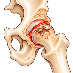
- 1This condition means bone cells in the femoral head, the ball of the joint, die because blood flow has been interrupted or reduced.
- 2The first symptom is often a deep groin ache that can arrive over days or weeks, and it commonly hurts at night or when you put weight on the leg.
- 3Common triggers include long-term steroid use, heavy alcohol intake, a hip fracture or dislocation, sickle cell disease and some other medical conditions, but about a quarter of cases have no clear cause.
- 4MRI is the most sensitive test and can find bone death in the hip before a normal X-ray shows anything.
- 5Small, early lesions may be managed with protected weight-bearing or joint-preserving surgery, while collapsed bone usually needs hip replacement.
- 6The earlier this condition is found, the more options you have, so unexplained hip pain in a person with a risk factor should never be ignored.
- 7Planned hip replacement for avascular necrosis in turkey can be an option once your scans have been reviewed, and fitness for travel and surgery has been confirmed.
Overview
What is avascular necrosis of the hip?
Avascular necrosis is a condition in which part of the ball of the hip joint loses its blood supply, the bone tissue dies and the surface can eventually cave in. It is also called osteonecrosis or aseptic necrosis. This page explains the causes, the stages, the tests and the treatments, including care in turkey.
What is avascular necrosis of the hip?
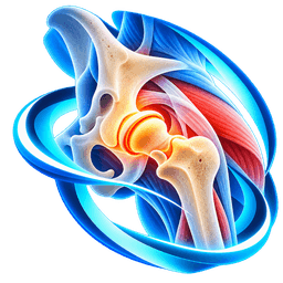
Bone is living tissue that depends on tiny blood vessels. At the top of the thigh bone, the femoral head has an unusually fragile blood supply. If those vessels are blocked, squeezed or damaged, the bone beneath the cartilage starves and part of it dies.
Dead bone cannot repair itself properly under load. Small cracks form beneath the cartilage, and in many people the rounded surface flattens, collapses and then wears the socket. That chain of events turns a bone problem into a joint problem.
Who gets this condition?
It often affects adults between 30 and 60 years of age, which is younger than typical hip osteoarthritis. Both men and women develop it. Roughly half of people with it have both hips involved, either at the same time or over the following years.
Because patients are younger, the choice of treatment carries extra weight. A long working life lies ahead, and an implant must last.
How serious is it?
It can be serious. Without treatment, many hips with a large area of dead bone collapse within 1 to 3 years, though small lesions sometimes stay stable. The outcome depends on size, location and stage, which is why scanning and specialist advice matter early.
Why do people use different names for it?
You may see osteonecrosis, aseptic necrosis, ischaemic necrosis or femoral head osteonecrosis in reports. All describe the same process of bone death from poor blood flow. If your scan report uses one of these terms, you are reading about the same problem as this page.
How is this page organised?
We start with how the hip is supplied with blood, then move through symptoms, causes, staging, diagnosis, treatment and recovery. Joint-saving surgery and hip replacement are both covered, and so is the pathway for travelling for surgery.
Anatomy
What happens in the body with avascular necrosis of the hip
The femoral head depends on a few small arteries that wrap around the neck of the thigh bone, and this slender supply explains why the hip is a favourite site for bone death. A closer look at that arrangement makes the condition easier to follow.
How is the hip supplied with blood?
The main arteries are the medial and lateral circumflex branches of the deep femoral artery. They form a ring around the base of the neck and send small vessels up along the neck to the ball. A very small vessel in the ligament of the head adds a little supply in adults.
Because the vessels run on the surface of the neck, a displaced fracture or dislocation can tear or kink them in one moment. Other causes act more slowly from inside the bone.
What happens when the supply fails?
Bone marrow and bone cells die within hours of losing blood flow, but the picture only appears over weeks to months. The body tries to heal by clearing dead tissue and laying new bone at the edge of the affected zone. This produces a rim, which MRI shows as a characteristic line.
The dead segment is weaker. The weight-bearing top of the ball is usually the part involved, so it is also the part under the most load. Microfractures join up into a crescent-shaped crack, known as the crescent sign, and the cartilage above loses its support.
How does the joint end up damaged?
Once the surface sags, the ball is no longer round. It grinds inside the socket, cartilage is stripped away and secondary arthritis follows. At that point the problem is no longer only about blood flow, and joint reconstruction becomes the main question.
Why are some areas worse than others?
A small lesion away from the weight-bearing zone behaves very differently from a large one at the top of the ball. Surgeons measure the percentage of the femoral head involved and its position, because these two features predict collapse better than any symptom score.
Symptoms & causes
Avascular necrosis of the hip symptoms and causes
Common symptoms
- Avascular necrosis hip symptoms usually begin with groin pain, the most typical sign, usually a deep ache that may be sharp on standing and can arrive over only a few days.
- Pain in the thigh, buttock or knee is common, because the nerves that serve the hip also supply these areas.
- Pain at night or at rest can occur even when early scans look mild, and it often makes patients check their symptoms online.
- A limp develops as the bone weakens, and many people notice they cannot bear full weight without an ache or a sharp twinge.
- Reduced movement, particularly rotating the leg inwards, appears as the ball loses its shape or the joint lining swells.
- Pain on climbing stairs, getting out of a car or standing up from a low seat becomes more obvious as the head begins to flatten.
- Groin pain that comes with a catching or clicking feeling may mean a small piece of cartilage has lifted over the dead bone.
- Both hips may hurt, either together or one after another, so new pain on the opposite side deserves early attention.
- Some people have no symptoms at first, and the changes are found incidentally on a scan taken for another reason.
Causes and risk factors
- Long-term or high-dose corticosteroid treatment is a leading cause, because steroids can alter fat metabolism and blood vessel health in bone.
- Heavy alcohol use over years is strongly linked with avascular necrosis of the hip, probably through fat deposits in marrow that squeeze small vessels.
- A displaced femoral neck fracture or a hip dislocation can interrupt the blood supply directly, which is why trauma is a recognised cause.
- Sickle cell disease and some other blood disorders can block bone vessels with abnormal cells or clots.
- Autoimmune diseases such as lupus, and their treatments, can damage blood vessels and increase risk.
- Radiation or chemotherapy near the pelvis, organ transplantation and some HIV treatments are associated with bone death.
- Decompression illness in divers and tunnel workers can form gas bubbles that block small vessels in bone.
- Some patients have no identifiable cause, which doctors call idiopathic avascular necrosis, and genetic clotting tendencies may play a part.
Types
Types and stages of avascular necrosis of the hip
Doctors classify this condition by cause and by stage, and the stage is the single most useful guide to treatment. Two systems are in common use, and both depend on imaging.
What are the types?
Traumatic avascular necrosis follows a fracture or dislocation, and it often appears within 2 years of the injury. Non-traumatic avascular necrosis arises from steroids, alcohol, blood disorders or unknown factors. Non-traumatic cases tend to be bilateral and to affect a larger area.
How are the avascular necrosis hip stages described?
The Ficat and Arlet system and the ARCO (Association Research Circulation Osseous) system divide the avascular necrosis hip stages according to X-ray, MRI and joint surface changes. The table gives a simple summary.
| Stage | What imaging shows | Typical approach |
|---|---|---|
| Stage 1 | Normal X-ray, abnormal MRI | Protected weight-bearing, medicines, consider core decompression |
| Stage 2 | Changes in bone density but a round head | Joint-preserving surgery for selected patients |
| Stage 3 | Crescent sign, flattening or early collapse | Limited options, hip replacement often discussed |
| Stage 4 | Collapse with arthritis of the socket | Hip replacement is the usual treatment |
Why do size and location matter?
A lesion covering less than about 15% of the weight-bearing surface usually has a good outlook. Lesions covering more than 30% are much more likely to collapse. The position matters as well: a lesion over the central top of the head collapses more readily than one at the edge.
Why does staging change what you can choose?
Joint-preserving procedures work best before collapse. After the head has flattened, the aim shifts from saving the ball to restoring a smooth, pain-free-feeling joint by replacement. Knowing your stage helps you decide how long to try conservative treatment and when to stop waiting.
Diagnosis
How is avascular necrosis of the hip diagnosed?
Avascular necrosis is confirmed by MRI, and a good history of risk factors points the way. Because early X-rays may look normal, diagnosis depends on a high level of suspicion.
How is avascular necrosis of the hip diagnosed?
The doctor asks about steroid courses, alcohol, past injuries, blood disorders, diving and family history. They then examine walking, movement and pain on turning the leg. Pain on rotation, especially inward, is typical, and a limp may be obvious.
Which imaging do doctors choose?
Start with a pelvis X-ray plus a frog-leg lateral view, since the crescent sign is best seen from the side. If the X-ray is normal but suspicion is high, an MRI follows. MRI detects the problem at stage 1 and shows size and location, which direct treatment.
CT shows bone detail and may help with surgical planning. Doctors rarely order bone scans now, as MRI is more accurate.
Does the other hip need a scan?
Often yes. When one hip has non-traumatic bone death, the other has changes in up to half of people, even if it does not hurt. An MRI of both hips can find early, treatable disease and let you protect the second hip.
What tests look for the cause?
Blood tests can look for sickle cell disease, clotting abnormalities, lipid disorders, autoimmune activity and kidney or liver function. This investigation shapes advice on medicines, alcohol and other risks, and it helps prevent more bone death elsewhere.
What should you bring to a remote review?
Share the MRI files themselves, not only the report, and all earlier X-rays so the surgeon can see how fast things changed. Add dates and doses for steroid courses, a list of other illnesses and a note on whether the other hip hurts. A free case review is a convenient way to start.
Tests you may have
- Pelvis X-ray (anteroposterior and frog-leg lateral) shows sclerosis, cysts, the crescent sign and collapse, although it can look normal in stage 1.
- MRI of both hips is the most sensitive test, detecting dead bone early and showing the size and position of the lesion.
- CT scan shows fine bone detail and the exact amount of collapse, and surgeons often rely on it when planning surgery.
- Blood tests check for sickle cell disease, clotting disorders, lipid problems, autoimmune markers and general fitness for anaesthesia.
- Bone scan can show reduced uptake in the centre of the head, though it is rarely needed once MRI is available.
- Bone marrow pressure measurement is a research and surgical tool used in a few centres and is not routine.
- Review of medicine history, including steroid dose and duration, helps to find the cause and the risk to other joints.
Look-alikes
Conditions that can feel like avascular necrosis of the hip
Several problems cause deep hip and groin pain with a sudden start, so the diagnosis rests on MRI and not on symptoms alone. The table sets bone death in the hip beside its common look-alikes.
| Condition | How it differs | How doctors tell |
|---|---|---|
| Hip osteoarthritis | Older patient, slow onset, joint space narrowing from the start | X-ray pattern and gradual history; see hip osteoarthritis |
| Transient bone marrow oedema | Fluid in bone that settles by itself within months | MRI lacks the rim of dead bone and repeats scan shows improvement |
| Stress fracture of the femoral neck | Athletes or people with bone thinning, pain with activity | MRI shows a fracture line instead of a rim |
| Femoroacetabular impingement | Younger, pinching pain on deep flexion | Hip shape on imaging; see femoroacetabular impingement |
| Labral tear | Catching and clicking, normal bone on MRI | MRI arthrogram shows labral damage |
| Septic arthritis or osteomyelitis | Fever, rapid severe pain, raised infection markers | Blood tests and urgent aspiration or imaging |
| Lumbar radiculopathy | Back pain and leg symptoms below the knee | Nerve examination and spine MRI |
Why is transient oedema the most important look-alike?
Bone marrow oedema syndrome causes similar pain and a hazy MRI, yet it often resolves with protected weight-bearing over 3 to 6 months. Misreading it as bone death leads to needless surgery, while missing true avascular necrosis allows collapse. Specialist MRI review resolves this.
Does infection look similar?
A hot, red and very painful hip with fever is more likely infection and needs urgent local assessment. Because steroids and immune problems raise infection risk, never assume that a steroid user's new pain is only bone death.
How do doctors confirm the final answer?
They combine the history, examination, MRI pattern and, when needed, blood tests or a follow-up scan after a few weeks. If the picture remains unclear, they treat cautiously and repeat imaging rather than operate on uncertainty.
Non-surgical
Non-surgical treatment for avascular necrosis of the hip
Non-surgical treatment of avascular necrosis of the hip can relieve pain and sometimes slow collapse, but it rarely makes dead bone live again, so it works best for very early and small lesions. Most patients with larger lesions eventually need surgery.
What does protected weight-bearing involve?
Using crutches or a walker to reduce the load on the hip may help early disease. The aim is to give the healing edge time to strengthen. Many specialists advise this for 6 to 12 weeks in stage 1 disease, although evidence that it prevents collapse is modest.
Which medicines help?
Simple pain relief and anti-inflammatory tablets reduce discomfort, though they do not repair bone. Doctors have tried bisphosphonates to slow collapse, and results vary, so they prescribe them selectively. Researchers have studied statins and blood thinners in people with specific risk factors, and their role remains unclear.
Any change to steroid treatment belongs to the prescribing doctor alone, because stopping them suddenly can be dangerous.
Does treating the cause help?
Yes, often. Reducing alcohol, lowering steroid doses where safe, managing cholesterol, and treating blood or autoimmune disease can lower the risk of further bone death, including in the other hip.
What about physical therapy?
Gentle range-of-movement work, pool exercise and strength training for the thigh and buttock muscles keep the joint mobile and the muscles ready for surgery or recovery. Avoid impact, such as jumping or running, while the head is at risk of collapse.
Are there shock wave or electrical treatments?
Researchers have tested extracorporeal shock wave therapy and pulsed electromagnetic fields in early disease. Some trials show pain relief, while the evidence for preventing collapse is limited. Ask for the research behind any such offer and be cautious of strong claims.
How long should you wait before deciding?
For a small, early lesion, 3 to 6 months of protected weight-bearing and repeat MRI is reasonable. For a large lesion with a crescent sign, delay rarely changes the outcome, and discussing surgery promptly is sensible.
Self-care
Exercises and self-care for avascular necrosis of the hip
Good self-care for avascular necrosis is about protecting the bone, keeping muscles strong and avoiding what made the condition worse. Always check with your surgeon or physiotherapist before starting a new exercise, because the safe load depends on your stage.
How should you manage weight-bearing?
Use crutches or a stick as advised, particularly on uneven ground or when carrying shopping. Keep your steps short and avoid pivoting on the affected leg. If a sharp pain returns suddenly, rest and call your clinic, since this could mean the surface has shifted.
Which movements are usually safe?
- Pool walking and gentle swimming: 20 to 30 minutes, three or four times a week.
- Seated knee extensions and ankle pumps: 2 sets of 15 repetitions.
- Supine hip abduction slides on a smooth floor: 10 repetitions each side.
- Stationary cycling at low resistance, with a high saddle, for 10 to 20 minutes.
- Upper-body and core work seated or lying down to maintain overall fitness.
What lifestyle habits matter?
Cut down or stop alcohol, stop smoking, and take cholesterol or blood disease treatment exactly as prescribed. Keep a healthy weight to lessen load on the hips. A diet with enough calcium, vitamin D and protein supports bone health.
What should you avoid?
Avoid running, jumping, heavy lifting and long periods of standing while the bone is weak. Do not start or increase steroid medicines yourself, and tell every doctor and dentist about your history so they take it into account.
How do you cope with pain and mood?
A sudden loss of mobility at a young age is hard. Pacing your day, using aids without embarrassment and speaking with others who share the condition can help. If low mood or sleep disturbance persists, mention it to your doctor.
Check with your doctor or physiotherapist before starting new exercises, and stop any movement that causes sharp pain.
Treatment
Avascular necrosis of the hip treatment options
Treatment for bone death in the hip depends on the stage. Joint-preserving surgery aims to save the natural ball before it collapses, while hip replacement is the standard answer once it has. Your surgeon should explain which route fits your age and scan.
What is core decompression?
Core decompression for hip necrosis drills one or more small channels from the outer thigh bone into the dead area. This lowers pressure inside the bone and may encourage new blood vessels. Surgeons sometimes add bone marrow cells or bone graft. It works best in stages 1 and 2 with small to moderate lesions, and results vary.
What are bone grafting and osteotomy?
A vascularised bone graft brings living bone with its blood supply into the weak area, and an osteotomy rotates the femoral head so healthy bone takes the load. Specialist centres perform these large operations in a small number of young patients. Recovery takes months, and success depends on lesion size.
When is total hip replacement the right choice?
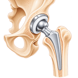
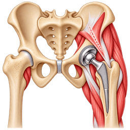
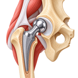
Total hip replacement removes the collapsed ball and replaces both sides of the joint. It reliably relieves pain in collapsed hips and gives good function. Younger patients should discuss implant bearings and long-term revision. You can also read about total hip replacement in turkey and the cost guide.
Is the anterior approach an option?
Yes, for suitable patients. The anterior hip replacement approach works between muscles at the front, which may speed early walking. Since many people with this condition are younger and active, some choose it for quicker recovery. See anterior hip replacement in turkey and its cost guide.
Does hip resurfacing fit?
Hip resurfacing needs enough healthy bone in the head to hold the metal cap. In avascular necrosis, large dead areas often make resurfacing unsuitable, so surgeons reserve it for small lesions in carefully selected people. Read about hip resurfacing in turkey and the cost guide.
Which operation do surgeons choose, and why?
The choice follows your stage, age, activity and the other hip. Early disease may justify a joint-saving attempt, with replacement kept in reserve. For collapse, replacement is the predictable option. Surgeons should be honest that joint preservation can fail, and that a later replacement remains possible after it.
When surgery is considered
Surgery should be discussed when imaging shows an expanding lesion, when the bone surface has started to collapse or when pain stops you walking or sleeping despite conservative care. Timing matters more here than in most forms of arthritis.
When is early surgery worth considering?
Consider a specialist opinion if MRI shows a lesion in the weight-bearing area, particularly one larger than about 30% of the head, or if pain is rising despite protected weight-bearing. Early joint-preserving surgery has the best chance when the head is still round.
When does replacement make sense?
- The head has flattened or collapsed on X-ray or CT.
- Pain limits walking, sleep or work in spite of 3 months of conservative treatment.
- Cartilage loss or secondary arthritis is visible in the socket.
- A previous joint-saving operation has failed and pain has returned.
- You are fit for anaesthesia and have support for recovery at home.
When is it reasonable to wait?
If the lesion is small, off the weight-bearing area and not causing pain, your doctor may simply monitor with repeat scans every 6 to 12 months. Treat the cause at the same time, for instance by reducing alcohol or steroid exposure.
How do work and family life affect timing?
Many patients are in the middle of careers and caring roles. Surgeons often agree a date that allows time for recovery and for cover at work. Delaying a few months is usually acceptable if the bone has not yet collapsed, though a collapsing head is better treated sooner.
What should you ask your surgeon?
Ask about the stage and lesion size, the chance that joint preservation will work, how long recovery takes and what the implant will mean for your job and sport. Ask what happens to the other hip, and what follow-up scans are planned.
Procedures
Procedures that may treat avascular necrosis of the hip
Costs
Avascular necrosis of the hip treatment cost in Turkey
| Procedure | Turkey package | US self-pay | Saving |
|---|---|---|---|
| Total Hip Replacement | $8,000 – $13,000 | $38,438 | ~73% |
| Anterior Hip Replacement | $9,000 – $14,000 | — | — |
| Hip Resurfacing | $9,000 – $14,000 | $43,325 | ~73% |
Packages are contracted partner prices with validity dates; benchmarks are source-backed estimates. Open a cost guide for the full country comparison.
In Turkey
Treating avascular necrosis of the hip in Turkey
Planned treatment of this condition in turkey can be reasonable when a surgeon has reviewed your MRI and X-rays, your general health is stable and the surgical plan is clear. Many younger patients in this group compare waiting times and surgeon experience across countries before they decide.
When is care in turkey a sensible choice?
Hip replacement for avascular necrosis in turkey tends to suit people with collapsed or advanced disease, who need a definite operation and can travel for 2 to 3 weeks. Avascular necrosis of the hip treatment in turkey may also include a second opinion on whether joint preservation is still possible. Read why patients travel in our guide to turkey.
If you have a new fracture, a dislocation, fever or very sudden severe pain, urgent local care comes first. Travel is only considered after you are medically stable and your treating team agrees.
What does the pathway look like?
- Send your MRI, X-rays and medicine history through our medical record review.
- A surgeon reviews the scans, asks for any missing items and states a staged recommendation.
- You receive a written plan with the operation, implant choice, expected stay and risks.
- Blood tests, a heart check and a review of any steroid or blood-thinning medicine are completed.
- After surgery you walk early with a physiotherapist and stay in hospital briefly.
- You remain nearby for wound checks and fitness-to-fly clearance, then arrange follow-up at home.
What extra checks matter for this condition?
Because avascular necrosis often involves steroids, sickle cell disease, lupus or alcohol, ask the team how they will manage these before anaesthesia. Bleeding, infection and healing risks differ. Confirm that the other hip has been scanned, and what the plan is if it also collapses.
How do you judge a hospital and surgeon?
Look for recognised international accreditation, a national licence, an orthopedic team that does hip replacements every week and clear policies on infection control and implant records. Ask for the written implant sticker details. Our list of questions to ask before surgery abroad is a good start.
What are the practical limits?
You will usually need 10 to 14 days nearby before flying, plus clot prevention for the journey. Read our advice on flying after surgery. Make sure a doctor at home will review you. Osteonecrosis of the hip surgery in turkey is available at many hospitals, so explore orthopedics in turkey or request a free case review.
Complications
Complications of avascular necrosis of the hip
Bone death in the hip can lead to joint collapse and arthritis, and its treatments carry their own risks. Knowing both sides helps you choose with confidence.
What happens if it is left alone?
Large lesions usually progress. The head flattens, the socket cartilage wears and pain limits walking within a few years. Muscle wasting, a shortened leg and a permanent limp may follow. The other hip may also be affected, and persistent pain can lead to low mood and poor sleep.
What are the risks of joint-preserving surgery?
Core decompression is a small operation, but it carries risks such as infection, bleeding, a stress fracture along the drill track and failure to prevent collapse. Larger grafting or osteotomy procedures have a longer recovery and a higher chance of complications.
What are the risks of hip replacement here?
The usual risks apply: infection, clots, dislocation, leg length difference, nerve irritation and loosening or wear over time. Younger patients, and people with steroid use, sickle cell disease or heavy alcohol history may have slightly higher rates of problems and revision. Honest counselling about this is part of good care.
Will an implant need replacing?
Possibly, particularly if you are under 50 or very active. Modern bearings last well, and many implants function for 15 years or more. A later revision is a bigger operation, so it is wise to plan long-term follow-up from the start.
How do you lower the risks?
Yes. Stopping smoking, limiting alcohol, optimising blood sugar and blood disorders, completing clot prevention and attending every follow-up reduce risk. Report any new pain early, rather than waiting for the next appointment.
Urgent care
When to seek urgent care for avascular necrosis of the hip
- Sudden severe hip or groin pain with inability to bear weight after a fall needs emergency assessment for a fracture or dislocation.
- Fever, shaking chills and a hot, swollen hip need same-day medical assessment, as infection must be excluded quickly.
- New hip or groin pain in someone on long-term steroids needs a call to your doctor within days, not weeks, so they can arrange an MRI.
- Pain that wakes you at night or does not ease with rest in a person with sickle cell disease needs prompt specialist review.
- Pain in the second hip after diagnosis in the first deserves early imaging, because early joint-saving treatment works best.
- Calf pain, swelling or breathlessness after an operation or a flight can mean a clot and requires emergency care.
- A sudden clunk, sharp pain and a leg that will not bear weight in someone with known disease may mean the bone has collapsed, so contact your surgeon at once.
Prevention
How to lower your risk of avascular necrosis of the hip
Not every case of this condition is preventable, but reducing the major triggers lowers risk significantly. Prevention focuses on medicines, alcohol, injury care and monitoring.
How do you lower steroid-related risk?
Steroids are often essential, so never stop them without advice. Doctors aim to use the lowest effective dose for the shortest time and may choose alternatives. If you must take them for a long time, ask whether hip symptoms should trigger an early MRI.
How does alcohol fit in?
Heavy, sustained drinking is a well-recognised risk. Keeping within national low-risk limits, or stopping if you already have bone changes, reduces the chance of more damage. Support from a family doctor is available for people finding this difficult.
How can you reduce risk after a hip injury?
After a displaced fracture or dislocation, urgent treatment to restore alignment within hours gives the blood vessels the best chance. Your surgeon may advise follow-up imaging for up to 2 years to look for late changes. Our page on hip and femoral neck fracture explains injuries in more detail.
What about divers and other special groups?
Divers and compressed-air workers can reduce risk by following decompression tables carefully. People with sickle cell disease benefit from regular specialist care, hydration and prompt treatment of crises. Those with lupus should keep their disease controlled.
What stays outside your control?
Some cases appear with no clear cause, and genes may play a part. Sensible monitoring is still useful. If you have risk factors and develop groin pain, early imaging is the best protection against late collapse.
Outlook
Living with avascular necrosis of the hip: outlook and recovery
The outlook for avascular necrosis depends mainly on the size of the lesion and the stage at diagnosis, and most people with advanced disease do well after hip replacement. Early detection widens the range of choices.
What is the natural course?
Small lesions may remain stable for years, and some heal. Large lesions in the weight-bearing zone commonly progress to collapse in 1 to 3 years. Because the pattern is so variable, repeat imaging and symptom review guide decisions rather than a single scan.
What are the results of joint preservation?
Core decompression and related procedures can delay or avoid replacement in many people with early disease, particularly those with small lesions. Rates of success differ between studies, so treat promises of reliable cure with scepticism. Even when it fails, a hip replacement can usually follow.
What is recovery like after hip replacement?
Most patients walk with support on the day after surgery and leave hospital in 2 to 4 days. Crutches or a stick are normally used for about 2 to 6 weeks. Desk work often resumes at 4 to 6 weeks, and manual jobs take 8 to 12 weeks or more. Strength continues to improve for up to 12 months. Our rehabilitation guide describes the process.
What about sport and work?
Walking, swimming, cycling and golf are generally accepted. High-impact sport can shorten implant life, so discuss it with your surgeon. Many younger people return to physical jobs with some adjustments.
How should you plan the long term?
Plan regular X-ray reviews, look after the other hip and keep the underlying cause under control. If you also have arthritis changes elsewhere, see the post-traumatic hip arthritis page, and see the hip area page for related problems.
Surgeons
Specialists who treat avascular necrosis of the hip
FAQ
Avascular necrosis of the hip: frequently asked questions
What is avascular necrosis of the hip?
What are the early signs of bone death in the hip?
What causes it to happen?
Can bone death in the hip heal on its own?
Does avascular necrosis always need surgery?
What is core decompression?
How long does recovery take after hip replacement for avascular necrosis?
Is avascular necrosis of the hip treatment in turkey safe?
How long do I need to stay in turkey for hip surgery?
Will both hips be affected?
Can I exercise with a collapsing femoral head?
Is hip resurfacing suitable for avascular necrosis?
Sources
Sources for this avascular necrosis of the hip guide
Peer-reviewed guidance and institutional sources used to write and review this page.
- 01Osteonecrosis of the Hip
American Academy of Orthopaedic Surgeons (OrthoInfo), 2022
https://orthoinfo.aaos.org/en/diseases--conditions/osteonecrosis-of-the-hip/
- 02Osteonecrosis
Mayo Clinic, 2023
https://www.mayoclinic.org/diseases-conditions/osteonecrosis/symptoms-causes/syc-20352302
- 03Osteonecrosis
National Institute of Arthritis and Musculoskeletal and Skin Diseases, 2023
https://www.niams.nih.gov/health-topics/osteonecrosis
- 04
- 05
- 06Total Hip Replacement
American Academy of Orthopaedic Surgeons (OrthoInfo), 2023
https://orthoinfo.aaos.org/en/treatment/total-hip-replacement/