Key takeaways
- 1Knee osteotomy realigns the leg to redistribute load away from an overloaded or damaged portion of the knee while preserving the natural joint.
- 2High tibial osteotomy is commonly used for varus alignment and medial compartment overload, while distal femoral osteotomy is commonly used for valgus alignment and lateral compartment overload.
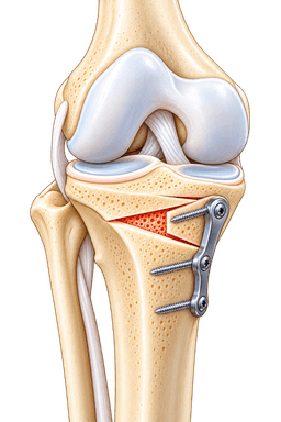
- 3Osteotomy does not replace the knee joint. The surgeon cuts and repositions bone, then stabilizes the correction with a plate and screws or another fixation system.
- 4The operation is particularly relevant to younger and active patients with localized joint damage, malalignment or certain instability patterns who are not ideal candidates for early knee replacement.
- 5Knee osteotomy can be performed together with cartilage restoration, meniscus repair, meniscal transplantation or ligament reconstruction when abnormal alignment would otherwise overload those procedures.
- 6Recovery includes both bone healing and rehabilitation, so return to demanding sport generally takes several months.
- 7The objective is joint preservation and load redistribution, not permanent immunity from osteoarthritis or a guarantee that knee replacement will never be needed.
Overview
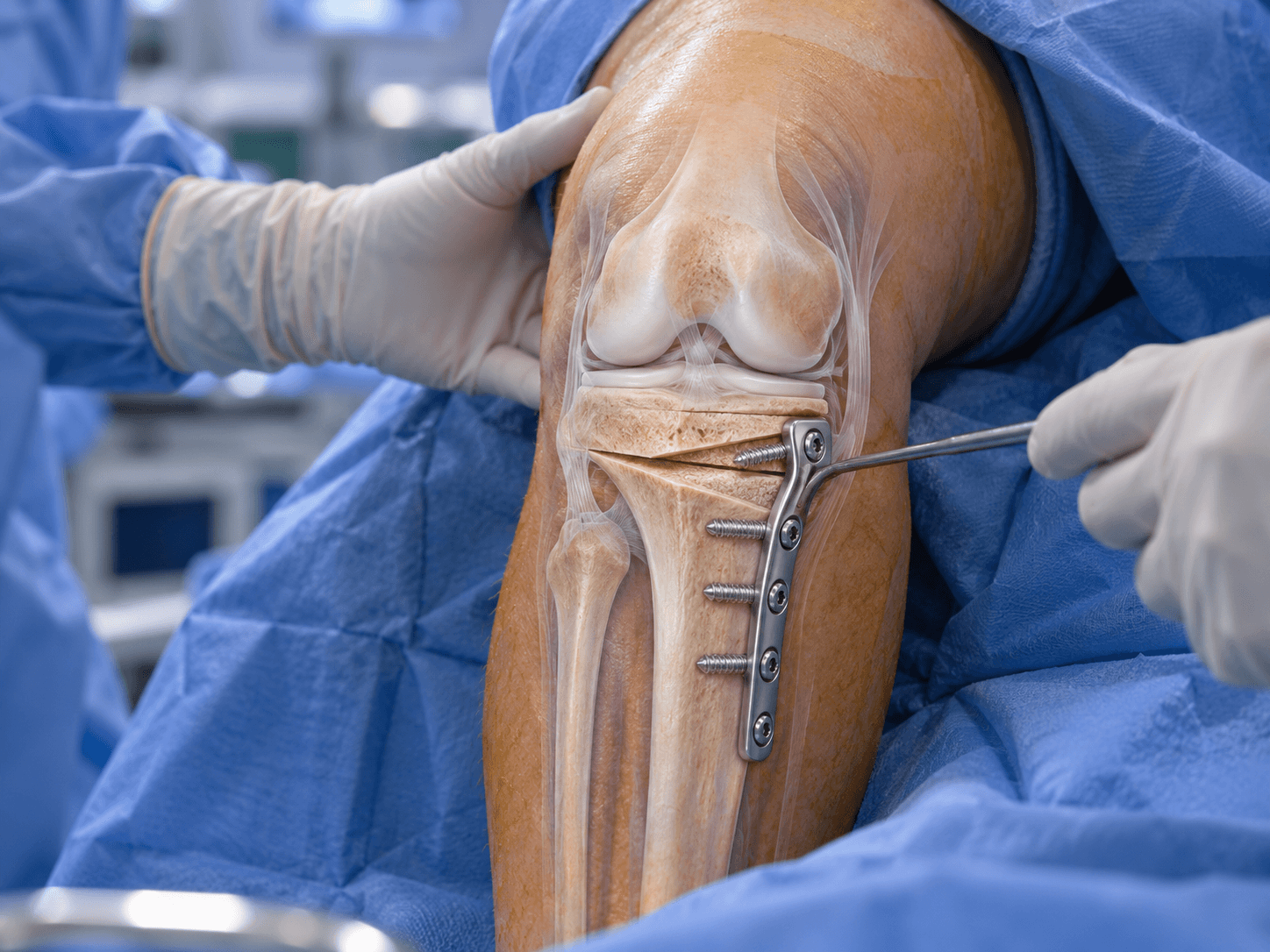
Knee osteotomy is a joint-preserving operation in which the surgeon cuts and realigns the tibia, femur or occasionally both bones to change how forces pass through the knee. The corrected bone position is stabilized while it heals, usually using a strong plate and screws.
The operation is based on a mechanical principle. If one compartment of the knee receives too much load because the leg is abnormally aligned, that overloaded region can become painful and progressively damaged. By changing the alignment, the surgeon shifts part of the body's weight toward a healthier region of the joint.
Unlike total knee replacement, osteotomy does not remove the entire joint surface and replace it with metal and polyethylene. The patient's own knee, ligaments, cartilage and bone remain in place except for the controlled bone cut required to create the correction.
This makes knee osteotomy particularly relevant in joint-preservation surgery. It can reduce symptoms and extend the useful life of the natural knee in appropriately selected patients.
Why Alignment Matters
When a person stands, body weight passes from the hip through the knee and toward the ankle along a mechanical axis.
In a normally aligned limb, load is distributed relatively evenly across the knee, although the medial compartment naturally carries a substantial share of weight.
When the leg becomes markedly varus, or bow-legged, the mechanical axis shifts farther toward the inner side of the knee. This increases medial compartment loading.
When the limb has valgus, or knock-kneed, alignment, the mechanical axis shifts toward the outer side. Lateral compartment structures then receive more stress.
These changes can become self-reinforcing. Cartilage and meniscal loss can increase deformity, and the deformity can then increase compartment loading.
What Does an Osteotomy Actually Change?
The surgeon changes the angle of the bone.
The goal is not merely cosmetic straightening. The correction is calculated to alter the weight-bearing line through the knee.
In medial compartment osteoarthritis associated with varus alignment, a valgus-producing high tibial osteotomy shifts load laterally.
In lateral compartment disease associated with valgus alignment, a varus-producing distal femoral osteotomy can shift load toward the healthier medial side.
The amount of correction is planned before surgery using standing radiographs and measurements of the entire limb.
High Tibial Osteotomy
A high tibial osteotomy, commonly abbreviated HTO, is performed through the upper tibia below the knee.
It is one of the most established forms of knee realignment surgery.
HTO is especially associated with varus knees in which the medial compartment is overloaded. The operation changes the relationship between the tibia and the mechanical axis so that less force passes through the damaged medial compartment.
The procedure can be performed using an opening-wedge or closing-wedge technique.
Modern medial opening-wedge HTO is widely used because it allows fine adjustment of the correction and preserves the proximal tibiofibular relationship, although closing-wedge techniques remain appropriate in selected cases.
Distal Femoral Osteotomy
A distal femoral osteotomy, or DFO, changes alignment through the lower femur just above the knee.
It is commonly considered when the deformity originates primarily in the femur rather than the tibia.
A classic indication is a valgus knee with symptomatic lateral compartment overload.
A varus-producing distal femoral osteotomy can shift load toward the healthier medial compartment.
DFO can also be used to correct femoral rotational abnormalities in selected patients with patellar instability or complex lower-limb malalignment.
Why the Deformity Should Be Corrected at Its Source
A patient may appear bow-legged or knock-kneed, but the deformity can arise from the tibia, femur or both.
Correcting the wrong bone can produce an abnormal joint-line orientation even when the overall leg looks straighter.
Modern planning therefore evaluates where the deformity actually originates.
A tibial deformity is generally corrected in the tibia.
A femoral deformity is generally corrected in the femur.
When both bones contribute substantially, a double-level osteotomy can sometimes provide a more anatomical correction than making a very large adjustment through only one bone.
Double-Level Osteotomy
A double-level osteotomy corrects both the distal femur and proximal tibia.
It is reserved for selected complex deformities.
The technique allows the mechanical axis to be corrected without creating an excessively tilted knee joint line.
This is particularly valuable when a large deformity is distributed across both bones.
Because two bones are cut and fixed, the operation and rehabilitation are more involved than a standard isolated HTO or DFO.
Opening-Wedge Osteotomy
During an opening-wedge osteotomy, the surgeon creates a controlled cut through the bone while preserving a hinge on the opposite side.
The osteotomy is gradually opened until the planned correction is achieved.
A wedge-shaped gap is created.
A plate and screws stabilize the new position.
Depending on correction size, surgeon preference and patient factors, the gap may be left to fill with new bone or supported using bone graft or a substitute.
Closing-Wedge Osteotomy
A closing-wedge procedure removes a wedge of bone.
The remaining bone surfaces are then brought together, changing alignment.
Fixation maintains the new position while the osteotomy heals.
Closing-wedge HTO has a long history and can provide reliable correction.
It changes bone geometry differently from opening-wedge surgery and can influence factors such as limb length and tibial slope.
The surgeon selects the technique according to deformity, desired correction and experience.
High Tibial Osteotomy for Medial Knee Arthritis
Medial compartment osteoarthritis is one of the best-known indications for HTO.
These patients usually have pain concentrated on the inner side of the knee and varus alignment that repeatedly increases load through that compartment.
By shifting the mechanical axis laterally, HTO reduces the proportion of body weight crossing the damaged medial region.
The operation does not remove the existing arthritis.
Instead, it changes the mechanical environment.
This distinction helps patients understand why improvement can occur even though X-rays still show cartilage damage after surgery.
Distal Femoral Osteotomy for Lateral Compartment Arthritis
Lateral compartment osteoarthritis is less common than medial compartment disease.
When it occurs with valgus alignment, the outer part of the knee can receive excessive load.
Distal femoral varus osteotomy repositions the femur so the weight-bearing line shifts toward the medial side.
The natural knee remains intact.
DFO can therefore be an attractive option for younger active patients with lateral disease who would otherwise face partial or total knee replacement relatively early.
Knee Osteotomy for Cartilage Restoration
Cartilage restoration depends on a favorable mechanical environment.
A sophisticated cartilage graft placed in an overloaded compartment can continue receiving excessive pressure.
For this reason, osteotomy is frequently combined with cartilage procedures when malalignment contributes to the defect.
For example, a medial femoral condyle cartilage restoration in a significantly varus knee may be combined with high tibial osteotomy.
The osteotomy unloads the reconstructed region while the cartilage procedure treats the structural lesion.
Knee Osteotomy for Meniscus Surgery
The meniscus distributes load across the knee.
A repaired meniscus root or transplanted meniscus can remain overloaded if substantial malalignment is left untreated.
HTO can therefore accompany medial meniscus root repair in selected varus knees.
Realignment can reduce medial compartment pressure while the meniscal tissue heals.
Similarly, meniscal transplantation can be combined with osteotomy when abnormal alignment would otherwise overload the graft.
The treatment plan should address both tissue damage and the mechanical environment.
Knee Osteotomy for Ligament Instability
Osteotomy is not used only for arthritis.
Bone geometry affects ligament forces.
In selected chronically unstable knees, altering tibial slope or coronal alignment can reduce stress on reconstructed ligaments.
For example, excessive posterior tibial slope can increase forces on the ACL.
A slope-reducing osteotomy may be considered in carefully selected patients with repeated ACL graft failure and abnormal anatomy.
Conversely, tibial slope can influence PCL biomechanics.
These are specialized procedures rather than routine treatment for every ligament tear.
Knee Osteotomy for Patellar Instability
Distal femoral osteotomy can play a role in selected patients with recurrent patellar instability associated with significant valgus deformity or excessive femoral rotation.
Correcting the femoral deformity changes the forces acting on the kneecap.
The operation may be combined with medial patellofemoral ligament reconstruction or another patellar stabilization procedure.
This is another example of why knee osteotomy should be understood as deformity correction rather than simply arthritis surgery.
Osteotomy Versus Knee Replacement
Both operations can relieve symptoms, but they solve different problems.
A knee replacement resurfaces damaged portions of the joint with artificial components.
An osteotomy preserves the joint and changes load distribution.
Younger patients with localized compartment damage, significant malalignment and relatively preserved cartilage elsewhere can be particularly attractive osteotomy candidates.
Patients with widespread end-stage arthritis across several compartments, severe stiffness or extensive joint destruction are generally more likely to benefit from arthroplasty.
Does Knee Osteotomy Prevent Knee Replacement?
Not necessarily.
One purpose of osteotomy is to delay the need for arthroplasty in patients for whom replacing the knee at a young age would be undesirable.
Many successful osteotomies continue functioning for a decade or longer.
However, osteoarthritis can continue progressing.
Some patients eventually undergo partial or total knee replacement.
The operation should therefore be viewed as joint preservation rather than a guarantee that replacement will never be necessary.
Can Knee Replacement Still Be Done After Osteotomy?
Yes.
Total knee replacement can be performed after a previous HTO or DFO if arthritis eventually progresses.
The surgery can be technically more complex because the bone has been realigned and previous plates, screws or scars may need to be considered.
Hardware can sometimes remain in place if it does not interfere with the replacement.
In other cases, removal is performed before or during arthroplasty.
A previous osteotomy therefore does not close the door to future replacement.
Conditions treated
Who it's for
- Medial compartment knee osteoarthritis associated with varus or bow-legged alignment
- Lateral compartment knee osteoarthritis associated with valgus or knock-kneed alignment
- Symptomatic unicompartmental cartilage damage with correctable malalignment
- Young or middle-aged active patients seeking joint preservation
- Patients considered relatively young for knee replacement
- Persistent compartment-specific pain despite appropriate nonsurgical treatment
- Focal cartilage restoration performed in an overloaded compartment
- Medial meniscus root repair associated with clinically important varus alignment
- Meniscal allograft transplantation requiring correction of compartment overload
- Selected chronic ligament instability associated with abnormal coronal alignment
- Selected recurrent ACL reconstruction failure associated with excessive tibial slope
- Selected PCL-related instability associated with unfavorable bony anatomy
- Recurrent patellar instability associated with significant valgus deformity
- Patellar instability associated with excessive femoral rotation requiring derotational osteotomy
- Post-traumatic malalignment affecting knee loading
- Symptomatic congenital or developmental lower-limb deformity
- Complex deformity involving both the femur and tibia that may require double-level osteotomy
- Patients with preserved cartilage in the compartment that will receive increased load after correction
Good candidates
The classic osteotomy candidate is relatively young, active and has disease concentrated in one part of the knee rather than across the entire joint.
The patient's alignment should contribute meaningfully to the problem.
A person with severe medial pain, varus alignment and relatively preserved lateral cartilage is a very different candidate from someone with diffuse tricompartmental osteoarthritis.
The operation is designed to redistribute load. There needs to be a reasonably healthy area toward which that load can be shifted.
Age
There is no absolute age cutoff.
Historically, high tibial osteotomy was often associated with patients younger than approximately 60 years.
Modern decision-making is more individualized.
A highly active patient in their early 60s with localized disease and good motion can occasionally be more appropriate for osteotomy than a younger patient with widespread cartilage destruction.
Age therefore contributes to decision-making but does not replace assessment of joint condition, activity and goals.
Activity Level
Osteotomy is particularly attractive to patients who want to remain physically active.
An artificial knee can provide excellent function, but high-impact repetitive activity is generally approached more cautiously after arthroplasty.
An osteotomy preserves the native joint surfaces and can allow active patients to continue a broader range of activities when recovery is successful.
The expected sport should still be discussed with the surgeon.
Returning to recreational activity is more predictable than returning to elite competition.
Range of Motion
Good preoperative knee motion is generally favorable.
A patient with relatively preserved flexion and minimal fixed flexion contracture is typically a stronger osteotomy candidate than someone with a severely stiff knee.
Marked motion loss can indicate more advanced joint disease.
Rehabilitation after osteotomy also requires restoring motion while the bone heals.
Compartment-Specific Disease
Osteotomy works best when one compartment is primarily responsible for symptoms.
For varus knees, this is commonly the medial compartment.
For valgus knees, it is commonly the lateral compartment.
The opposite compartment should remain relatively preserved.
Diffuse cartilage loss substantially reduces the logic of transferring weight from one diseased area onto another diseased area.
Patellofemoral Disease
Patellofemoral cartilage damage does not automatically exclude osteotomy.
The importance depends on severity, symptoms and osteotomy type.
Certain changes in tibial slope and patellar height can affect patellofemoral mechanics.
The surgeon evaluates the entire joint rather than deciding based solely on one radiographic compartment.
Severe symptomatic patellofemoral arthritis can make joint-preserving realignment less attractive.
Body Weight
Body weight influences forces across the osteotomy and knee.
Higher body mass can also be associated with more advanced osteoarthritis and increased complication risk.
There is no universal BMI threshold that applies to every patient.
The surgeon considers body composition, bone quality, metabolic health and activity together.
Weight optimization can improve both surgical safety and the mechanical environment after correction.
Smoking and Nicotine
Nicotine is particularly important in osteotomy because a controlled bone cut must heal.
Smoking and nicotine exposure can interfere with bone formation and increase the risk of delayed union or nonunion.
Patients are commonly advised to stop smoking and nicotine before surgery and during bone healing.
The exact cessation requirements depend on the surgical program.
Diabetes
Well-controlled diabetes does not automatically exclude osteotomy.
Poor glycemic control can increase infection and wound-healing risk and may negatively affect bone healing.
Diabetes should therefore be optimized before elective surgery.
The patient's broader vascular and metabolic health also matters.
Bone Quality
Stable fixation requires sufficient bone quality.
Osteoporosis or other metabolic bone disorders can influence fixation and healing.
The surgeon may investigate bone health when risk factors are present.
Severe bone deficiency can affect whether osteotomy is the most appropriate treatment.
Patients With Previous Surgery
Prior arthroscopy, meniscus surgery or ligament reconstruction does not automatically prevent osteotomy.
In fact, osteotomy is commonly integrated into revision sports-knee surgery.
The important issues are previous tunnels, hardware, scars and the current mechanical problem.
CT may be needed in complex revision cases to understand previous bone work.
When Knee Osteotomy May Not Be Appropriate
Advanced tricompartmental osteoarthritis generally reduces the value of realignment.
Severe inflammatory arthritis, major uncorrectable instability and substantial stiffness can also favor other strategies.
Active infection must be treated before elective osteotomy.
A patient unwilling or unable to follow weight-bearing restrictions and prolonged rehabilitation may also be a poor candidate.
The operation requires meaningful participation in recovery.
Before surgery
Identifying the Source of the Deformity
The most important planning principle is identifying where the malalignment originates.
The surgeon assesses whether the deformity is primarily in the tibia, femur or both.
Correcting the wrong bone can restore the mechanical axis while creating an abnormal joint-line angle.
Modern deformity planning therefore examines both limb alignment and individual bone geometry.
This determines whether the patient needs HTO, DFO or double-level correction.
Medical History
The surgeon asks where pain occurs and how long symptoms have been present.
Activity-related medial pain suggests a different mechanical pattern from lateral or patellofemoral symptoms.
Swelling, stiffness and instability are also documented.
Previous fractures, ligament reconstructions, meniscus surgery and cartilage procedures are particularly important because they can alter bone geometry and joint mechanics.
Patients should provide previous operative reports whenever possible.
Physical Examination
The examination begins with standing alignment and gait.
The surgeon observes whether the legs appear varus or valgus.
Knee range of motion, swelling, tenderness and stability are assessed.
The hip and ankle are also considered because apparent knee alignment can be influenced by the entire lower limb.
Rotational alignment may require more detailed examination.
The patella is assessed when instability or anterior knee pain contributes to the treatment plan.
Weight-Bearing Knee X-Rays
Standing radiographs show joint-space narrowing and compartment-specific osteoarthritis.
They help determine whether the medial, lateral and patellofemoral surfaces remain suitable for preservation.
Non-weight-bearing imaging can underestimate compartment narrowing.
This is why standing films are particularly important when deciding between osteotomy and arthroplasty.
Full-Length Alignment X-Rays
A long-leg standing radiograph from hip to ankle is one of the most important tests before knee osteotomy.
It shows the mechanical axis of the entire limb.
The surgeon can determine where the weight-bearing line crosses the knee.
Several angles can then be measured to determine whether deformity originates in the femur, tibia or both.
The planned correction is calculated from this image.
Mechanical Axis
The mechanical axis represents the load-bearing relationship from the center of the hip toward the ankle.
Varus alignment shifts this line medially.
Valgus alignment shifts it laterally.
The surgeon plans to move this line toward a more favorable position across the knee.
The exact target depends on the indication.
A patient undergoing osteotomy for established medial arthritis may require a different correction from someone undergoing realignment primarily to protect a meniscus or ligament reconstruction.
Joint-Line Orientation
Correcting alignment is not simply about placing the ankle under the hip.
The knee joint line should also remain anatomically reasonable.
A very large correction through only the tibia can produce excessive joint-line obliquity.
This can alter joint mechanics despite improving the overall mechanical axis.
Double-level osteotomy can sometimes distribute a large correction between the femur and tibia to maintain a more physiological joint line.
MRI
MRI helps evaluate cartilage, menisci and ligaments.
For an osteotomy performed because of unicompartmental osteoarthritis, the surgeon needs to understand the condition of the compartment receiving the redistributed load.
MRI is particularly useful when cartilage restoration, meniscal repair or ligament reconstruction is also planned.
Bone-marrow lesions and subchondral pathology can provide additional information.
MRI does not replace weight-bearing alignment radiographs because it is usually performed lying down.
CT
CT is used selectively.
Rotational deformities require accurate assessment of femoral and tibial torsion.
Three-dimensional CT can help plan derotational osteotomy.
Revision cases can also benefit from CT when previous bone tunnels or hardware are present.
Routine straightforward medial opening-wedge HTO does not necessarily require CT.
Assessing Cartilage
The amount and distribution of cartilage damage influence whether osteotomy is likely to work.
The overloaded compartment can have substantial disease.
The compartment receiving additional load after correction should remain reasonably preserved.
The surgeon may use MRI, radiographs and previous arthroscopy findings.
Occasionally, arthroscopy is performed during the same operation to evaluate and treat the joint.
Meniscus Evaluation
The menisci are assessed because they strongly influence compartment loading.
A medial meniscus root tear can allow extrusion and rapidly increase medial compartment stress.
In a significantly varus knee, simply repairing the root without addressing malalignment may leave the repair exposed to excessive load.
The treatment plan can therefore combine root repair with HTO in selected cases.
Previous meniscectomy is also important because loss of meniscal tissue can accelerate cartilage overload.
Ligament Evaluation
ACL, PCL and collateral ligament stability are examined.
Coronal and sagittal bone alignment influence ligament forces.
In chronic ligament-deficient knees, osteotomy may be part of a staged or combined reconstruction.
The surgeon needs to determine whether instability comes primarily from soft-tissue failure, bony alignment or both.
Planning the Correction
The correction is calculated rather than estimated visually during surgery.
Digital planning software or calibrated radiographs can be used.
The surgeon identifies the desired weight-bearing line and determines the required opening or closing angle.
The hinge location, osteotomy plane and fixation strategy are then planned.
The goal is precise correction without damaging the joint or creating secondary deformity.
Overcorrection and Undercorrection
Both can compromise results.
Insufficient correction may leave the diseased compartment overloaded.
Excessive correction can overload the opposite side and alter joint-line mechanics.
Precision is therefore central to osteotomy surgery.
Intraoperative fluoroscopy and alignment methods help confirm that the target has been achieved.
Tibial Slope
High tibial osteotomy can change posterior tibial slope.
This matters because slope influences both knee motion and cruciate ligament forces.
Opening-wedge and closing-wedge techniques can affect slope differently depending on surgical execution.
In a patient with ACL or PCL problems, slope may even be the primary target of the osteotomy.
The surgical plan should therefore consider both coronal and sagittal alignment.
Patellar Height
Some osteotomy techniques can influence patellar height.
A standard medial opening wedge can produce a relative lowering of the patella in certain circumstances.
Specialized biplanar or distal tuberosity techniques can be considered when patellar mechanics are important.
This becomes particularly relevant when the patient already has patellofemoral symptoms or may need future arthroplasty.
Prehabilitation
The knee should ideally enter surgery with good motion and controlled swelling.
Quadriceps and hip strength can be improved beforehand.
The patient should learn crutch use.
This is especially useful because postoperative weight-bearing may be restricted.
Prehabilitation also gives the physiotherapist a baseline against which postoperative progress can be measured.
Bone Health
Vitamin D deficiency, osteoporosis and other bone-health issues can be investigated when clinically appropriate.
The osteotomy must heal through new bone formation.
Nutrition and metabolic health therefore matter.
Severe nutritional deficiency should be corrected.
Patients taking medications that affect bone metabolism should discuss them with the surgical team.
Smoking Cessation
Nicotine cessation deserves specific attention.
Bone healing depends on blood supply and cellular activity.
Smoking can impair both.
Some surgeons require biochemical nicotine testing before elective osteotomy.
Patients should clarify the program's requirements well before the operation.
Medication Review
The surgical team should receive a complete list of medications and supplements.
Anticoagulants and antiplatelet drugs require individualized management.
Certain diabetes medications may need temporary adjustment.
Patients should not stop prescribed drugs independently.
The anaesthetist, surgeon and prescribing clinician should coordinate the plan.
Planning Work and Home Recovery
The patient will commonly use crutches for several weeks.
A desk worker and construction worker therefore need very different return-to-work plans.
Home stairs should be considered.
Transport arrangements are important because driving may not be possible during early recovery.
International patients should arrange physiotherapy in advance and plan to remain near the surgical center long enough for early wound and X-ray assessment.
How the operation is performed
Knee osteotomy surgery involves making a carefully planned cut through the tibia, femur or both bones, correcting the alignment to a predetermined angle and stabilizing the bone with internal fixation while it heals.
The operation can be performed using opening-wedge, closing-wedge, dome, rotational or other specialized osteotomy patterns depending on the deformity.
Modern knee-preservation surgery most commonly uses high tibial or distal femoral osteotomy.
Anaesthesia and Positioning
The operation is performed under general or spinal/regional anaesthesia.
Peripheral nerve blocks can be used for postoperative pain control.
The patient lies on the operating table so the surgeon can image the entire correction with fluoroscopy.
A tourniquet may be used.
The leg is prepared from the thigh to the ankle to allow alignment assessment during the operation.
Arthroscopy Before Osteotomy
Some surgeons perform knee arthroscopy at the beginning of the procedure when cartilage or meniscal pathology needs direct assessment.
A meniscus repair or cartilage procedure can be completed at the same time.
Arthroscopy is not mandatory for every osteotomy.
Its value depends on the diagnosis and associated treatment.
The main realignment procedure is performed through a separate bone approach.
Medial Opening-Wedge High Tibial Osteotomy
How the Procedure Works
A medial opening-wedge HTO is performed through the upper tibia below the knee.
The surgeon makes a controlled bone cut extending across most of the tibia while preserving a lateral cortical hinge.
The osteotomy is gradually opened on the medial side.
This rotates the lower leg into the planned valgus correction.
The opening is measured until the desired mechanical axis is achieved.
A plate and screws then secure the new position.
Preserving the Lateral Hinge
The opposite cortex acts as a hinge while the correction is opened.
Preserving this hinge adds stability.
If it fractures, the osteotomy can still heal, but fixation and rehabilitation may need to be adjusted depending on the fracture pattern.
Careful saw depth and controlled distraction help reduce hinge injury.
Opening the Osteotomy
Specialized wedges or spreaders gradually open the bone.
The surgeon does not simply force the correction in one movement.
Gradual opening protects the hinge and surrounding tissues.
Fluoroscopy checks the angle.
Alignment rods, cables or navigation can help determine where the mechanical axis passes through the knee.
Bone Graft
Small opening-wedge corrections may heal without additional graft material.
Larger gaps can sometimes be filled with autograft, donor bone, synthetic substitute or another biologic material.
Practice varies substantially.
Stable fixation and healthy bone biology are essential regardless of whether graft is used.
Plate Fixation
A rigid plate is positioned along the medial tibia.
Locking screws can provide angular stability.
Modern dedicated HTO plates are designed to maintain correction while allowing bone healing.
The construct must resist walking and muscle forces during recovery.
Weight-bearing recommendations depend partly on the stability of this fixation.
Lateral Closing-Wedge High Tibial Osteotomy
How the Procedure Works
A closing-wedge HTO removes a wedge-shaped section from the lateral side of the upper tibia.
The remaining bone surfaces are brought together.
Closing the wedge creates the desired valgus correction.
The bone is then fixed with a plate, staples or another appropriate construct.
Potential Advantages
The bone surfaces are directly apposed, which can support healing.
The technique does not create a large open gap.
It also affects tibial slope differently from medial opening-wedge procedures.
Potential Limitations
The approach involves the lateral side of the tibia and can require management of the proximal tibiofibular region.
Bone removal can slightly shorten the limb.
Modern practice has shifted substantially toward opening-wedge HTO in many centers, but closing-wedge surgery remains a valid technique.
Recent comparative evidence does not establish universal superiority of one method for every patient.
Distal Femoral Osteotomy
How DFO Works
A distal femoral osteotomy is made through the femur just above the knee.
The correction changes the relationship between the femoral shaft and joint surface.
For a valgus knee with lateral compartment overload, the operation commonly creates a varus correction.
The osteotomy can be performed using an opening- or closing-wedge technique.
A strong plate and screws stabilize the femur.
Medial Closing-Wedge DFO
A wedge of bone can be removed from the medial side of the distal femur.
The osteotomy is closed to create the planned varus correction.
Direct bone contact supports healing.
The plate secures the correction.
This is one established technique for valgus deformity.
Lateral Opening-Wedge DFO
The femur can alternatively be opened on the lateral side.
A controlled hinge remains on the medial cortex.
The osteotomy gap is opened until the planned correction has been reached.
A plate stabilizes the new alignment.
Technique selection depends on deformity geometry and surgeon preference.
DFO for Patellar Instability
A valgus deformity can contribute to lateral forces on the patella.
In selected patients, DFO reduces these abnormal forces.
When excessive femoral anteversion is also present, a derotational component can correct torsion.
The operation may be combined with MPFL reconstruction.
The goal is to correct the underlying bone deformity rather than relying only on soft-tissue stabilization.
Double-Level Osteotomy
Why Two Osteotomies May Be Needed
Large deformities sometimes arise partly from the femur and partly from the tibia.
Correcting all of the deformity through one bone can create an abnormal knee joint-line angle.
Double-level osteotomy distributes the correction.
One osteotomy is performed through the distal femur and another through the proximal tibia.
Each correction is smaller than it would be if one bone had to compensate for the entire deformity.
Advantages
The mechanical axis can be restored while maintaining a more physiological joint-line orientation.
This can be particularly important for active patients and large deformities.
Limitations
Two osteotomies mean two bone-healing sites.
The operation is longer and technically more complex.
Rehabilitation may be more cautious.
The procedure should therefore be reserved for patients whose deformity pattern justifies the additional surgery.
Slope-Changing Osteotomy
Why Tibial Slope Matters
Posterior tibial slope affects the tendency of the tibia to translate forward or backward under load.
Excessive slope can increase stress on the ACL.
Reduced slope influences the PCL differently.
In selected revision ligament cases, changing slope can reduce forces on a reconstructed graft.
Slope-Reducing Osteotomy
A slope-reducing procedure removes or closes a wedge in a way that decreases posterior tibial slope.
It may be considered after repeated ACL graft failure when excessive slope is believed to be an important mechanical contributor.
This is specialized revision surgery.
It is not routinely recommended after a first ACL injury.
Slope-Increasing Osteotomy
Increasing posterior tibial slope may be considered in very selected situations involving chronic PCL deficiency or other specific mechanics.
Again, this is uncommon specialist surgery.
The correction needs precise planning because excessive change can destabilize other structures.
Derotational Osteotomy
Coronal alignment is not the only type of deformity.
Excessive femoral anteversion or tibial torsion can affect patellar tracking and lower-limb mechanics.
A derotational osteotomy allows the bone to be rotated around its long axis.
The corrected position is then fixed with a plate or intramedullary device.
CT-based rotational measurements often contribute to planning.
Intraoperative Alignment Checking
Fluoroscopy provides real-time X-ray guidance.
The surgeon can assess osteotomy position and hardware.
A long alignment rod or cable can be placed from the hip toward the ankle to estimate the mechanical axis.
Navigation or other computer-assisted systems can provide additional information in some centers.
The final correction should match the preoperative plan rather than relying purely on visual judgment.
Checking Tibial Slope
Lateral fluoroscopic imaging can assess sagittal alignment.
During HTO, the surgeon controls the anterior and posterior opening of the wedge because unequal opening can unintentionally alter tibial slope.
This is particularly important when ligament biomechanics matter.
Precise fixation maintains the planned relationship as the osteotomy heals.
Protecting the Neurovascular Structures
Important nerves and blood vessels lie around the knee.
HTO places structures such as the peroneal nerve and popliteal vessels within the broader operative region.
DFO has its own surrounding neurovascular anatomy.
The surgeon uses controlled dissection, retractors and imaging to protect these structures.
Major injury is uncommon but potentially serious.
Combined Meniscus Repair
If a medial meniscus root tear is being repaired with HTO, arthroscopy is usually performed first.
The root is reattached using sutures and fixation.
The osteotomy then unloads the medial compartment.
The rehabilitation plan protects both the bone correction and meniscus repair.
Restrictions may therefore be greater than after isolated HTO.
Combined Cartilage Restoration
Cartilage restoration can be performed before or after the osteotomy during the same surgical session depending on technique.
The restored cartilage surface benefits from reduced compartment loading after correction.
Recovery follows the most protective aspect of both procedures.
A patient undergoing MACI plus HTO therefore should not use a standard isolated HTO rehabilitation schedule.
Combined Ligament Reconstruction
The surgeon plans bone tunnels and osteotomy hardware carefully so they do not conflict.
In some revision ACL cases, the osteotomy is performed alone first and the ligament reconstruction follows later.
In other cases, correction and reconstruction can be completed together.
The choice depends on tunnel anatomy, graft needs, deformity and surgeon strategy.
Wound Closure
Once alignment and hardware position are confirmed, the wound is irrigated and closed in layers.
A sterile dressing is applied.
A drain is used selectively.
Some patients receive a brace.
The patient then moves to postoperative recovery.
Hospital stay
Immediately After Surgery
The patient is monitored while anaesthesia wears off.
Circulation, sensation and muscle function are checked.
Pain is managed using a multimodal approach.
Regional nerve blocks, local anaesthetic, paracetamol or acetaminophen, anti-inflammatory medication when appropriate and limited stronger medication can all be used.
Blood tests may be repeated after larger procedures or when blood loss is clinically relevant.
Typical Hospital Stay
Many isolated HTO and DFO procedures require approximately one or two nights in hospital.
Some programs discharge selected patients sooner.
Complex combined reconstruction can require a longer stay.
An overnight policy should not be treated as an indicator of surgical quality.
Discharge is based on medical stability, mobility and pain control.
Walking After Knee Osteotomy
Mobilization begins early, usually with physiotherapy.
The patient uses crutches.
The amount of weight placed through the operated leg depends on fixation strength, osteotomy type and surgeon protocol.
Some modern locking-plate constructs allow relatively early progressive weight-bearing.
Other situations require partial or minimal loading until X-rays show sufficient healing.
Combined cartilage, meniscus or ligament procedures can also change the rules.
Why Weight-Bearing Protocols Vary
Osteotomy rehabilitation has changed as fixation technology has improved.
Older constructs frequently required prolonged restricted weight-bearing.
Modern angular-stable plates can provide considerably stronger fixation.
Evidence increasingly supports earlier loading in selected patients.
However, this does not mean every osteotomy patient should immediately fully weight-bear.
Correction size, bone quality, hinge integrity and associated procedures still matter.
Brace
A brace is used selectively.
Some surgeons do not routinely brace an isolated stable HTO.
Others use a hinged brace during early walking.
DFO, combined ligament surgery or additional soft-tissue procedures can make bracing more likely.
The patient should receive specific instructions about whether the brace is required during sleep, exercises and walking.
Early Physiotherapy
Ankle pumps begin immediately to support circulation.
Quadriceps activation is encouraged.
Knee extension is an early priority.
Controlled flexion begins as tolerated or according to associated-procedure restrictions.
The physiotherapist also teaches safe transfers and stair technique.
Thrombosis Prevention
Lower-limb surgery and reduced mobility increase the risk of venous thrombosis.
Patients may receive anticoagulant medication according to the hospital's protocol and individual risk.
Mechanical compression can also be used.
Early safe movement is encouraged.
Patients should understand the warning signs of deep-vein thrombosis and pulmonary embolism.
Before Discharge
The patient should be able to mobilize safely using crutches.
Weight-bearing instructions must be clear.
Medication and wound care are reviewed.
The physiotherapy plan should identify permitted exercises and restrictions.
Follow-up appointments and the timing of the first postoperative X-ray should already be arranged.
International patients need written documentation that can be shared with their rehabilitation team at home.
Recovery
Major functional recovery after knee osteotomy often takes approximately three to six months, while return to demanding sport commonly takes six to twelve months. Bone healing usually becomes sufficiently advanced much earlier than final muscle and sports recovery, but the exact timeline depends on osteotomy type and associated procedures.
Recovery occurs in stages.
First the bone must unite.
Then strength, movement and walking mechanics need to normalize.
Later, impact and sports loading can be reintroduced.
A patient can feel much better before the osteotomy has completely remodeled.
Bone Healing
An osteotomy creates a controlled fracture-like environment.
New bone forms across the correction site.
X-rays monitor this process.
Early healing can be visible within several weeks.
More complete union generally develops over the following months.
Smoking, metabolic health, correction size and fixation stability can influence the speed of healing.
High Tibial Osteotomy Recovery
HTO recovery depends on opening versus closing wedge, fixation and correction size.
Modern locking plates can allow progressive loading earlier than older fixation systems.
The surgeon still confirms healing radiographically.
Patients commonly need several weeks of crutch use.
By three to six months, many routine daily activities are substantially improved.
High-impact sport generally takes longer.
Distal Femoral Osteotomy Recovery
DFO also requires bone union before unrestricted loading.
The femur experiences substantial mechanical forces.
Crutches are typically used during early recovery.
Recent evidence suggests that appropriately selected patients can progress weight-bearing without clearly increasing major union or fixation complications, but protocols remain individualized.
The patient's own surgeon should determine timing.
Recovery After Double-Level Osteotomy
Two osteotomy sites need to heal.
Rehabilitation can therefore be more cautious.
Muscle weakness may also be greater because surgery involves both femur and tibia.
Return to demanding sport can take considerably longer than after an isolated correction.
X-rays monitor both sites.
Recovery After HTO With Cartilage Restoration
The cartilage procedure can be the limiting factor.
Even when the osteotomy fixation would permit faster loading, the cartilage restoration may require more protection.
For MACI, OCA or another major restoration, the rehabilitation programme follows cartilage biology as well as bone healing.
Running and high-impact activity are usually delayed accordingly.
Recovery After HTO With Meniscus Root Repair
Meniscus root repair often requires protected early weight-bearing and restrictions on deep flexion.
These restrictions can overlap naturally with osteotomy rehabilitation.
The root must heal to the tibia while the osteotomy also unites.
The patient should follow a combined protocol rather than one designed for isolated HTO.
Return to Work
Desk workers can sometimes return within approximately four to eight weeks.
Remote work may be possible earlier.
The main limitations are crutches, swelling and transportation.
Occupations involving prolonged standing usually require longer.
Heavy physical work can require three to six months or more.
The exact timing should reflect the job's real demands.
Driving
Driving resumes only when the patient can safely operate the vehicle and perform an emergency stop.
Crutches, brace use and limited weight-bearing can delay driving.
Pain medication that impairs alertness is another contraindication.
Right-sided surgery generally affects pedal control more directly.
Individual medical clearance is recommended.
Return to Sport
Many patients return to recreational sport after knee osteotomy.
Low-impact activities generally return earlier than running, football or other pivoting sports.
Historical and contemporary studies report high rates of return to some level of sport, but not every patient returns to the exact same level.
The underlying arthritis and associated procedures also influence the result.
Osteotomy should therefore support continued activity without promising a guaranteed return to elite competition.
Flying After Knee Osteotomy
International travel should be planned carefully.
Osteotomy is major lower-limb bone surgery.
Thrombosis risk is temporarily elevated, and long flights add prolonged immobility.
Patients should not assume they can fly as soon as they leave hospital.
The surgeon considers wound condition, mobility, anticoagulation and flight length before providing clearance.
Recovery timeline
- Protect the correction and control pain and swelling.1Protect the correction and control pain and swelling.
Weeks 0–2
Use crutches according to the prescribed weight-bearing level, maintain knee extension, begin gentle flexion and activate the quadriceps. Wound healing and safe mobility are the priorities.
- Maintain motion and begin progressive strengthening.2Maintain motion and begin progressive strengthening.
Weeks 2–6
Continue protected weight-bearing as instructed. Improve knee motion, quadriceps control and hip strength without exposing the healing osteotomy to unapproved impact.
- Progress toward normal walking.3Progress toward normal walking.
Weeks 6–12
Follow-up X-rays guide progression. Gradually increase weight-bearing and reduce crutch use when bone healing and gait control are adequate.
- Restore functional strength and endurance.4Restore functional strength and endurance.
Months 3–5
Progress resistance training, cycling, balance and single-leg control. Routine activities become easier while high-impact activity remains controlled.
- Prepare for running and impact.5Prepare for running and impact.
Months 5–7
Introduce higher-level strength and selected impact activity after adequate bone union and clinical clearance. Combined procedures may delay this phase.
- Restore running and sports-specific movement.6Restore running and sports-specific movement.
Months 6–9
Progress jogging, running, jumping and controlled agility while monitoring symptoms and movement quality.
- Return to unrestricted demanding sport.7Return to unrestricted demanding sport.
Months 9–12+
Return depends on bone union, strength, knee symptoms, movement control and sport-specific testing rather than calendar time alone.
Outcomes and success rates
Knee osteotomy can significantly reduce pain and improve function when symptoms arise from compartment overload associated with correctable malalignment.
The operation is particularly effective when the patient has a clear mechanical problem and a relatively preserved compartment toward which load can be shifted.
The objective is not to make an arthritic knee completely normal.
The operation redistributes load.
Many patients become more active and postpone arthroplasty for years.
The durability of the result depends on disease severity, correction accuracy, age, weight, smoking, meniscus status and the condition of the remaining cartilage.
What Is the Success Rate of High Tibial Osteotomy?
There is no single high tibial osteotomy success rate that applies to every patient.
Studies define success differently.
Some measure pain and knee scores.
Others define success as avoiding total knee replacement.
A patient can experience major functional improvement while still showing arthritis on X-rays.
Long-term studies consistently demonstrate that many well-selected patients remain free of arthroplasty for a decade or longer.
Survival decreases as follow-up extends because osteoarthritis can continue progressing.
Ten-Year Joint Preservation
Modern systematic evidence shows that a substantial majority of patients undergoing appropriately selected HTO can avoid conversion to arthroplasty for at least ten years.
Reported figures differ between studies because patient age, severity of arthritis, surgical technique and definition of failure vary.
Contemporary medial opening-wedge series generally show durable mid- and long-term results.
Patients should therefore be given a range rather than a guaranteed percentage.
Twenty-Year Outcomes
Some osteotomies remain functional for decades.
Long-term studies also demonstrate that the probability of eventually needing arthroplasty increases with time.
Patient age at surgery matters.
A 40-year-old whose osteotomy postpones knee replacement for 15 years has achieved an important joint-preservation benefit even if arthroplasty eventually becomes necessary.
This is why conversion to knee replacement many years later should not automatically be interpreted as failure of the original strategy.
Pain Relief
Pain improvement generally results from reducing load through the diseased compartment.
The patient may still experience symptoms from cartilage degeneration, patellofemoral disease or meniscal damage.
The best pain outcomes therefore occur when symptoms truly match the overloaded compartment.
Diffuse pain without a clear mechanical pattern is less predictable.
Functional Improvement
Patient-reported knee scores usually improve after successful osteotomy.
Walking, stairs and recreational activity can become easier.
Many patients return to work and sport.
High-impact athletic performance is less predictable than routine daily function.
The patient's preoperative activity level and the amount of existing arthritis influence the ceiling of recovery.
Return to Work
Research has generally shown high rates of return to employment after osteotomy.
Desk-based workers return earlier.
Manual workers need more time because the bone and muscles must tolerate lifting, climbing and prolonged standing.
Some patients modify their workload after surgery.
The return-to-work percentage alone therefore does not capture the difference between returning to an office and returning to heavy construction.
Return to Sport
Most appropriately selected patients can return to some level of sporting activity.
A proportion return at the same or higher level.
Others move toward lower-impact activity.
This is not necessarily a poor outcome because many patients undergo osteotomy after years of progressive knee symptoms.
Age and underlying osteoarthritis also influence sporting expectations.
High Tibial Osteotomy Versus Unicompartmental Knee Replacement
Both can treat isolated medial compartment disease in selected patients.
HTO preserves the native joint and is often favored for younger active individuals with malalignment.
Unicompartmental knee replacement resurfaces the diseased compartment and can provide rapid pain relief in appropriately selected patients.
There is substantial overlap.
Activity goals, deformity, age, ligament stability and cartilage distribution all influence the decision.
Neither operation should be described as universally superior.
HTO With Advanced Medial Osteoarthritis
Advanced radiographic arthritis was historically considered a poor indication for osteotomy.
More recent evidence shows that selected patients with relatively advanced medial compartment disease can still obtain meaningful outcomes.
Results become less predictable as joint damage becomes more extensive.
Patient selection remains essential.
A preserved lateral compartment and appropriate motion continue to matter.
Opening-Wedge Versus Closing-Wedge Outcomes
Both techniques can correct varus malalignment successfully.
Modern comparative evidence does not show a universal clinical advantage for one approach in every patient.
Opening-wedge surgery has become particularly popular because of correction flexibility and preservation of the proximal tibiofibular region.
Closing-wedge surgery remains useful and can provide reliable long-term results.
The surgeon should choose the technique according to deformity and mechanical objectives.
Distal Femoral Osteotomy Outcomes
DFO can provide durable improvement in patients with valgus alignment and lateral compartment overload.
Long-term series and systematic reviews show favorable functional outcomes in properly selected patients.
Like HTO, survivorship declines over time because underlying arthritis can progress.
The operation can still provide many years of native-joint function before arthroplasty becomes necessary.
DFO for Patellar Instability
When clinically important valgus or femoral rotational deformity contributes to patellar instability, correcting the bone can reduce the pathological lateral forces.
Systematic evidence shows meaningful improvement in stability and patient-reported function in appropriately selected patients.
The procedure often works as part of a larger patellofemoral reconstruction rather than as an isolated treatment in every case.
Alignment Accuracy and Outcome
Accurate correction is one of the most important determinants of success.
Persistent undercorrection can leave the diseased compartment overloaded.
Excessive correction can overload the opposite compartment.
The target should therefore be individualized.
Modern imaging, planning software and intraoperative alignment techniques are intended to improve reproducibility.
Effect of Smoking
Smoking has particular relevance because the osteotomy must heal.
Delayed union and nonunion can compromise outcome.
Nicotine also affects wound healing.
Patients who stop smoking improve the biological environment for healing.
Smoking cessation therefore forms part of outcome optimization rather than being a minor lifestyle recommendation.
Effect of Body Weight
Higher body weight increases force through the knee and fixation construct.
It can also influence osteoarthritis progression.
Many higher-BMI patients still improve after osteotomy, but results can be less predictable at extremes.
Weight management is therefore valuable both before and after surgery.
Conversion to Knee Replacement
Some patients eventually progress to partial or total knee replacement.
The timing can range from a few years to decades.
An osteotomy that provides ten or fifteen years of useful native-joint function can be particularly meaningful for a younger patient.
When conversion becomes necessary, the arthroplasty surgeon needs to account for previous alignment correction and hardware.
Knee Replacement After HTO
Total knee replacement after previous HTO can achieve good outcomes.
The procedure can be more technically demanding than primary replacement.
Scarring, altered tibial anatomy, previous hardware and changes in patellar height can complicate exposure and component positioning.
Recent large systematic analyses suggest that long-term replacement survival can remain comparable while certain complications are more frequent than after uncomplicated primary TKA.
This possibility should be discussed before osteotomy without presenting later arthroplasty as inevitable.
Implants and technology
Osteotomy Plates
The most important implant in modern knee osteotomy is usually a strong fixation plate.
The plate maintains the corrected bone position while healing occurs.
Dedicated HTO and DFO systems are shaped for the anatomy of the proximal tibia or distal femur.
Multiple screw holes allow fixation on both sides of the osteotomy.
The implant generally remains in place after the bone heals unless it becomes symptomatic.
Locking Plates
Locking plate technology has significantly influenced osteotomy rehabilitation.
A locking screw threads into the plate itself, creating a fixed-angle construct.
This can provide strong stability even when the osteotomy contains an opening gap.
Modern angular-stable fixation has contributed to increased confidence in earlier progressive weight-bearing in selected patients.
The plate does not replace bone healing. It holds alignment while healing develops.
Conventional Screws
Non-locking screws can also be incorporated into fixation constructs.
They compress the plate against bone.
Some systems combine locking and conventional screws.
The choice depends on plate design and surgeon strategy.
Accurate screw length is important to avoid irritation or penetration into unwanted areas.
Opening-Wedge Spacers
Some osteotomy systems include wedges, blocks or spacers to help maintain the planned opening.
These can be metallic or made from another biocompatible material.
Not every opening-wedge osteotomy uses a permanent spacer.
Modern plate constructs can maintain many corrections without one.
The role depends on the system and correction size.
Bone Graft
Bone graft is a biological rather than mechanical implant.
Autograft can be harvested from the patient's pelvis or another source.
Allograft comes from screened donor bone.
Synthetic bone substitutes are another option.
Grafting practices vary because many osteotomies can heal without additional material, particularly with stable fixation and modest correction.
Calcium-Phosphate and Synthetic Substitutes
Synthetic bone substitutes can fill an opening-wedge defect.
They provide a scaffold through which new bone can grow.
Different materials have different resorption characteristics and mechanical properties.
A substitute does not eliminate the need for biological healing.
Its use should be based on defect size, patient bone quality and surgeon preference.
Fluoroscopy
Fluoroscopy is one of the core technologies used during osteotomy.
It provides real-time X-ray images.
The surgeon confirms saw position, hinge location, correction angle, screw length and final hardware placement.
Fluoroscopy also helps avoid penetration into the knee joint.
Alignment Rods and Cables
A radiopaque rod or cable can be positioned from the hip toward the ankle while the patient lies on the operating table.
The surgeon observes where the line crosses the knee.
This provides an intraoperative estimate of the corrected mechanical axis.
The method is widely used because it is simple and does not require expensive navigation equipment.
Computer Navigation
Navigation systems can provide real-time measurements of alignment.
Trackers are attached to the bones and the system calculates limb position.
This can help the surgeon quantify correction.
Navigation may reduce reliance on repeated manual alignment checks.
However, it adds equipment and procedural complexity and is not essential for every successful osteotomy.
Patient-Specific Cutting Guides
CT or other imaging can be used to create a three-dimensional model of the patient's bone.
A custom guide can then be designed to fit the anatomy and direct the osteotomy or drill holes.
The objective is to translate the preoperative plan accurately into surgery.
Patient-specific instrumentation is particularly attractive for complex multiplanar or rotational corrections.
Clinical adoption varies.
3D Surgical Planning
Three-dimensional planning software can simulate the correction before surgery.
The surgeon can assess coronal alignment, sagittal alignment and rotation.
This is especially helpful when several deformity components coexist.
3D models can also predict where plates and screws will sit after correction.
Complex double-level and rotational osteotomies may benefit most.
3D-Printed Models
A physical model of the patient's femur or tibia can be manufactured from CT data.
The surgeon can inspect the deformity before surgery and rehearse the correction.
Models can be particularly useful for unusual post-traumatic anatomy.
Routine straightforward HTO usually does not require this technology.
Intraoperative Navigation Versus Conventional Planning
Navigation can improve measurement precision, but good conventional osteotomy surgery remains possible with calibrated radiographs, fluoroscopy and careful surgical technique.
Technology should support rather than replace deformity analysis.
A surgeon who does not identify the correct source of deformity cannot compensate simply by using navigation.
Accurate planning remains the foundation.
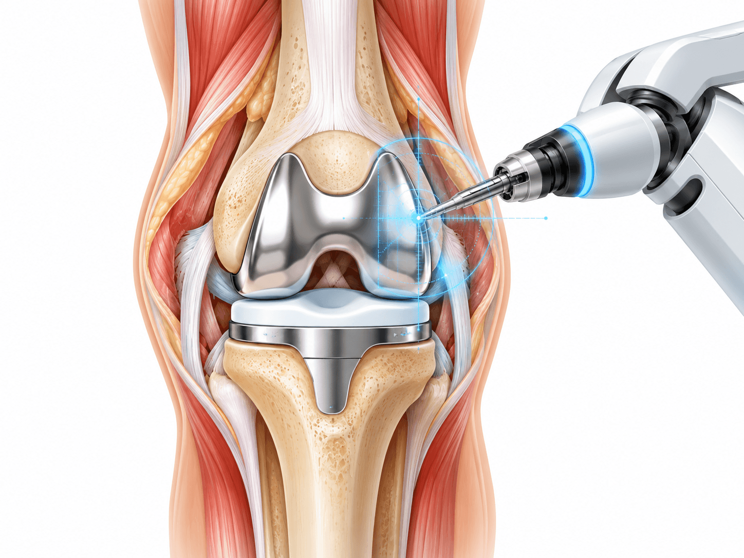
Robotic Osteotomy
Robotic technology is developing in orthopedic deformity surgery but is not standard for routine knee osteotomy.
Most widely deployed orthopedic robots remain focused on joint replacement.
Research platforms may eventually assist with precise bone cutting or real-time correction.
At present, robotic branding should not be presented as essential to a high-quality HTO or DFO.
Digital Planning Software
Modern software can calculate mechanical axis deviation, joint orientation angles and correction magnitude.
The surgeon can simulate different osteotomy locations.
This helps determine whether HTO, DFO or double-level correction will preserve a physiological joint line.
Digital planning also improves documentation and communication.
Fixation for Double-Level Osteotomy
Double-level correction typically requires separate fixation for the femoral and tibial osteotomies.
This means two plates or another combination of fixation devices may be necessary.
Hardware placement is planned so the implants do not interfere with each other or future surgery.
The greater implant burden contributes to the complexity and cost of the procedure.
Intramedullary Fixation
Certain deformity corrections can be stabilized with an intramedullary nail rather than a plate.
The nail travels through the central canal of the bone.
This can be useful for selected femoral or tibial deformities.
Standard medial opening-wedge HTO is usually treated with plate fixation, but intramedullary technology has a role in broader deformity correction.
Fixation and Future Knee Replacement
Implant position can influence later arthroplasty.
Hardware that lies away from the future replacement components can sometimes remain.
Other plates need removal.
Surgeons performing osteotomy in younger patients often consider the possibility of future knee replacement during initial hardware placement.
Smart and Sensor-Based Alignment
Experimental systems can measure forces or alignment intraoperatively.
Wearable sensors are also being studied during rehabilitation.
These technologies may eventually improve individualized correction and recovery monitoring.
They are not currently required for standard evidence-based knee osteotomy.
Risks and how they are managed
All surgery carries risk. Partner hospitals follow enhanced-recovery and infection-prevention protocols, and your surgeon will discuss the risks specific to your case before consent.
- Delayed bone healing: The osteotomy can take longer than expected to unite, requiring prolonged protection and additional imaging.
- Nonunion: The bone may fail to unite completely. Smoking, metabolic problems, large corrections and poor bone biology can increase the risk, and additional bone grafting or fixation may occasionally be required.
- Loss of correction: The bone can shift before healing if fixation fails or the osteotomy is overloaded prematurely.
- Hinge fracture: The intact cortical hinge can fracture during an opening-wedge osteotomy. Many hinge fractures still heal successfully, but some require modified fixation or rehabilitation.
- Hardware irritation: Plates and screws can become prominent or uncomfortable, particularly in thin patients. Hardware removal may be considered after full bone healing.
- Infection: Superficial infection may respond to antibiotics, while deeper infection involving the plate can require surgical washout and occasionally hardware management.
- Blood clots: Deep-vein thrombosis can occur after lower-limb surgery, with pulmonary embolism being a rare but serious complication.
- Bleeding or haematoma: Blood can accumulate around the surgical site and occasionally require treatment.
- Knee stiffness: Swelling, pain and scar tissue can temporarily reduce motion. Persistent stiffness occasionally requires additional treatment.
- Persistent pain: Osteotomy cannot guarantee complete symptom resolution, particularly when cartilage damage is more extensive than anticipated.
- Undercorrection: Too little realignment can leave the damaged compartment overloaded and reduce the expected benefit.
- Overcorrection: Excessive realignment can overload the opposite compartment and create new symptoms.
- Joint-line obliquity: A large correction through the wrong bone can create an abnormal joint-line angle, which is why accurate deformity analysis is important.
- Unintended change in tibial slope: HTO can alter sagittal alignment and thereby influence cruciate ligament forces and knee mechanics.
- Change in patellar height: Certain tibial osteotomy techniques can alter the relative position of the kneecap.
- Nerve injury: Nerve damage is uncommon but can produce numbness, weakness or neuropathic pain.
- Blood-vessel injury: Major vascular injury is rare but potentially serious because important vessels lie behind the knee.
- Compartment syndrome: Excessive pressure within the leg muscles is uncommon but requires urgent treatment.
- Fracture: The bone can fracture outside the planned osteotomy line during surgery or recovery.
- Delayed return to activity: Bone healing can progress more slowly than expected even without complete nonunion.
- Progression of osteoarthritis: Osteotomy redistributes load but does not eliminate the biological process of arthritis.
- Need for future knee replacement: Some patients eventually progress to partial or total knee arthroplasty.
- Complexity of later knee replacement: Conversion to arthroplasty after osteotomy can be technically more demanding than primary replacement.
- Complications from associated surgery: Meniscus repair, cartilage restoration or ligament reconstruction adds its own procedure-specific risks.
- Anaesthetic and medical complications: Cardiovascular, respiratory, urinary or medication-related problems can occur after major surgery, particularly in patients with significant medical conditions.
Alternatives
- Structured physiotherapy: Strengthening the quadriceps, hips and core can improve function and reduce symptoms without changing the bony alignment.
- Activity modification: Reducing repetitive high-impact loading can help control compartment-specific knee pain.
- Weight management: Reducing excess body weight decreases force passing through the knee and can improve symptoms.
- Pain medication: Appropriate analgesics or anti-inflammatory medication may help manage symptoms when medically suitable.
- Unloader knee brace: A valgus or varus-producing brace can temporarily shift load away from the painful compartment without surgery.
- Injections: Corticosteroid, hyaluronic acid or selected biological injections may provide symptom relief but do not permanently correct structural malalignment.
- Meniscus repair: A symptomatic meniscus tear may require repair when alignment does not create substantial compartment overload.
- Cartilage restoration: A focal cartilage defect can sometimes be treated without osteotomy when limb alignment is already acceptable.
- Ligament reconstruction: Knee instability can sometimes be addressed through ligament reconstruction alone when bony alignment is not an important contributor.
- Distal femoral osteotomy: DFO may be a better alternative to HTO when the deformity originates primarily in the femur.
- High tibial osteotomy: HTO may be preferred when the deformity originates primarily in the tibia.
- Double-level osteotomy: Large deformities involving both femur and tibia can be corrected at both levels rather than creating an excessive correction in one bone.
- Unicompartmental knee replacement: Selected patients with isolated medial or lateral compartment arthritis may be better candidates for partial knee replacement than osteotomy.
- Total knee replacement: Patients with widespread end-stage osteoarthritis, major stiffness or multicompartment cartilage loss are generally more appropriate for total knee arthroplasty.
- Continued observation: Patients with manageable symptoms and preserved function may choose to delay surgery while monitoring progression.
What Knee Osteotomy costs
The contracted Turkey partner package next to approved self-pay benchmarks. Benchmarks are 20th–80th percentile ranges of approved records, normalised to USD.
Turkey package
$5,000 – $8,500
United Kingdom self-pay
$8,000 – $21,150
Germany self-pay
$6,750 – $18,150
Typical self-pay range by country
Surgeons who perform Knee Osteotomy
All surgeonsHospitals offering this procedure
Antalya Yaşam Hospital
Private hospital in Muratpaşa, Antalya, with an orthopedics and traumatology department
BHT Clinic Istanbul Tema Hospital
Large private hospital in Atakent, Küçükçekmece, with an orthopedics and traumatology unit
Biruni University Hospital
University hospital in Küçükçekmece, Istanbul, with a published orthopedics and traumatology department
Çankaya Hospital for Orthopedic Care
Çankaya Hospital for Orthopedic Care is an orthopedic hospital in central Ankara with robotic-arm assisted surgery
Sources and references
Peer-reviewed guidance and institutional sources used to write and review this page.
- 01Definition of HTO, indications, opening- and closing-wedge concepts, joint preservation and general recovery.
Hospital for Special Surgery, 2024
https://www.hss.edu/health-library/conditions-and-treatments/high-tibial-osteotomy-knee-surgery
- 02Contemporary patient selection, medial compartment disease, joint preservation, activity and factors affecting outcomes
Pubmed, 2025
https://pubmed.ncbi.nlm.nih.gov/40514158/
- 03Contemporary long-term HTO survivorship and avoidance of knee arthroplasty. The pooled non-conversion proportion in this systematic review was 86%. PubMed
Pubmed, 2026
https://pubmed.ncbi.nlm.nih.gov/41696927/
- 04Outcomes and survivorship in patients with advanced radiographic medial compartment osteoarthritis.
Pubmed, 2024
https://pubmed.ncbi.nlm.nih.gov/38430233/
- 05Very long-term survival and predictors of HTO durability. PubMed
American Journal of Sports Medicine / PubMed, 2024
https://pubmed.ncbi.nlm.nih.gov/38243788/
- 06Large systematic review comparing alignment correction, complications, reinterventions and conversion to arthroplasty after opening- and closing-wedge HTO.
Knee Surgery, Sports Traumatology, Arthroscopy / PubMed, 2026
https://pubmed.ncbi.nlm.nih.gov/42024297/
- 07Comparative evidence for osteotomy technique, locking plate fixation and postoperative weight-bearing. PubMed
Knee Surgery, Sports Traumatology, Arthroscopy / PubMed
https://pubmed.ncbi.nlm.nih.gov/36473985/