Key takeaways
- 1Hip arthroscopy is minimally invasive surgery performed through small portals using a camera and specialized instruments to diagnose and treat structural hip problems.
- 2Common reasons for surgery include femoroacetabular impingement, labral tears, selected cartilage injuries, loose bodies and certain synovial conditions.
- 3Hip arthroscopy labral repair aims to preserve and reattach a repairable labrum rather than routinely removing damaged tissue.
- 4When hip impingement caused the labral tear, repairing the labrum without correcting the underlying cam or pincer deformity can leave the mechanical cause untreated.
- 5Hip arthroscopy is generally most successful in appropriately selected patients with limited or no advanced osteoarthritis.
- 6Arthroscopy does not regenerate an extensively arthritic hip and is usually a poor substitute for hip replacement when major joint-space loss is already present.
- 7Most procedures are performed as outpatient surgery, with patients beginning assisted walking shortly afterward.
- 8Recovery typically requires structured physiotherapy and usually lasts several months rather than a few weeks, especially for athletes.
- 9Temporary numbness or discomfort related to the traction used to open the hip joint can occur, although modern traction and postless techniques aim to reduce these problems.
- 10Return to sport, work, driving, sleeping positions and intimacy after hip arthroscopy should be based on healing, pain and the exact procedure rather than one universal timetable.
Overview
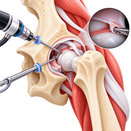
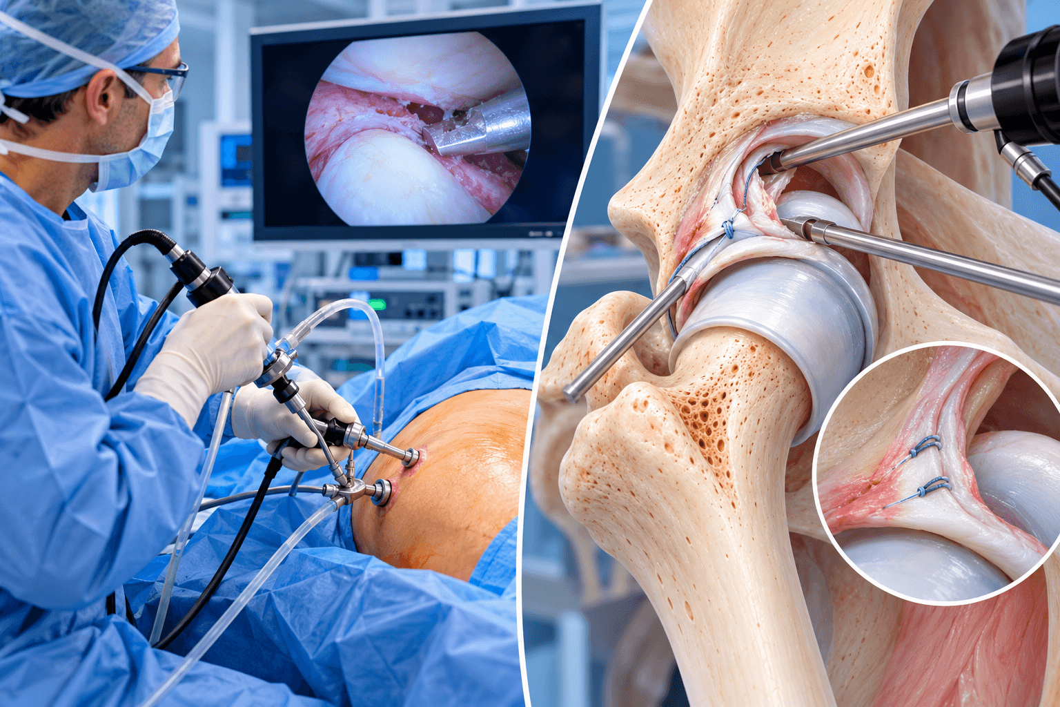
Hip arthroscopy is a minimally invasive operation in which an orthopedic surgeon inserts a small camera into the hip joint through a small incision and uses additional small instruments through other portals to diagnose and treat structural problems. The camera sends a magnified image to a monitor, allowing the surgeon to inspect the labrum, cartilage, femoral head, acetabulum, capsule and other structures without using the larger exposure required for traditional open hip surgery.
The procedure is sometimes called hip scope surgery or arthroscopic hip surgery. Although the incisions are small, the operation can involve substantial reconstruction inside the joint. The surgeon may repair torn tissue, remove damaged fragments, reshape abnormal bone and restore the seal of the labrum around the socket.
Hip arthroscopy has developed into an important component of modern hip-preservation surgery, particularly for younger or middle-aged patients whose hip pain is caused by mechanical abnormalities but whose joint has not yet progressed to advanced osteoarthritis.
What Does an Arthroscope Do?
An arthroscope is a narrow instrument containing a camera and light source. Once inserted into the hip, it provides a magnified view of structures that cannot otherwise be seen directly without opening the joint.
Sterile fluid is circulated through the hip during surgery to create a clear working environment. The surgeon views the procedure on high-definition monitors and introduces small instruments through separate portals.
Depending on the operation, these instruments can include suture passers, graspers, radiofrequency probes, burrs for reshaping bone, anchors for repairing the labrum and devices for treating selected cartilage damage.
Why Is Hip Arthroscopy More Challenging Than Knee Arthroscopy?
The hip is a deep ball-and-socket joint surrounded by powerful muscles and a strong capsule. Unlike the knee, there is very little natural working space between the femoral head and acetabulum.
To safely insert the arthroscope into the central part of the joint, the surgeon usually needs to create temporary separation between the femoral head and socket. This is achieved through controlled traction.
The depth of the joint and proximity of nerves and blood vessels make hip arthroscopy technically demanding. Surgeon experience is therefore particularly important, especially when complex labral or bony reconstruction is required.
What Conditions Can Hip Arthroscopy Treat?
The most common contemporary indication is femoroacetabular impingement syndrome, often combined with a labral tear.
Hip arthroscopy can also treat selected isolated labral tears, loose bodies, some cartilage lesions, synovial disorders and certain abnormalities around the joint. In specialist centers, arthroscopic or endoscopic techniques can also address extra-articular problems such as snapping tendons or selected tendon disorders.
Not every hip problem can be treated arthroscopically. Severe dysplasia, extensive osteoarthritis, major deformity and certain tendon or bone conditions can require different operations.
Femoroacetabular Impingement
Femoroacetabular impingement, or FAI, occurs when the femoral head-neck junction and acetabular rim contact each other abnormally during hip motion.
Repeated contact can damage the labrum and articular cartilage.
FAI is broadly described as cam, pincer or mixed morphology. Cam impingement involves excess bone or loss of the normal round contour at the femoral head-neck junction. Pincer morphology involves excessive or unfavorable acetabular coverage or rim morphology.
Many symptomatic patients have a combination.
The presence of cam or pincer morphology on an X-ray alone does not mean surgery is required. Many people have these shapes without symptoms. A diagnosis of FAI syndrome requires the clinical picture, examination and imaging to fit together.
Cam Impingement
Cam morphology creates a prominent or insufficiently concave region at the femoral head-neck junction.
During flexion and rotation, this region can enter the acetabulum abnormally and shear the cartilage or pull on the labrum.
Arthroscopic femoroplasty reshapes this area and restores a smoother femoral head-neck offset.
The surgeon must remove enough abnormal bone to reduce impingement without weakening the femoral neck through excessive resection.
Pincer Impingement
Pincer-type impingement occurs when the acetabular rim provides excessive or unfavorably oriented coverage.
The labrum can become compressed between the acetabulum and femoral neck.
Arthroscopic acetabuloplasty or rim trimming removes a controlled amount of excess acetabular bone when clinically appropriate.
The amount removed must be carefully planned. Excessive rim resection can reduce coverage too much and contribute to instability.
Mixed FAI
Many symptomatic patients have both cam and pincer features.
During surgery, the surgeon can treat both sides of the mechanical conflict.
The labrum is commonly repaired at the same time if it is detached or torn.
This comprehensive approach is important because treating the damaged labrum without addressing the bone repeatedly causing impingement can compromise the repair.
Hip Labral Tear
The acetabular labrum is a ring of fibrocartilage around the edge of the hip socket.
It deepens the socket and helps create a fluid seal around the femoral head. This seal contributes to joint stability, lubrication and distribution of load.
Labral tears can develop from FAI, dysplasia, trauma, repetitive athletic activity or degeneration.
Symptoms often include groin pain, clicking, catching, stiffness or pain with rotation.
A tear visible on MRI does not automatically require surgery. Labral abnormalities can also appear in people without symptoms. Clinical correlation is essential.
Hip Arthroscopy Labral Repair
Hip arthroscopy labral repair aims to preserve a torn but viable labrum and reattach it to the acetabular rim using small suture anchors.
The surgeon first identifies the tear and prepares the underlying bone to encourage healing. Small anchors are inserted around the rim. Sutures are passed through or around the labral tissue and tied or secured so the labrum returns to the edge of the acetabulum.
The goal is more than closing a tear. A good repair attempts to restore the labrum's suction seal and functional relationship with the femoral head.
Modern hip-preservation surgery generally favors preservation and repair of viable labral tissue when practical rather than unnecessarily removing it.
Labral Debridement
Not every labrum can be repaired.
Some tears involve small unstable flaps, severely degenerative tissue or areas that are not structurally suitable for fixation.
In selected situations, the surgeon can trim unstable tissue while preserving as much healthy labrum as possible.
This is known as labral debridement.
Extensive historical removal of labral tissue has become less desirable as understanding of the labrum's biomechanical role has improved.
Labral Reconstruction
Sometimes the labrum is too damaged, too small or previously removed to provide a useful repair.
In these cases, the surgeon can reconstruct the deficient segment using graft tissue.
The graft may come from donor tissue or another suitable tendon source depending on the technique.
Labral reconstruction is more complex than routine repair and is frequently performed in revision cases or severe labral deficiency.
Labral Augmentation
Augmentation preserves part of the patient's native labrum while adding graft tissue to improve its size or function.
This can be considered when the labrum remains partially viable but is too thin or damaged to create an adequate seal on its own.
The decision between repair, augmentation and reconstruction is made according to tissue quality rather than keyword preference or implant marketing.
Cartilage Damage
Articular cartilage covers the femoral head and acetabulum.
Once damaged, its ability to heal is limited.
Hip arthroscopy can treat selected focal cartilage defects by stabilizing loose edges, removing unstable fragments or performing a cartilage procedure.
The prognosis depends heavily on the extent of cartilage damage. A small localized defect in an otherwise preserved joint is very different from widespread cartilage loss caused by advanced osteoarthritis.
Microfracture
Microfracture is a marrow-stimulation technique used for selected full-thickness cartilage defects.
The damaged area is prepared and small openings are created in the underlying bone. Marrow elements enter the defect and form repair tissue.
This repair tissue is not identical to native hyaline cartilage.
Microfracture also changes rehabilitation. Patients commonly need more restrictive weight bearing to protect the healing cartilage surface.
Newer cartilage-restoration strategies can be considered in selected cases, and microfracture is no longer automatically the answer for every cartilage defect.
Loose Bodies
Small pieces of cartilage, bone or other tissue can become free within the joint.
These loose bodies can cause catching, locking and pain.
Arthroscopy provides an effective minimally invasive method for locating and removing them.
The surgeon should also determine why they developed. Treating only the loose body without addressing an underlying structural problem can allow symptoms to recur.
Synovial Disorders
The synovium is the tissue lining the joint.
Inflammatory or proliferative synovial conditions can occasionally produce pain, swelling or loose bodies.
Arthroscopic synovectomy can remove abnormal synovial tissue in selected cases.
These disorders require accurate diagnosis because their biology and recurrence risks differ substantially from routine FAI.
Ligamentum Teres Problems
The ligamentum teres lies within the hip joint.
Partial tears can occur with trauma or instability.
Arthroscopic debridement, thermal treatment or reconstruction can be considered in carefully selected patients.
Isolated ligamentum teres pathology is less common than FAI and labral injury, and the surgeon must evaluate whether underlying instability or dysplasia is contributing.
Snapping Hip
Snapping hip can be internal, external or intra-articular.
Some forms respond well to physiotherapy and do not require surgery.
Persistent internal snapping related to the iliopsoas tendon can occasionally be treated endoscopically, although surgeons are increasingly cautious about releasing important stabilizing structures in patients who may already have subtle instability.
External snapping related to the iliotibial band can also be treated surgically in selected refractory cases.
These procedures are related to hip endoscopy but are not identical to central-compartment arthroscopy.
Hip Arthroscopy and Dysplasia
Hip dysplasia means the acetabulum does not provide enough coverage of the femoral head.
A dysplastic hip can develop labral tears because the labrum is carrying abnormal loads.
Repairing that labrum through arthroscopy without correcting significant structural instability can fail.
In some patients, periacetabular osteotomy is the more appropriate operation because it reorients the socket and addresses the cause of instability.
Hip arthroscopy can occasionally be used alongside corrective surgery or for carefully selected borderline cases, but significant dysplasia requires specialist evaluation.
Hip Arthroscopy and Osteoarthritis
Advanced osteoarthritis is one of the strongest reasons not to expect a good result from hip arthroscopy.
Arthroscopy can repair labral tissue and reshape bone, but it cannot recreate an extensively worn joint surface.
When cartilage loss has become widespread and the joint space is substantially narrowed, pain is often generated by the arthritic joint itself rather than by an isolated repairable lesion.
In those circumstances, total hip replacement may provide a much more predictable treatment.
Can Hip Arthroscopy Prevent Hip Replacement?
Hip arthroscopy is a joint-preservation operation, but it should not be advertised as a guaranteed way to prevent future hip replacement.
Correcting symptomatic FAI and repairing the labrum can improve pain and function and may remove mechanical factors associated with cartilage damage.
However, osteoarthritis is influenced by many variables, and current research does not establish that arthroscopy reliably prevents future arthritis in every patient.
The more cartilage damage already present at surgery, the less realistic it is to expect arthroscopy to permanently prevent progression.
Hip Arthroscopy vs Hip Replacement
Hip arthroscopy preserves the native joint.
Total hip replacement removes damaged joint surfaces and replaces them with prosthetic components.
These operations are therefore intended for different biological situations.
A younger patient with symptomatic cam impingement, a repairable labral tear and preserved cartilage may be an excellent arthroscopy candidate.
A patient with severe joint-space narrowing and widespread bone-on-bone arthritis usually requires a completely different strategy.
Choosing correctly between preservation and replacement is more important than trying to make every patient eligible for the less invasive procedure.
Conditions treated
Who it's for
- Femoroacetabular impingement syndrome that remains symptomatic despite appropriate nonsurgical treatment
- Symptomatic acetabular labral tears associated with a treatable structural problem
- Repairable labral tears requiring arthroscopic fixation
- Cam impingement requiring femoroplasty
- Selected pincer impingement requiring acetabular rim correction
- Combined cam and pincer femoroacetabular impingement
- Selected focal cartilage lesions
- Loose bodies within the hip joint
- Selected synovial disorders requiring arthroscopic synovectomy
- Selected ligamentum teres injuries
- Certain mechanical symptoms following previous hip surgery
- Selected extra-articular hip conditions treated endoscopically
- Revision treatment after previous hip arthroscopy when a correctable structural problem remains
- Selected athletes with mechanical hip pathology and preserved joint cartilage
Good candidates
A good candidate for hip arthroscopy has a structural problem that matches their symptoms and is realistically correctable through minimally invasive surgery.
This distinction is important because MRI abnormalities alone are not enough. Labral tears and cam morphology can exist in people who have no hip pain.
The surgeon considers the pain pattern, examination findings, radiographs, MRI findings and response to previous treatment. Diagnostic injections can sometimes help confirm that pain is truly originating from inside the hip.
Joint preservation is most attractive when the articular cartilage is still reasonably healthy.
Patients With FAI
Patients with symptomatic femoroacetabular impingement often report groin pain during deep hip flexion, sitting, squatting or rotational sport.
They may feel restricted when tying shoes, getting into a low car or performing athletic movements.
When examination and imaging confirm clinically meaningful cam or pincer morphology and conservative treatment has failed, arthroscopy can correct the bone abnormality and treat associated labral damage.
The objective is to restore functional clearance rather than remove bone simply because it appears prominent on an X-ray.
Patients With Labral Tears
A symptomatic labral tear can produce deep groin pain, clicking or catching.
Many tears are initially treated without surgery.
Physiotherapy can improve hip mechanics and strengthen surrounding muscles even though it does not physically stitch a torn labrum back together.
If symptoms remain substantial and the tear appears to be a meaningful pain generator, arthroscopy can be considered.
The surgeon also needs to identify why the labrum tore. Mechanical abnormalities should be addressed during the same procedure where appropriate.
Athletes
Athletes frequently develop symptoms from FAI and labral pathology because their sports repeatedly place the hip through demanding ranges.
Hip arthroscopy can allow many appropriately selected athletes to return to competitive activity.
However, return to sport is not guaranteed, and the timeline depends on sport, position, cartilage health, procedure complexity and rehabilitation.
A professional football player and a recreational cyclist should not receive identical return-to-sport expectations.
Younger Adults
Hip arthroscopy is often performed in young and middle-aged adults because preserving the natural joint has particular value when significant arthritis is absent.
Chronological age alone is not the main determinant.
A healthy 45-year-old with good cartilage can be a stronger candidate than a 30-year-old with advanced degenerative change.
Biological joint condition matters more than simply fitting into a young age category.
Older Adults
Some older adults remain candidates when arthritis is limited and a clearly correctable problem exists.
However, the probability of cartilage degeneration increases with age.
The surgeon should carefully evaluate joint-space preservation and cartilage condition.
In an older patient with substantial osteoarthritis, arthroscopy may provide temporary or inadequate relief and can simply delay the more appropriate hip replacement.
Patients With Early Arthritis
Mild degenerative changes do not always rule out arthroscopy.
The decision depends on how much cartilage remains, how much joint-space narrowing exists and whether symptoms appear to come predominantly from treatable impingement or labral pathology.
Expectations should become more cautious as degenerative change increases.
A patient should understand that arthroscopy can treat mechanical pathology but cannot restore a globally arthritic joint to normal cartilage.
Patients With Advanced Arthritis
Advanced osteoarthritis generally predicts poor results.
When substantial joint-space loss, large areas of cartilage destruction and major arthritic change are present, total hip replacement is usually more appropriate.
Performing arthroscopy simply because the patient wants to avoid replacement can expose them to surgery, rehabilitation and expense without addressing the fundamental cause of pain.
Good hip-preservation surgery includes knowing when not to perform arthroscopy.
Patients With Hip Dysplasia
Significant dysplasia deserves particular caution.
The labrum often becomes enlarged and overloaded because it is helping stabilize an undercovered femoral head.
Removing or even repairing labral tissue without correcting the bony instability may not solve the problem.
Patients with dysplasia should be evaluated by a surgeon familiar with both hip arthroscopy and periacetabular osteotomy rather than being assessed solely through an arthroscopy-focused lens.
Patients With Generalized Ligamentous Laxity
Some patients naturally have greater joint flexibility.
This can influence capsular stability.
If arthroscopy is performed, capsular management may become especially important because leaving the capsule excessively open can contribute to postoperative instability.
The surgeon evaluates Beighton-type hypermobility findings, hip anatomy and symptoms when planning the procedure.
Patients With Obesity
Hip arthroscopy can technically be performed in patients with higher body weight, but positioning, traction and instrument access can become more challenging.
Obesity can also affect rehabilitation and overall surgical risk.
The decision is individualized.
A patient's body weight should be considered alongside joint damage, symptoms and expected benefit rather than functioning as the only selection criterion.
Revision Hip Arthroscopy Candidates
A patient with persistent symptoms after previous arthroscopy requires detailed reassessment.
Common reasons include residual cam morphology, inadequate acetabular correction, recurrent labral damage, adhesions, capsular insufficiency or progressive arthritis.
Revision surgery should not simply repeat the original operation without identifying why the first procedure failed.
CT imaging, old operative records and previous arthroscopy photographs can be especially valuable.
Before surgery
Confirming the Diagnosis
Hip pain has many possible sources.
The lumbar spine, sacroiliac joint, abdominal wall, adductor muscles, iliopsoas tendon and surrounding nerves can all cause symptoms that patients describe as hip pain.
A successful arthroscopy begins with establishing that the joint itself is an important pain generator.
The surgeon reviews symptom location, activities that trigger pain and mechanical symptoms such as catching or clicking.
The entire clinical picture needs to match the proposed operation.
Physical Examination
The examination includes gait, hip range of motion and strength.
Provocative maneuvers place the hip into positions that reproduce impingement or labral symptoms.
The surgeon assesses rotational range, flexion and abductor strength.
Hypermobile patients are evaluated for generalized laxity.
The spine and adjacent joints are also assessed because treating an incidental labral tear will not help if the primary pain source lies elsewhere.
X-Rays
High-quality radiographs are fundamental in hip-preservation assessment.
They show joint-space width, cam morphology, acetabular coverage, pelvic orientation and arthritic changes.
Specialized views can demonstrate the femoral head-neck junction more clearly.
X-rays are often more useful than patients expect because they reveal the bone mechanics driving labral injury.
MRI demonstrates soft tissues but should not replace careful radiographic analysis of hip shape.
MRI
MRI helps evaluate the labrum, cartilage and surrounding soft tissues.
MR arthrography, in which contrast is placed inside the joint, was historically used extensively and remains useful in selected settings.
Modern high-resolution MRI can visualize labral pathology well without intra-articular contrast in many centers.
The choice depends on scanner quality, local expertise and the clinical question.
CT Scan
CT is useful when the surgeon needs detailed three-dimensional information about bony morphology.
It can show femoral version, acetabular version and the exact distribution of cam bone.
Three-dimensional models can assist planning in complex FAI, revision surgery or unusual deformity.
CT is not required for every simple labral tear.
Radiation exposure and clinical usefulness should both be considered.
Diagnostic Injection
An image-guided injection of local anaesthetic into the hip joint can sometimes help determine whether pain is truly intra-articular.
Substantial temporary relief suggests that structures within the hip are an important pain source.
A lack of response does not absolutely exclude hip disease, but it can prompt the surgeon to reconsider the diagnosis.
Corticosteroid can sometimes be included for therapeutic relief depending on the clinical context.
Conservative Treatment Before Surgery
Many patients first undergo activity modification and physiotherapy.
Rehabilitation focuses on hip and core strength, movement patterns and avoiding repetitive provocative positions while symptoms settle.
Anti-inflammatory medication or other pain treatment can be used when medically appropriate.
Some patients improve enough that surgery becomes unnecessary.
Hip arthroscopy is generally considered when meaningful symptoms persist despite a reasonable nonsurgical programme and the anatomy suggests a surgically correctable problem.
Understanding the Labral Plan
Patients should ask whether the surgeon expects to repair, debride, augment or reconstruct the labrum.
The decision sometimes changes during surgery once tissue quality is directly visualized.
However, the general treatment philosophy should be discussed beforehand.
A surgeon who preserves viable labral tissue whenever reasonable may approach the same MRI differently from a surgeon whose routine practice is extensive debridement.
Understanding the Bone Correction
When FAI is present, the patient should understand which bone abnormality is being corrected.
Cam morphology is treated on the femoral side.
Pincer pathology is treated on the acetabular side.
Mixed impingement can require both.
The operation should not be described merely as “fixing a labral tear” if substantial bone reshaping is also planned, because bony correction affects rehabilitation and long-term mechanics.
Cartilage Assessment
Cartilage health is one of the most important predictors of whether joint-preservation surgery makes sense.
MRI provides useful information, but the true severity of cartilage damage can occasionally become fully apparent only during arthroscopy.
Patients with suspected cartilage injury should understand that unexpected severe arthritis can worsen prognosis.
The surgeon should discuss how intraoperative findings could change the procedure.
Evaluating Dysplasia
Acetabular coverage must be assessed carefully.
Borderline coverage creates one of the more complex areas of modern hip preservation because some patients behave like impingement patients while others have instability.
Capsular laxity, femoral version and symptoms all contribute.
If structural instability is substantial, periacetabular osteotomy may provide a more logical mechanical correction than arthroscopy alone.
Medical Assessment
Although hip arthroscopy patients are frequently young and medically healthy, routine preoperative assessment remains important.
The surgical team reviews cardiovascular and respiratory history, medication, allergies and previous anaesthesia.
Blood tests depend on patient age, hospital protocol and medical history.
The patient should disclose supplements as well as prescription medication.
Blood-Thinning Medication
Anticoagulants and antiplatelet medication require specific instructions.
The patient should never stop an important cardiovascular medication independently.
The surgeon, anaesthetist and prescribing physician coordinate interruption where needed.
The objective is to balance surgical bleeding with clotting risk.
Smoking and Nicotine
Smoking and nicotine can impair tissue healing.
Labral repair and bone remodeling both depend on a biological healing response.
Stopping before surgery is therefore strongly encouraged.
Nicotine avoidance also reduces general wound and cardiovascular risks.
Preparing for Crutches
Most patients use crutches after the operation.
Practicing beforehand can make the first postoperative days easier.
The patient should know whether weight bearing will be full, partial or limited.
A labral repair with straightforward FAI correction can have a different protocol from microfracture or another cartilage-restoration procedure.
Preparing the Home
Frequently used items should be placed within easy reach.
A stable chair is useful.
The patient may temporarily have difficulty bending deeply to put on socks or shoes.
A backpack can make carrying objects easier while using crutches.
Patients should also arrange transportation because they will not drive immediately after surgery.
Planning Work Leave
The type of work matters.
Someone working remotely at a computer may return relatively quickly.
A nurse, construction worker, fitness professional or warehouse employee places much greater demands on the hip.
Patients should plan leave based on their actual occupational tasks rather than comparing themselves with general internet timelines.
Planning Rehabilitation
Physiotherapy is not an optional afterthought for most hip arthroscopy patients.
The surgeon and therapist need a procedure-specific protocol.
A repaired labrum, reshaped femoral neck and healing capsule each have biological limits.
The programme gradually restores motion while avoiding excessive irritation, then builds strength and finally introduces impact, agility and sport-specific loading.
Discussing Expectations
One of the most useful preoperative conversations concerns what surgery can and cannot accomplish.
Arthroscopy can correct certain mechanical problems and repair soft tissues.
It cannot guarantee that the hip will never develop arthritis.
It cannot make severely damaged cartilage normal again.
Realistic expectations improve decision-making and help the patient judge recovery according to meaningful functional goals rather than simply waiting for every sensation to disappear.
How the operation is performed
Hip arthroscopy surgery is performed through several small incisions around the hip. The surgeon gently distracts the femoral head from the socket, inserts a camera into the joint and uses specialized instruments to repair the labrum, treat cartilage damage and reshape abnormal bone where necessary.
The exact sequence varies according to the planned procedures.
Most operations involve work in the central compartment, where the labrum and weight-bearing cartilage are inspected, followed by work in the peripheral compartment around the femoral head-neck junction.
Patient Positioning
Hip arthroscopy can be performed with the patient lying on the back or on the side.
The surgeon selects the position according to training, equipment and preferred technique.
The foot is placed in a specialized boot or traction setup.
The leg needs to remain carefully padded because the operation requires controlled distraction of the joint.
Modern operating tables and postless systems have changed how traction can be applied.
Traction
The normal hip joint has almost no working space.
Traction gently separates the femoral head from the acetabulum by several millimeters so instruments can enter safely.
Historically, many systems used a padded post against the perineal region to provide countertraction.
Pressure from the post and traction itself could temporarily irritate nerves or soft tissues.
Modern techniques aim to minimize traction force and duration, and many surgeons now use postless distraction systems that rely on patient positioning and table friction instead of a perineal post.
Postless Hip Arthroscopy
Postless arthroscopy eliminates the traditional perineal post.
The operating table is positioned in a way that uses gravity and friction to counter traction.
This approach is intended to reduce pressure-related complications involving the groin and pudendal nerve.
Not every hospital uses postless equipment.
Excellent hip arthroscopy can still be performed with carefully controlled conventional traction, but the method of traction is a reasonable topic for patients to discuss with the surgeon.
Creating the Portals
The surgeon uses anatomical landmarks, a needle and usually fluoroscopic imaging to establish safe entry into the joint.
A guidewire and cannula create the pathway for the arthroscope.
Additional portals are then established for surgical instruments.
The skin incisions are generally small.
The location and number of portals vary according to the work required.
Diagnostic Arthroscopy
The surgeon first systematically inspects the joint.
The labrum is examined for tearing, detachment and tissue quality.
The acetabular and femoral cartilage are inspected.
The ligamentum teres and synovium are evaluated.
Unexpected findings can modify the planned procedure.
Arthroscopy provides direct visualization that can reveal damage not fully visible on MRI.
Capsulotomy
The hip capsule is a strong layer surrounding the joint.
A controlled incision in the capsule provides access for instruments.
Different capsulotomy patterns exist.
Capsular management has become increasingly important in modern hip-preservation surgery because excessive disruption can contribute to instability in susceptible patients.
The surgeon balances adequate working access with preservation of stability.
Labral Repair
During hip arthroscopy labral repair, the damaged labrum is assessed for viability.
The acetabular rim is prepared while avoiding unnecessary removal of supportive bone.
Small anchors are inserted into the rim.
Sutures are passed through or around the labrum and secured to restore its position.
The exact repair construct varies according to tear shape, labral size and surgeon technique.
Restoring the Suction Seal
The labrum contributes to a fluid seal around the femoral head.
One objective of repair is to restore this relationship.
After the sutures are secured, the surgeon can inspect how the labrum contacts the femoral head when traction is released.
A labrum that maintains a functional seal may contribute to joint lubrication and stability.
This biomechanical concept is one reason modern surgery emphasizes preservation rather than indiscriminate labral removal.
Acetabuloplasty
If pincer morphology requires correction, the surgeon uses a motorized burr to remove a carefully controlled amount of acetabular rim bone.
The aim is to eliminate pathological overcoverage while preserving enough socket coverage for stability.
Labral tissue may be temporarily elevated from the rim and then repaired after the bone correction.
The procedure requires precise planning because both inadequate and excessive rim trimming can create problems.
Femoroplasty
Cam correction is performed by reshaping the femoral head-neck junction.
Traction is usually released and the hip is moved so the surgeon can visualize different regions of the cam deformity.
A burr removes the abnormal prominence and creates a smoother transition between the femoral head and neck.
Fluoroscopy and dynamic examination help confirm that enough bone has been removed to improve clearance.
Dynamic Examination
The surgeon can move the hip through flexion and rotation during arthroscopy.
This allows direct assessment of whether the reshaped femur still contacts the acetabular rim abnormally.
Dynamic testing helps complement static preoperative imaging.
The objective is functional clearance, not simply making the X-ray look different.
Labral Reconstruction
When native labral tissue is irreparable, a graft can recreate the deficient segment.
The damaged labrum is prepared and anchors are placed along the acetabular rim.
The graft is sized and secured so it can contact the femoral head and contribute to a new seal.
Reconstruction takes longer than simple repair and is particularly relevant in complex primary cases and revision arthroscopy.
Cartilage Debridement
Unstable cartilage flaps can be trimmed to create stable edges.
The goal is not to remove normal cartilage.
Debridement can reduce mechanical catching from unstable tissue, but it does not regenerate the missing articular surface.
The size and location of the cartilage defect remain important prognostic factors.
Microfracture and Cartilage Procedures
For selected full-thickness defects, the surgeon can perform marrow stimulation or another cartilage-restoration strategy.
The damaged area is prepared until stable cartilage margins remain.
Small openings allow marrow elements to enter the defect.
These procedures often require a more protective rehabilitation programme because excessive early weight bearing can disrupt the healing surface.
Loose-Body Removal
Loose fragments are identified with the camera and removed using graspers.
Larger fragments occasionally require enlargement of a portal.
The rest of the joint is inspected because multiple loose bodies can be present.
The surgeon also treats the underlying disorder where possible.
Synovectomy
Abnormal or inflamed synovial tissue can be removed with arthroscopic shavers or radiofrequency instruments.
The extent depends on the diagnosis.
Diffuse synovial disease requires a different strategy from a small localized inflamed area.
Patients with proliferative synovial disorders may require longer-term surveillance because recurrence can occur.
Capsular Closure
At the end of surgery, the capsule can be repaired with sutures.
Capsular closure is particularly important in patients at increased risk of instability, including those with laxity or borderline structural coverage.
Some surgeons routinely close most capsulotomies.
Others tailor treatment according to anatomy and access.
Modern hip preservation increasingly recognizes the capsule as a functional stabilizer rather than merely a structure that blocks access to the joint.
Capsular Plication
Plication tightens the capsule more than simple closure.
It can be useful in selected patients with instability or generalized laxity.
The surgeon removes slack by overlapping or tensioning capsular tissue.
Overtightening should also be avoided because excessive restriction can reduce range of motion.
How Long Does a Hip Arthroscopy Take?
A hip arthroscopy commonly takes around one to two hours, although combined procedures can take two to three hours or longer.
This directly answers the searches “how long does a hip arthroscopy take,” “how long does hip arthroscopy take,” and “how long does hip arthroscopy surgery take.”
A short loose-body removal may be relatively quick. A complex operation combining labral reconstruction, acetabuloplasty, extensive femoroplasty, capsular work and cartilage restoration takes significantly longer.
Surgical quality should not be judged by speed alone. Precise bone correction and tissue preservation are more important than completing the operation within a particular number of minutes.
Fluoroscopy
Fluoroscopy provides real-time X-ray images during the procedure.
It can help establish joint distraction, guide portal entry and assess bone correction.
The surgeon uses radiation-protection measures.
Some modern navigation or imaging approaches can supplement fluoroscopy, but it remains widely used because it provides immediate information about bone anatomy.
Closing the Portals
After the instruments are removed, the small incisions are closed using sutures, adhesive strips or another closure method.
Dressings are applied.
The patient then moves to the recovery area.
Although the external wounds are small, the internal labral, capsular and bony work still requires meaningful healing time.
Hospital stay
Recovery Room
After the hip arthroscopy operation, the patient is monitored while anaesthesia wears off.
Blood pressure, oxygen level, pain and neurological function are assessed.
The surgical team checks movement and sensation in the leg and foot.
Temporary numbness can occur because traction and positioning place pressure on nerves during the procedure.
Most patients become medically ready to leave hospital later the same day.
Same-Day Discharge
Hip arthroscopy is usually an outpatient operation.
The patient typically does not need an overnight hospital stay unless medical circumstances, postoperative symptoms or logistical factors justify observation.
Before discharge, the patient should be able to tolerate fluids, control pain adequately and mobilize safely with crutches.
An adult should normally accompany the patient home after anaesthesia.
Pain After Surgery
Pain varies according to the work performed.
A combination of local anaesthetic, oral medication and sometimes a regional block is used to improve comfort.
The hip can feel sore and swollen.
Muscles around the joint can also feel tight from positioning and traction.
Pain should generally become progressively more manageable rather than worsen significantly after an initial period of improvement.
Swelling
Sterile fluid is used during arthroscopy to expand and wash the joint.
Some of this fluid can temporarily track into tissues around the hip and upper thigh.
The area can feel swollen or full during the first day.
The body gradually absorbs this fluid.
Bruising can also develop around the portals.
Temporary Numbness
Traction can temporarily irritate nerves supplying the leg, foot or perineal region.
Patients can experience numbness or tingling.
Most traction-related neurological symptoms reported after modern hip arthroscopy are temporary.
Persistent weakness, severe sensory loss or worsening neurological symptoms should be assessed promptly.
Crutches
Most patients leave the surgical center using crutches.
The amount of weight permitted through the operated leg depends on the exact procedure.
Some surgeons allow near-full or weight bearing as tolerated relatively early after routine labral repair and FAI correction.
Cartilage procedures can require more restrictive loading.
The patient should receive written instructions rather than rely on generic online advice.
Hip Brace
Some surgeons use a brace after labral repair or capsular surgery.
The brace can limit excessive extension, rotation or flexion during the early healing period.
Other surgeons do not routinely prescribe one.
The absence of a brace does not mean the operation was less substantial.
Rehabilitation philosophies vary.
Blood-Clot Prevention
Hip arthroscopy patients generally have a lower thrombotic risk than many hip replacement patients, but venous thrombosis can still occur.
Early mobility is important.
Medication may be prescribed according to the patient's individual risk and surgical protocol.
Patients with previous blood clots or other risk factors should discuss prevention specifically.
Before Going Home
The patient should understand how to use crutches, how much weight can be placed on the leg and how to manage the dressing.
Medication instructions should be clear.
The patient should also know when physiotherapy begins and when the first follow-up appointment is scheduled.
Emergency symptoms such as chest pain, shortness of breath, uncontrolled pain or neurological deterioration should be clearly explained.
Recovery
Arthroscopy hip recovery is a staged rehabilitation process that usually takes several months. The small incisions can heal quickly, but the repaired labrum, capsule, reshaped bone and surrounding muscles require substantially longer to recover.
Early rehabilitation focuses on protecting the repair and restoring gentle motion. The middle phase rebuilds walking mechanics and strength. Later rehabilitation adds impact, agility and sport-specific activity.
The patient should therefore not judge recovery simply by when the small portal scars look healed.
Hip Arthroscopy Recovery Time
For many patients, hip arthroscopy recovery time is approximately 3–6 months for broad functional recovery, while high-level athletes can require longer before full competition.
Basic household independence returns much sooner.
Desk work may be possible in one to three weeks.
Crutches are frequently used during the first few weeks.
Running and high-impact sport generally come later because strength, tissue healing and movement quality must all recover first.
Patients undergoing major cartilage restoration, reconstruction or revision surgery can have substantially longer rehabilitation.
Day of Surgery
The patient generally goes home using crutches.
Pain medication and ice are used according to instructions.
Short walking distances around the home help circulation.
The operated leg can feel heavy or temporarily numb.
The patient should not attempt unnecessary exercise on the first evening simply because the incisions appear small.
Days 1–3
Soreness, stiffness and swelling are common.
The patient performs basic exercises prescribed by the rehabilitation team.
These can include ankle pumps and gentle muscle activation.
Range-of-motion work begins according to the surgical protocol.
The main objective is controlled recovery rather than aggressively stretching the hip.
Week 1
Patients become more comfortable with crutch walking.
Portal wounds are monitored.
Sitting for long periods can be uncomfortable because hip flexion compresses the front of the joint.
Frequent position changes are useful.
Some patients begin formal physiotherapy during the first days, while other programmes start after the first postoperative review.
Weeks 2–4
Walking gradually improves.
The amount of weight placed through the leg increases according to the surgeon's protocol.
Patients who had routine FAI correction and labral repair may begin reducing crutch use if gait is controlled.
Those who had microfracture or extensive cartilage work frequently remain protected longer.
The goal is to avoid limping as support is reduced.
Weeks 4–6
Daily function usually becomes much easier.
Patients can often walk more normally and perform routine activities with less discomfort.
Strengthening becomes more active.
The therapist works on gluteal muscles, core stability and controlled hip mechanics.
Cycling on a stationary bike may already be part of rehabilitation depending on the protocol.
Weeks 6–12
Strength and endurance increase.
Patients can usually tolerate longer periods of walking and more demanding exercises.
Movement should become symmetrical.
Running is not introduced merely because pain is low.
The patient must demonstrate sufficient strength, balance and control before impact activity begins.
Months 3–4
Suitable patients can begin a graded return to running once they meet rehabilitation criteria.
This usually starts with short intervals and low total volume.
Agility and directional changes are added later.
The hip's response over the following day is important; increasing pain or prolonged soreness suggests the workload progressed too quickly.
Months 4–6
Many recreational athletes return progressively to sport during this period.
Sport-specific movements become more demanding.
Cutting, jumping and high-speed acceleration require substantially more hip control than straight-line walking.
Athletes should therefore complete a functional progression rather than using the calendar alone.
Beyond Six Months
Complex repairs and high-performance sports can require more than six months.
Some athletes continue improving for nine to twelve months.
Residual muscular asymmetry can persist even after pain has disappeared.
The objective is not merely to enter a competition but to return with enough strength and movement quality to tolerate repetitive loading.
Crutches After Hip Arthroscopy
Crutch duration depends on the procedure.
A patient undergoing limited debridement may progress quickly.
Labral repair often requires a period of protected or assisted walking.
Cartilage procedures can extend restrictions.
The surgeon's protocol should specify whether the patient is non-weight bearing, toe-touch weight bearing, partial weight bearing or weight bearing as tolerated.
The terminology needs to be understood correctly because patients frequently interpret it differently.
When Can I Walk Normally?
Many patients progress toward unassisted walking within the first several weeks.
The correct milestone is not simply walking without crutches but walking without a significant limp.
If the patient drops the pelvis, shortens the step or leans to one side, continued assistance can produce better mechanics.
A cane or one crutch can be useful during the transition.
Physiotherapy
Physiotherapy plays a central role.
Early treatment controls swelling and restores safe motion.
Later phases strengthen the gluteal muscles, hip rotators, core and lower limb.
Neuromuscular training helps the patient control hip position during squatting, stepping and sport-specific movement.
An athlete who restores strength but continues moving into repeated impingement positions may remain symptomatic.
Range of Motion
The hip should regain motion gradually.
Aggressive stretching early after labral repair or capsular surgery can irritate healing tissues.
Certain movements may initially be limited according to the surgical approach.
As healing progresses, the therapist restores extension, rotation and flexion while monitoring symptoms.
The goal is functional motion rather than forcing the joint into extreme positions.
Cycling
A stationary bicycle is frequently used relatively early because it encourages controlled hip motion with low impact.
The seat is positioned to avoid excessive hip flexion.
Resistance begins low.
Outdoor cycling returns later because balance, sudden stops and fall risk create additional demands.
Swimming
Pool-based exercise can be useful once the incisions are completely healed.
Walking in water reduces body-weight load.
Swimming provides conditioning without repetitive impact.
Breaststroke kicking can place the hip through substantial abduction and rotation, so stroke choice should be discussed during early rehabilitation.
Return to Running
Running typically begins only after walking is normal, hip strength is sufficiently restored and the patient can perform controlled single-leg tasks.
The precise timing varies.
A routine uncomplicated case can begin much earlier than a patient who underwent microfracture.
Distance and speed are increased gradually.
Return to running is a rehabilitation milestone, not the end of recovery.
Return to Sport
Recent sports literature supports a high potential for return among appropriately selected athletes, but reported percentages vary substantially because studies define “return” differently.
Returning to any training is not the same as returning to a previous competitive level.
The type of sport also matters.
Straight-line endurance activities, rotational sports and contact sports place very different loads on the hip.
A patient should ask about returning to their exact sport rather than relying on one global statistic.
Return to Work
Office employees can sometimes resume work within one to three weeks.
Sitting comfort and transportation are common limiting factors.
Jobs involving prolonged standing, lifting, squatting or climbing require considerably longer.
Heavy manual workers may need two to three months or more depending on what was repaired.
A staged return with temporary restrictions can sometimes shorten total absence from work.
Driving
Driving should wait until the patient can enter and exit the vehicle safely, is no longer taking medication that impairs alertness and can perform an emergency stop without hesitation.
Right-sided surgery tends to affect braking more directly.
The patient also needs adequate hip flexion to sit comfortably.
Crutch dependence or weight-bearing restrictions commonly delay return.
How to Sleep After Hip Arthroscopy
Patients frequently search how to sleep after hip arthroscopy because night positioning can be uncomfortable during the first few weeks.
Sleeping on the back is often the easiest early position. A pillow beneath or between the knees can reduce tension around the hip according to the surgeon's instructions.
Some patients can sleep on the non-operated side with pillows supporting the operated leg once it is comfortable. Sleeping directly on fresh portals is generally uncomfortable initially.
The most important rule is to respect any specific movement restrictions associated with capsular or labral repair. A patient does not need to remain in one rigid position all night unless their surgeon has given a specific reason.
Sleeping on the Operated Side
The operated side is usually tender early.
There is no benefit to forcing side sleeping simply because the patient technically can.
Once the wounds have healed and pressure is comfortable, side sleeping can often return gradually.
A pillow between the knees can support alignment.
Persistent severe night pain should be discussed with the surgical team rather than attributed automatically to normal recovery.
Intimacy After Hip Arthroscopy
Intimacy after hip arthroscopy can usually resume gradually once pain is controlled, the incisions are healed and the patient can move without placing excessive stress on the repaired hip. There is no universal date because labral repair, capsular work and cartilage procedures differ.
During the early phase, positions requiring deep hip flexion, wide abduction, forceful rotation or substantial weight through the operated leg may irritate healing tissues.
The patient and partner should choose comfortable positions in which the operated hip is supported and can remain within the movement limits given by the surgeon.
For uncomplicated recovery, intimacy commonly becomes practical within the first several weeks, but more demanding positions should return gradually. If a specific movement causes sharp groin pain, it should be stopped rather than pushed through.
Sexual Function and Traction Symptoms
Temporary perineal numbness or altered genital sensation has been reported after hip arthroscopy because traction can affect the pudendal nerve and surrounding tissues.
Modern techniques aim to minimize these problems, and postless systems avoid direct pressure from a perineal post.
Most temporary traction-related symptoms resolve.
Persistent sexual dysfunction, numbness, severe genital pain or bladder changes should be reported to the surgeon rather than assumed to be an expected part of recovery.
Returning to the Gym
Upper-body training can resume relatively early when the patient can safely position themselves and protect weight-bearing restrictions.
Lower-body exercise progresses according to rehabilitation.
Deep loaded squats, heavy deadlifts and explosive movements should not be among the first exercises resumed.
The therapist gradually restores strength before introducing high-load hip flexion and rotational demands.
Squatting
Deep squatting often reproduces the same flexion position that triggered preoperative FAI symptoms.
After corrective surgery, the patient eventually needs to regain functional squat mechanics without impingement.
Depth returns progressively.
Early aggressive deep squatting can irritate the labral repair and anterior capsule.
Movement quality matters more than reaching maximum depth quickly.
Stairs
Most patients can use stairs immediately with crutches and the correct technique.
Repeated stair climbing as exercise is unnecessary in the earliest phase.
Later, controlled step-ups and step-downs become useful rehabilitation exercises.
Single-leg pelvic control is particularly important.
Travel and Flying
Short travel can resume when the patient can sit safely and move around.
Long flights create additional issues because prolonged sitting can increase stiffness and venous thrombosis risk.
International patients should remain near the treating center long enough for initial postoperative assessment.
The return journey should accommodate crutches and allow frequent movement.
A patient who has undergone cartilage repair or another procedure with strict weight-bearing restrictions may need additional airport assistance.
Scar Recovery
Hip arthroscopy generally leaves several small portal scars.
Early redness and firmness gradually improve.
The scars should be protected from excessive sun while immature.
Scar massage can be considered after complete wound closure if recommended.
The cosmetic scars may become barely noticeable, but their size should not be confused with the amount of internal reconstruction performed.
Recovery timeline
- Protect the repair, control symptoms and establish safe assisted walking.1Protect the repair, control symptoms and establish safe assisted walking.
Days 0–14
The patient uses crutches according to weight-bearing instructions while pain and swelling gradually decrease. Gentle range-of-motion exercises and muscle activation begin. Excessive hip flexion and rotation are avoided according to the surgical protocol.
- Restore walking mechanics and early mobility.2Restore walking mechanics and early mobility.
Weeks 2–6
Crutch use decreases when permitted and when the patient can walk without a significant limp. Physiotherapy focuses on controlled motion, gluteal activation and core stability. Patients with cartilage procedures can remain protected longer.
- Rebuild strength and normal daily function.3Rebuild strength and normal daily function.
Weeks 6–12
Resistance training progresses. Balance and single-leg control improve. Most patients become considerably more independent, but high-impact exercise is generally still limited until strength and movement quality are adequate.
- Begin higher-level functional rehabilitation.4Begin higher-level functional rehabilitation.
Months 3–4
Suitable patients can start a graded running programme once objective criteria are met. More demanding strengthening, lateral movement and controlled agility are introduced according to the patient's goals.
- Progress toward return to sport and unrestricted recreation.5Progress toward return to sport and unrestricted recreation.
Months 4–6
Running volume, cutting, jumping and sport-specific drills are increased gradually. Return to play depends on strength, pain, range of motion and functional testing rather than time alone.
- Reach mature athletic recovery where required.6Reach mature athletic recovery where required.
Months 6–12
Competitive athletes and patients who underwent complex reconstruction may continue improving beyond six months. The final phase focuses on workload tolerance, confidence and reducing recurrent movement patterns that overload the hip.
Outcomes and success rates
How Successful Is Hip Arthroscopy?
Hip arthroscopy can provide substantial improvements in pain and function when the correct patient is selected and the underlying mechanical problem is treated comprehensively.
The procedure has particularly established roles in symptomatic femoroacetabular impingement and associated labral pathology in patients without advanced arthritis.
Success should not be reduced to one percentage because studies include different diagnoses, procedures, ages and definitions of improvement.
A young athlete undergoing primary FAI correction with preserved cartilage has a very different prognosis from an older patient with significant degenerative disease.
Pain Relief
Pain generally improves gradually over several months.
Some patients notice that the deep preoperative impingement pain feels different almost immediately, but surgical soreness replaces it.
Muscle irritation can persist during rehabilitation.
The hip can temporarily flare as activity increases.
The overall trend should be toward improved function and tolerance rather than expecting every day to be better than the previous one.
Labral Repair Outcomes
Labral repair can restore stability and the suction seal when viable tissue remains.
Modern practice has increasingly emphasized labral preservation.
Repair outcomes are influenced by tissue quality, associated FAI correction, cartilage condition and capsular management.
A perfectly repaired labrum can still fail to provide relief if substantial arthritis or uncorrected instability remains.
The labrum should therefore be considered one component of the hip-preservation strategy rather than an isolated structure.
FAI Outcomes
Appropriately selected patients with symptomatic FAI generally demonstrate meaningful improvements after arthroscopic correction.
Removing a cam deformity or correcting clinically significant pincer morphology reduces pathological contact.
The procedure is most predictable when degenerative changes remain limited.
Advanced cartilage damage is associated with less favorable outcomes and a greater chance that hip replacement may eventually be required.
Hip Arthroscopy and Osteoarthritis Progression
Whether arthroscopy definitively prevents later osteoarthritis remains uncertain.
FAI is associated with cartilage injury, and correcting abnormal mechanical contact is biologically logical.
However, arthritis progression is multifactorial and long-term comparative evidence does not justify guaranteeing prevention.
Patients should view arthroscopy primarily as treatment for current symptomatic mechanical pathology, with preservation of future joint health as an important but less predictable objective.
Return to Sport
Many recreational and high-level athletes return to sport after hip arthroscopy.
The literature generally reports favorable return rates, but definitions vary substantially.
Some studies count any return to participation. Others require return to the same competitive level.
The average time also varies according to sport and procedure.
For patient counseling, it is more useful to discuss the demands of the specific sport and objective rehabilitation milestones than to quote one universal return percentage.
Return to Previous Performance
Returning to competition does not automatically mean the athlete performs at exactly the same level as before symptoms.
Age, time away from sport, cartilage damage and overall conditioning can influence performance.
Professional athletes sometimes return relatively quickly because rehabilitation resources are intensive.
That timeline should not automatically be applied to recreational patients.
Risk of Further Surgery
Some patients eventually undergo another arthroscopy because of residual impingement, recurrent labral problems, adhesions, instability or another issue.
Others progress to total hip replacement if arthritis develops.
The probability varies greatly according to baseline cartilage condition, age, hip morphology and quality of the original correction.
Patients with substantial arthritis before arthroscopy are more likely to progress toward replacement.
Revision Hip Arthroscopy
Revision arthroscopy can still improve symptoms when a specific correctable problem is found.
Residual cam morphology is one recognized cause.
Capsular insufficiency, adhesions or recurrent labral pathology can also contribute.
Revision outcomes are generally less predictable than uncomplicated primary surgery.
The surgeon should carefully determine whether the remaining joint remains worth preserving before offering another scope.
Cartilage Damage and Prognosis
Cartilage condition strongly influences long-term outcome.
The labrum can be repaired and bone can be reshaped, but widespread cartilage loss continues to generate pain.
Patients with focal defects can sometimes benefit from targeted cartilage treatment.
Diffuse degeneration is a different biological problem.
This distinction should be explained clearly before surgery.
Satisfaction
Patient satisfaction is generally strongest when the diagnosis is accurate and expectations are realistic.
Patients who understand that rehabilitation takes months are less likely to interpret early stiffness as treatment failure.
Clear preoperative counseling about potential arthritis progression and return to sport also helps patients evaluate the result according to realistic goals.
Implants and technology
Arthroscope and Camera Systems
Modern arthroscopes provide high-definition visualization inside the deep hip joint.
Different viewing angles allow the surgeon to inspect the acetabular rim, cartilage and peripheral compartment.
The camera connects to large monitors so subtle tissue damage can be seen more clearly.
Improved imaging technology has contributed substantially to the development of complex arthroscopic hip reconstruction.
Labral Suture Anchors
Labral repair commonly uses small anchors inserted into the acetabular rim.
Modern anchors can be extremely small.
The anchor secures suture to bone while the suture holds the labrum in the desired position.
Different implants use different materials and fixation mechanisms.
The most important issue is secure placement without entering the articular cartilage or weakening the rim.
Knotless Anchors
Knotless systems secure the labrum without requiring a traditional arthroscopic knot.
They allow the surgeon to control tension and position.
Knotted systems also remain effective.
There is no universal evidence that a patient should select their surgeon based on whether the repair is knotless.
The quality of tissue preservation, anchor placement and overall mechanical correction matters more.
All-Suture Anchors
Some anchors consist primarily of high-strength suture material that expands within a small bone tunnel.
Their small diameter allows multiple fixation points while reducing the amount of acetabular bone removed.
The surgeon selects anchor type according to bone quality, labral anatomy and preferred technique.
Radiofrequency Devices
Radiofrequency instruments can control bleeding and treat selected soft tissues.
They provide precise energy but must be used carefully because excessive thermal exposure can damage cartilage.
Experienced arthroscopic technique involves maintaining visualization and controlling both device contact and fluid temperature.
Motorized Burrs
Burrs reshape cam and pincer bone.
Different burr sizes allow gradual contouring.
Fluoroscopy and direct visualization help guide resection.
The surgeon aims to correct the mechanical deformity without removing excessive bone.
The quality of bony correction can be one of the most technically important parts of FAI surgery.
Shavers
Arthroscopic shavers remove damaged tissue, loose fragments and selected synovium.
They use suction and a rotating cutting surface.
Because the hip cartilage can be close to the instrument, careful control is essential.
Iatrogenic cartilage damage is a recognized arthroscopy complication that experienced technique aims to avoid.
Labral Grafts
Labral reconstruction uses donor or autograft tissue.
Common graft choices depend on surgeon preference and available tissue banks.
The graft is sized to recreate the missing labral segment.
Modern reconstruction techniques aim to restore functional contact around the femoral head rather than simply filling an anatomical gap.
Fluoroscopy
Fluoroscopy is commonly used during hip arthroscopy.
It assists with joint access and can show how much cam or pincer bone has been removed.
Surgeons use radiation protection and minimize unnecessary exposure.
Fluoroscopy complements rather than replaces visual and dynamic assessment.
Postless Traction Systems
Postless equipment allows the surgeon to distract the hip without placing a padded post against the perineum.
This technology is particularly relevant because traditional traction-related complications can involve perineal pressure and temporary pudendal nerve symptoms.
Postless systems are increasingly adopted, although clinical technique and careful traction remain important regardless of the equipment used.
Three-Dimensional Planning
CT-based three-dimensional planning can map complex cam morphology and acetabular orientation.
It is especially useful in revision cases or unusual anatomy.
The surgeon can visualize regions that might be under-resected through standard imaging.
Not every patient needs CT-based planning, and unnecessary radiation should be avoided.
Computer Navigation
Navigation can assist with bony correction and provide quantitative information.
Its use in hip arthroscopy is less standardized than navigation in joint replacement.
Technology can improve information but cannot compensate for an incorrect diagnosis or poor candidate selection.
The procedure remains highly dependent on surgeon understanding of hip-preservation biomechanics.
Biologic Treatments
Platelet-rich plasma and other biologic products are sometimes discussed alongside arthroscopy.
Evidence differs according to the specific product and indication.
They should not be presented as proven replacements for structural repair.
A labrum detached from the acetabulum or a major cam deformity is primarily a mechanical problem.
Biological adjuncts cannot substitute for appropriate structural treatment when surgery is indicated.
Risks and how they are managed
All surgery carries risk. Partner hospitals follow enhanced-recovery and infection-prevention protocols, and your surgeon will discuss the risks specific to your case before consent.
- Temporary traction-related nerve symptoms: Controlled distraction can irritate nerves and cause temporary numbness or tingling in the leg, foot or perineal region. Modern traction techniques aim to minimize force and duration.
- Pudendal nerve irritation: Perineal pressure during traditional traction can cause temporary genital or perineal numbness and, rarely, sexual or urinary symptoms. Postless techniques avoid direct perineal-post pressure.
- Sciatic or other nerve injury: Neurological injury is uncommon but can cause sensory changes or weakness. Persistent deficits require specialist assessment.
- Perineal soft-tissue injury: A traction post can create pressure-related soreness or skin injury. Careful padding and postless systems reduce this concern.
- Infection: Infection after hip arthroscopy is uncommon but can involve the portal wounds or the joint itself. Deep infection may require additional surgery and antibiotics.
- Blood clots: Deep-vein thrombosis can occur after arthroscopic surgery, although overall risk is lower than after major joint replacement. Prevention is individualized.
- Bleeding or hematoma: Small amounts of bleeding and bruising are expected. A clinically significant collection is less common.
- Cartilage injury during surgery: Instruments can inadvertently damage articular cartilage during access or treatment. Accurate portal placement and experienced technique reduce the risk.
- Labral repair failure: A repaired labrum can fail to heal or can tear again, particularly if significant mechanical abnormalities or cartilage disease remain.
- Residual impingement: Insufficient cam or pincer correction can leave the original mechanical conflict and contribute to persistent symptoms.
- Over-resection of bone: Excessive femoral or acetabular bone removal can weaken the femoral neck or create instability.
- Femoral neck fracture: Rare excessive femoral resection or premature high-impact loading can increase fracture risk after femoroplasty.
- Hip instability: Excessive capsular disruption, untreated dysplasia or generalized laxity can contribute to postoperative instability.
- Capsular stiffness: Scar formation can limit motion and contribute to pain.
- Adhesions: Internal scar tissue can form between structures and occasionally require further treatment.
- Heterotopic ossification: Bone can develop within soft tissues around the hip and, in more severe cases, restrict motion.
- Persistent hip pain: Surgery does not guarantee symptom relief. Pain can persist if the original diagnosis was incomplete, cartilage damage is extensive or another structure is responsible.
- Progression of arthritis: Hip arthroscopy cannot guarantee prevention of osteoarthritis. Some patients eventually require total hip replacement.
- Need for revision arthroscopy: Residual impingement, recurrent labral damage, adhesions or instability can occasionally require another arthroscopic procedure.
- Conversion to total hip replacement: Patients whose degenerative disease progresses may eventually need THA despite previous arthroscopy.
- Fluid extravasation: Irrigation fluid can temporarily enter surrounding tissues. Severe fluid-related complications are uncommon but possible.
- Instrument breakage: Arthroscopic instruments can rarely malfunction or break and require retrieval.
- Anaesthetic complications: Cardiovascular, respiratory, medication-related and other anaesthetic complications can occur with any surgery.
Alternatives
- Activity modification: Avoiding repetitive deep flexion, prolonged provocative positions or specific athletic movements can reduce symptoms in some patients with FAI or labral pathology.
- Physiotherapy: Strengthening the gluteal muscles, core and surrounding hip musculature can improve movement mechanics and pain even when a labral tear remains structurally present.
- Anti-inflammatory or pain medication: Medication can reduce pain and inflammation when medically appropriate, although it does not correct cam or pincer bone morphology.
- Image-guided corticosteroid injection: An injection can provide temporary symptom relief and can sometimes help determine whether pain is coming from inside the joint.
- Continued observation: Imaging abnormalities do not require treatment when symptoms remain manageable.
- Periacetabular osteotomy: Significant acetabular dysplasia may require socket realignment rather than arthroscopy alone.
- Open hip-preservation surgery: Selected complex deformities may require open surgical dislocation or another reconstructive approach rather than arthroscopy.
- Cartilage-restoration procedures: Selected focal cartilage defects can require specific restorative techniques either arthroscopically or through another approach.
- Total hip replacement: Advanced osteoarthritis with substantial joint-space loss is generally treated more predictably with hip replacement than with arthroscopy.
- No surgery: Patients whose symptoms improve with rehabilitation or whose imaging findings are incidental do not need an operation simply because a labral tear or cam morphology exists.
What Hip Arthroscopy costs
The contracted Turkey partner package next to approved self-pay benchmarks. Benchmarks are 20th–80th percentile ranges of approved records, normalised to USD.
Turkey package
$4,500 – $7,500
United States self-pay
$16,500 – $38,500
United Kingdom self-pay
$6,500 – $17,900
Germany self-pay
$5,800 – $16,250
Typical self-pay range by country
Surgeons who perform Hip Arthroscopy
All surgeonsHospitals offering this procedure
Antalya Yaşam Hospital
Private hospital in Muratpaşa, Antalya, with an orthopedics and traumatology department
BHT Clinic Istanbul Tema Hospital
Large private hospital in Atakent, Küçükçekmece, with an orthopedics and traumatology unit
Biruni University Hospital
University hospital in Küçükçekmece, Istanbul, with a published orthopedics and traumatology department
Çankaya Hospital for Orthopedic Care
Çankaya Hospital for Orthopedic Care is an orthopedic hospital in central Ankara with robotic-arm assisted surgery
Sources and references
Peer-reviewed guidance and institutional sources used to write and review this page.
- 01Procedure overview, common indications, traction, arthroscopic instrumentation, outpatient pathway and broad recovery expectations.
Hospital for Special Surgery
https://www.hss.edu/health-library/conditions-and-treatments/list/hip-arthroscopy
- 02Arthroscopic technique, same-day discharge, common procedures, crutch use, rehabilitation and expected recovery over several months.
Johns Hopkins Medicine
https://www.hopkinsmedicine.org/health/treatment-tests-and-therapies/hip-arthroscopy
- 03Nonsurgical management, arthroscopic repair versus debridement, crutch duration and return-to-sport principles.
https://www.hss.edu/health-library/conditions-and-treatments/list/hip-labral-tear?utm_source=chatgpt.com