Key takeaways
- 1ACL reconstruction replaces a torn anterior cruciate ligament with a tendon graft rather than simply stitching most ACL tears back together.
- 2The operation is usually arthroscopic, commonly takes around 1–3 hours and is often performed as day-case surgery.
- 3Autograft is generally preferred over allograft for young or highly active patients because graft failure risk is lower.
- 4Rehabilitation begins immediately, but return to pivoting or competitive sport usually requires many months and should be based on strength, movement quality, knee symptoms and functional testing—not time alone.
- 5ACL reconstruction can restore stability and improve knee function, but it cannot guarantee return to the same sport or eliminate the future risk of graft rupture, opposite-knee ACL injury or osteoarthritis.
Overview
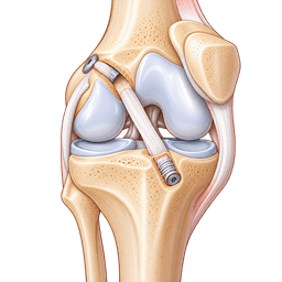
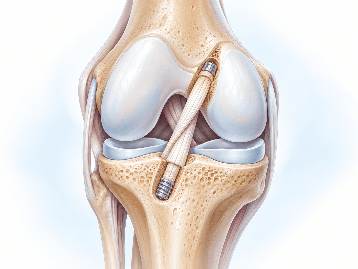
ACL reconstruction is an arthroscopic knee operation that replaces a torn anterior cruciate ligament with a tendon graft. The goal is to restore rotational and forward-backward stability of the knee so that patients can walk, work, exercise and, when appropriate, return safely to activities that involve running, jumping, cutting or pivoting.
The anterior cruciate ligament, usually shortened to ACL, runs diagonally through the center of the knee between the femur and tibia. Together with the posterior cruciate ligament and the collateral ligaments, it helps control the movement of the knee. The ACL is especially important when the body changes direction while the foot is planted.
An ACL can tear during football, soccer, basketball, skiing, rugby, handball and other sports that involve sudden deceleration, landing or pivoting. Many ACL tears occur without another person striking the knee. AAOS describes the ACL as an important restraint against the tibia moving too far forward relative to the femur and as an important contributor to rotational stability.
Not every ACL tear requires surgery. Some people can function well after structured physiotherapy, particularly if they have a partial tear, do not experience repeated episodes of the knee giving way and do not need to return to high-demand pivoting activities. Other patients continue to feel instability despite rehabilitation. For them, ACL reconstruction surgery may provide a more stable platform for work, daily life or sport.
The word reconstruction matters. In conventional ACL reconstruction, the torn ligament is not simply sewn together. A new ligament is created from a tendon graft. That graft may come from the patient's own patellar tendon, hamstring tendons or quadriceps tendon. In selected circumstances, donor tissue may be used instead.
During arthroscopic ACL reconstruction, a small camera is introduced into the knee through a portal. The surgeon examines the joint, confirms the ligament injury and assesses the menisci and articular cartilage. Additional portals allow small instruments to enter the knee. The surgeon prepares the chosen graft, creates accurately positioned bone tunnels or sockets in the femur and tibia, passes the graft through them and secures it using fixation devices.
Associated injuries are important. ACL tears may occur with meniscus tears, cartilage damage, medial collateral ligament injury or other structural damage. AAOS notes that roughly half of ACL injuries are associated with other knee injuries.
For this reason, ACL surgery is not identical for every patient. One person may need an isolated ACL reconstruction. Another may undergo ACL reconstruction together with meniscus repair. A third may require additional treatment for cartilage or another ligament.
These differences affect operating time, weight-bearing, rehabilitation and return-to-sport timing.
The goal of the procedure is functional stability, not simply creating an MRI image that looks normal. A technically successful graft still requires months of biological healing, muscle strengthening, neuromuscular retraining and progressive sports-specific rehabilitation.
Patients sometimes ask whether they will receive a “new ACL.” Biologically, the answer is more nuanced. The graft begins as tendon tissue. After implantation, it gradually remodels and adapts to its new role inside the knee. The process continues long after the surgical wounds have healed.
That is why an athlete can feel relatively normal several months after ACL reconstruction while the graft and entire lower limb are still adapting.
The rehabilitation phase therefore has the same importance as the operation itself.
Conditions treated
Who it's for
- Complete ACL tear with symptomatic knee instability
- Recurrent episodes of the knee giving way
- Desire to return to pivoting, cutting or high-demand sport
- Physically demanding occupation requiring a stable knee
- ACL tear associated with a repairable meniscus injury
- ACL tear with other ligament or cartilage injuries requiring surgery
- Persistent instability despite an appropriate rehabilitation programme
- Chronic ACL deficiency causing functional limitation
- Selected young active patients at risk of secondary meniscus or cartilage damage
- Selected revision cases after failure of a previous ACL reconstruction
Good candidates
A good candidate for ACL reconstruction is usually someone whose knee instability interferes with the activities that matter to them and who understands that surgery is only one part of the treatment.
Age alone does not determine candidacy. A fit adult in their 40s who plays tennis or skis may have a greater functional need for ACL reconstruction than a much younger person who is comfortable with low-demand activities and has no instability.
The clinical question is therefore not simply, “Is the ACL torn?” It is, “Does the ACL injury prevent this patient from doing what they need or want to do safely?”
Good candidates often have one or more of the following features:
repeated instability during turning or changing direction;
a goal of returning to football, basketball, skiing, rugby or another pivoting sport;
an occupation involving climbing, uneven surfaces, heavy lifting or rapid movement;
associated meniscus damage that may benefit from repair;
a complete tear accompanied by functional instability;
failure of a well-designed course of physiotherapy;
realistic expectations about rehabilitation.
AAOS emphasizes that activity level rather than age alone should influence whether ACL reconstruction is considered.
A patient also needs to be ready for rehabilitation. ACL surgery cannot substitute for physiotherapy. Someone who is unwilling or unable to participate in months of progressive exercises may achieve a technically stable graft but still have weakness, poor movement control or difficulty returning to sport.
The condition of the knee before surgery matters as well.
Many surgeons prefer the swelling to settle and knee motion to improve before reconstruction. A knee that is very swollen, painful and unable to straighten fully may need a period of “prehabilitation” first. The objective is to enter surgery with a calm joint, good quadriceps activation and as much normal range of motion as possible.
Other medical issues may need optimization before surgery. These include poorly controlled diabetes, active infection, significant cardiopulmonary disease, anaemia, smoking, obesity or medications that affect bleeding.
ACL reconstruction may not be the preferred treatment for every torn ACL.
For example, nonsurgical care may be reasonable for a patient with a partial tear and a stable knee, or for someone with a complete tear who does not experience instability and does not intend to return to pivoting activities. AAOS specifically describes these groups as potential candidates for nonsurgical management.
Before surgery
Preparing properly for ACL reconstruction surgery can affect both the early postoperative experience and the quality of rehabilitation.
Confirming the diagnosis
The process usually starts with a detailed history and knee examination. The surgeon asks how the injury happened, whether a pop was heard or felt, how quickly the knee swelled and whether the knee now gives way.
Clinical tests assess ACL stability and other knee structures.
MRI is commonly used to confirm the ACL injury and evaluate the menisci, cartilage, collateral ligaments and other structures. Plain X-rays may also be performed to assess the bones and exclude fracture or significant arthritis.
The decision to reconstruct the ACL should not be based on MRI alone. Symptoms, activity level, instability, associated damage and patient goals need to be considered together.
Prehabilitation before ACL surgery
Prehabilitation can begin before the operation.
Typical goals include reducing swelling, restoring full knee extension, improving flexion, normalizing walking and rebuilding quadriceps control.
Entering surgery with a stiff or significantly swollen knee can make the initial postoperative period more difficult. For that reason, some patients benefit from several weeks of physiotherapy before the operation.
Common preoperative exercises may include quadriceps activation, straight-leg raises, stationary cycling when tolerated, heel slides and progressive strengthening.
The exact programme should be prescribed by the treating physiotherapist or surgeon because associated meniscus or cartilage injuries can change what is safe.
Timing of ACL reconstruction
There is no single correct number of days between an ACL tear and surgery for every patient.
The knee needs to be ready, but unnecessary prolonged instability can expose the menisci and cartilage to additional injury.
AAOS's current clinical practice guideline states that when surgery is indicated for an acute isolated ACL tear, earlier reconstruction is preferred because the risk of additional meniscal and cartilage injury begins to increase within approximately three months.
This does not mean every ACL tear should be operated on immediately.
A severely swollen, painful knee with limited movement may need rehabilitation first. Associated injuries can also alter timing. The surgeon balances joint recovery against the risk created by ongoing instability.
Choosing the ACL graft
One of the most important discussions before surgery concerns the graft.
Common choices are:
Graft | Source | Potential advantages | Considerations |
|---|---|---|---|
Bone–patellar tendon–bone autograft | Middle portion of patellar tendon with bone blocks | Strong bone-to-bone fixation; extensive long-term evidence | More anterior or kneeling pain in some patients |
Hamstring autograft | Usually semitendinosus ± gracilis | Smaller harvest site; often less kneeling discomfort | Hamstring weakness and graft diameter can matter |
Quadriceps tendon autograft | Quadriceps tendon, with or without bone block | Large graft; increasingly used in primary and revision surgery | Temporary quadriceps weakness; technique dependent |
Allograft | Donor tendon | No autograft harvest; shorter graft-harvest time | Higher failure concerns in younger/high-demand patients |
Other grafts | Selected autograft/allograft sources | Useful in complex or revision cases | Highly case-specific |
The AAOS guideline recommends that surgeons consider autograft over allograft, particularly in young or active patients, because of better outcomes and a lower graft-failure risk.
When comparing patellar tendon with hamstring autograft in skeletally mature patients, AAOS states that patellar tendon graft may be favored when reducing graft failure is particularly important, whereas hamstring graft may be favored when minimizing anterior or kneeling pain is a priority.
Quadriceps tendon graft has become increasingly common and may be especially useful in certain primary and revision reconstructions. Graft selection should still be individualized according to age, sport, occupation, anatomy, previous surgery and surgeon experience.
Medical assessment
Before the operation, the hospital may arrange:
blood tests;
anaesthetic assessment;
medication review;
electrocardiogram when clinically indicated;
pregnancy testing when appropriate;
additional medical clearance for significant health conditions.
Patients taking anticoagulants, antiplatelet medication, diabetes medication or other important prescription drugs should receive individualized instructions. They should never stop these medications without medical advice.
Smoking and nicotine
Smoking and nicotine can interfere with tissue healing.
Patients who smoke or use nicotine products should discuss cessation with their healthcare team. Stopping before surgery and remaining nicotine-free during recovery is generally encouraged.
Planning the first postoperative week
International and local patients should plan the practical aspects of recovery before travelling to hospital.
Arrange:
transport home;
help with shopping or household responsibilities;
a comfortable place to elevate the leg;
prescribed medications;
ice or approved cold-therapy equipment;
crutches if recommended;
physiotherapy appointments;
a follow-up appointment;
wound-care supplies when required.
A patient travelling internationally should also know how to contact the treating team after leaving the hospital.
How the operation is performed
How is ACL reconstruction done?
ACL reconstruction is usually performed arthroscopically. The surgeon inspects the knee with a small camera, prepares a tendon graft, creates precisely positioned tunnels or sockets in the femur and tibia, passes the graft into the anatomical position of the torn ACL and fixes it securely to the bone.
NHS describes ACL surgery as keyhole surgery in which an arthroscope is introduced through a small incision, the damaged ACL is assessed, a graft is positioned in place of the torn ligament and fixation devices are used to secure it.
Step 1 — Anaesthesia and positioning
ACL reconstruction may be performed under general anaesthesia or spinal/regional anaesthesia.
A peripheral nerve block may also be offered to reduce postoperative pain.
The patient is positioned so that the surgeon can move the knee freely during the operation. A tourniquet may be used depending on surgeon preference.
Step 2 — Arthroscopic examination
The surgeon inserts the arthroscope into the knee.
Saline solution expands the joint and improves visibility.
The surgeon examines:
the torn ACL;
posterior cruciate ligament;
medial meniscus;
lateral meniscus;
articular cartilage;
patellofemoral joint;
other structures visible arthroscopically.
This examination is important because an MRI does not always identify every clinically relevant lesion.
If a meniscus tear is found, the surgeon may repair or selectively trim it depending on tear pattern, tissue quality and location.
Meniscus preservation is generally desirable whenever a repairable tear is present because the menisci play an important role in load distribution and joint stability.
Step 3 — Graft harvest and preparation
When an autograft is used, the selected tendon is harvested through an additional incision.
The graft is then prepared on a sterile table.
Its diameter and length are measured.
Strong sutures are placed through the ends of the graft so it can later be passed into the bone tunnels.
The graft choice determines exactly how this part is performed.
A bone–patellar tendon–bone graft includes small bone blocks.
A hamstring graft is commonly folded to form multiple strands.
A quadriceps graft may be used with or without a bone block.
Step 4 — Preparing the original ACL footprint
The damaged ACL tissue is assessed.
Enough tissue is removed to allow anatomical tunnel positioning while preserving useful landmarks when possible.
The objective is not simply to create “a ligament” inside the joint. The graft needs to reproduce the orientation and function of the native ACL as closely as possible.
Tunnel position is one of the important technical factors in ACL reconstruction.
Poorly positioned tunnels can alter graft tension and knee mechanics and are a recognized cause of reconstruction failure.
Step 5 — Femoral tunnel or socket
Using arthroscopic guides, the surgeon identifies the ACL attachment area on the femur.
A guidewire is placed.
A drill or reamer creates a tunnel or socket matched to the graft.
Different drilling techniques can be used.
The exact technique is less important to the patient than accurate anatomical positioning and secure graft fixation.
Step 6 — Tibial tunnel
A tibial guide is used to identify the native tibial ACL attachment.
A guidewire is passed through the tibia.
The tunnel is drilled to the planned size.
The position is checked arthroscopically to ensure the graft will not impinge on surrounding structures as the knee moves.
Step 7 — Passing the ACL graft
The prepared graft is passed through the tibial and femoral tunnels or sockets.
The surgeon checks its position and tension.
The knee may be moved repeatedly through flexion and extension to ensure the graft tracks correctly.
Step 8 — Fixation
Different fixation systems are available.
They include interference screws, cortical buttons, suspensory fixation and other orthopedic implants.
Some fixation devices remain inside the bone permanently.
The choice depends on the graft, bone quality, tunnel technique and surgeon preference.
Step 9 — Tensioning the graft
The graft is tensioned with the knee positioned according to the surgical technique.
The surgeon assesses stability and range of motion.
A correctly positioned ACL graft should control abnormal translation and rotational instability without preventing normal knee motion.
Step 10 — Associated procedures
If the patient also has a meniscus tear, cartilage lesion or another ligament injury, treatment may be performed in the same operation.
This is one reason why ACL reconstruction operating times differ substantially between patients.
An isolated ACL reconstruction may be relatively straightforward.
ACL reconstruction combined with meniscal root repair, multiple meniscus repairs or complex multiligament surgery can take longer and may require a more restrictive rehabilitation programme.
Step 11 — Closing the incisions
The arthroscopy portals and graft-harvest incision are closed.
Sterile dressings are applied.
A brace may be placed if required.
The patient then goes to the recovery area.
Single-bundle versus double-bundle ACL reconstruction
The normal ACL has functional bundles that behave differently as the knee moves.
This led surgeons to develop double-bundle reconstruction techniques intended to reproduce more of the native ligament anatomy.
However, AAOS reviewed the evidence and concluded that both single-bundle and double-bundle ACL reconstruction can be considered because measured clinical outcomes are broadly similar.
Single-bundle reconstruction remains widely performed.
The most appropriate technique depends on the surgeon's experience and individual anatomy.
ACL reconstruction versus ACL repair
These procedures are different.
ACL reconstruction replaces the torn ligament with graft tissue.
ACL repair attempts to preserve and reattach the original ACL.
Traditional repair has historically had a higher failure rate than reconstruction because the ACL has limited intrinsic healing capacity in the intra-articular environment. AAOS therefore describes reconstruction as the usual surgical treatment for most ACL tears.
Modern repair techniques and biologic approaches are being studied and may be reasonable in highly selected injury patterns, particularly some proximal tears with good-quality tissue.
They should not be presented as interchangeable with standard ACL reconstruction.
Hospital stay
Most patients having an uncomplicated primary ACL reconstruction can leave the hospital on the same day.
After surgery, the patient is transferred to a recovery area while the effects of anaesthesia wear off.
Nurses monitor blood pressure, pulse, pain control, wound dressings and circulation in the leg.
Pain is treated using a multimodal approach that may include local anaesthetic, a nerve block, acetaminophen/paracetamol, anti-inflammatory medication when appropriate and stronger pain medication for a limited period.
A physiotherapist or trained clinical team member may help the patient stand and walk.
NHS guidance states that most patients begin mobilizing soon after surgery and can usually return home on the day of the operation.
Crutches may be used initially.
Weight-bearing instructions depend on what was performed.
After an isolated ACL reconstruction, patients are often permitted to bear weight relatively early.
However, rehabilitation may be more protective when ACL reconstruction is combined with a meniscus root repair, complex meniscus repair, cartilage procedure or additional ligament reconstruction.
A knee brace is not required for every ACL reconstruction.
Some surgeons use one routinely in the early phase, while others use it selectively.
Before discharge, the patient should understand:
how much weight can be placed through the leg;
how to use crutches;
whether a brace is required;
wound-care instructions;
medication dosing;
exercises to begin immediately;
warning signs requiring medical attention;
the date of follow-up.
Patients should not drive themselves home after anaesthesia.
International patients should remain within practical reach of the treating team during the initial postoperative period according to the surgeon's travel protocol.
Recovery
ACL reconstruction recovery time
ACL reconstruction recovery is a gradual process that usually takes many months. Walking and basic daily activity return much earlier than competitive sport. Many patients are functioning independently within weeks, but running, cutting, pivoting and unrestricted sport require progressive rehabilitation and objective clearance.
NHS notes that patients may resume many activities within a few months but that returning to sport can take up to a year.
AAOS describes return to full sports participation as commonly occurring within approximately 6–12 months, depending on strength, mechanics, symptoms and individual progress.
Those ranges should not be interpreted as a guarantee.
A professional athlete, recreational skier and office worker have very different functional endpoints.
Why recovery takes so long
The wounds heal relatively quickly.
The deeper recovery is slower.
Three processes occur at the same time:
The graft must biologically remodel.
The tendon graft adapts to its ligament-like role inside the knee.
Muscles must recover.
Quadriceps weakness is common after ACL injury and surgery.
Movement patterns must be retrained.
Strength alone does not ensure safe cutting, landing or pivoting.
Return-to-sport planning therefore considers both biology and function.
Recovery timeline
- Control swelling and regain full extension1Control swelling and regain full extension
Days 0–14
Use crutches as instructed, begin quadriceps activation and range-of-motion work, manage pain and swelling, protect the wounds and aim for a fully straight knee. Weight-bearing depends on associated meniscus or cartilage procedures.
- Walk with improving control2Walk with improving control
Weeks 2–6
Progress knee bending, gait, quadriceps and hip strength. Crutches may be discontinued when walking is safe and controlled. Desk work may become possible during this period.
- Restore strength and single-leg control3Restore strength and single-leg control
Weeks 6–12
Progressive resistance training becomes more important. The knee should tolerate increased activity without recurrent swelling. Continue working toward normal range of motion.
- Meet criteria for running progression4Meet criteria for running progression
Months 3–5
Running starts only when knee symptoms, strength and movement quality are satisfactory. Introduce progressive impact, landing and plyometric work under supervision.
- Rebuild agility, power and sports-specific capacity5Rebuild agility, power and sports-specific capacity
Months 5–8
Add multidirectional drills, acceleration, deceleration, jumping, landing and sport-specific tasks. Continue objective strength and functional testing.
- Return to unrestricted sport after clearance6Return to unrestricted sport after clearance
Months 9–12+
Return should be criteria-based and consider symptoms, strength, hop testing, movement quality, sport demands and psychological readiness. Some athletes require longer than 12 months.
Outcomes and success rates
ACL reconstruction is generally effective at restoring mechanical knee stability and improving function in appropriately selected patients.
Long-term observational research from the MOON cohort has shown meaningful improvements in validated knee outcome scores after ACL reconstruction that were maintained through approximately 10 years, although activity level may decline over time and outcomes vary between patients.
“Success,” however, can mean several different things.
A stable knee is one outcome.
Returning to jogging is another.
Returning to competitive football at the same level is much more demanding.
For this reason, a single ACL reconstruction success-rate percentage can be misleading.
Return to sport after ACL reconstruction
Return-to-sport rates vary according to age, sport, competition level, associated injuries, rehabilitation and how “return” is defined.
A 2026 systematic review and meta-analysis of elite and professional athletes reported that approximately 85.8% returned to play, with a pooled average return time of approximately 292 days. The population was highly selected elite/professional athletes, so these figures should not be presented as the expected outcome for every ACL patient.
The same study estimated graft failure at about 7% in that athlete population, again emphasizing that failure rates depend strongly on population and activity exposure.
Will the knee be exactly the same as before injury?
Not necessarily.
Many patients regain high levels of function, but reconstruction cannot recreate a completely untouched knee.
The original injury may already have damaged the meniscus, cartilage or bone.
The graft is not biologically identical to the original ACL.
Some patients also experience persistent weakness, stiffness, altered sensation, kneeling pain or reduced confidence.
The realistic objective is a stable, functional knee—not a promise that the injury never happened.
Does ACL reconstruction prevent arthritis?
ACL reconstruction is performed primarily to restore stability and function.
It should not be promoted as a guaranteed method of preventing osteoarthritis.
ACL injury itself can initiate changes inside the knee, particularly when the meniscus or cartilage is also damaged.
Preventing repeated instability and preserving repairable meniscal tissue may be beneficial, but reconstructed knees can still develop degenerative changes later in life.
Implants and technology
ACL fixation technology includes:
interference screws;
cortical buttons;
suspensory devices;
adjustable-loop systems;
fixed-loop systems;
staples in selected techniques;
bioabsorbable or biocomposite fixation in some systems;
metallic fixation.
The “best” implant cannot be determined independently of the graft and surgical technique.
Correct tunnel position, graft preparation, fixation, tensioning and rehabilitation generally matter more than marketing language around a specific implant.
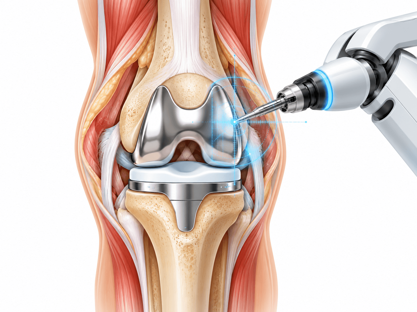
Computer navigation and robotic technology
Navigation and robotic assistance are increasingly visible across orthopaedic surgery.
ACL reconstruction may use digital planning, intraoperative measurement or navigation systems in selected centers, but conventional arthroscopic reconstruction remains established practice.
Technology should support anatomical accuracy rather than replace surgical expertise.
Patients should therefore ask what clinical benefit a proposed technology offers for their specific knee rather than choosing a surgeon solely because a robotic or computer system is advertised.
Risks and how they are managed
All surgery carries risk. Partner hospitals follow enhanced-recovery and infection-prevention protocols, and your surgeon will discuss the risks specific to your case before consent.
- Graft failure or recurrent instability
- Knee stiffness or arthrofibrosis
- Infection
- Blood clots
- Persistent pain
- Anterior knee or kneeling pain
- Numbness around the scars
- Donor-site weakness
- Neurovascular injury
- Fixation or hardware problems
- Meniscus or cartilage problems
- Contralateral ACL injury
- Need for revision surgery
- Anaesthetic complications
- Failure to return to the previous sport level
Alternatives
- Structured physiotherapy
- Activity modification
- Functional knee brace
- ACL repair
- Delayed reconstruction
What ACL costs
The contracted Turkey partner package next to approved self-pay benchmarks. Benchmarks are 20th–80th percentile ranges of approved records, normalised to USD.
Turkey package
$4,500 – $7,500
United States self-pay
$16,750 – $47,700
Germany self-pay
$7,500 – $22,700
Typical self-pay range by country
Surgeons who perform ACL
All surgeonsSurgeon profiles for this procedure are being added.
Hospitals offering this procedure
Antalya Yaşam Hospital
Private hospital in Muratpaşa, Antalya, with an orthopedics and traumatology department
BHT Clinic Istanbul Tema Hospital
Large private hospital in Atakent, Küçükçekmece, with an orthopedics and traumatology unit
Biruni University Hospital
University hospital in Küçükçekmece, Istanbul, with a published orthopedics and traumatology department
Çankaya Hospital for Orthopedic Care
Çankaya Hospital for Orthopedic Care is an orthopedic hospital in central Ankara with robotic-arm assisted surgery
Sources and references
Peer-reviewed guidance and institutional sources used to write and review this page.
- 01Management of Anterior Cruciate Ligament Injuries — Clinical Practice Guideline
American Academy of Orthopaedic Surgeons (AAOS), 2022
https://www.aaos.org/aclcpg
- 02ACL Injury: Does It Require Surgery?
OrthoInfo — American Academy of Orthopaedic Surgeons, 2026
https://www.orthoinfo.org/treatment/acl-injury-does-it-require-surgery/
- 03Anterior Cruciate Ligament (ACL) Injuries
OrthoInfo — American Academy of Orthopaedic Surgeons, 2026
https://www.orthoinfo.org/diseases--conditions/anterior-cruciate-ligament-acl-injuries/
- 04How ACL Surgery Is Done
NHS, 2025
https://www.nhs.uk/tests-and-treatments/acl-anterior-cruciate-ligament-surgery/how-its-done/
- 05Recovering from ACL Surgery
NHS, 2025
https://www.nhs.uk/tests-and-treatments/acl-anterior-cruciate-ligament-surgery/recovering/
- 06Return to Sport After Anterior Cruciate Ligament Reconstruction — Prognostic Factors and Prognostic
Annals of Physical and Rehabilitation Medicine / PubMed, 2025
https://pubmed.ncbi.nlm.nih.gov/39892026/