Key takeaways
- 1MPFL reconstruction is designed primarily for recurrent lateral patellar instability, especially when the kneecap repeatedly dislocates or subluxes despite appropriate rehabilitation.
- 2The MPFL is the main soft-tissue restraint against lateral movement of the kneecap during early knee flexion, so recurrent dislocation can leave the ligament stretched or functionally deficient.
- 3Not every patellar dislocation requires surgery. A first-time uncomplicated dislocation is often treated initially without reconstruction unless another surgical problem, such as an osteochondral fracture or major loose body, is present.
- 4MPFL reconstruction does not correct every cause of patellar instability. Patella alta, excessive tibial tubercle lateralization, severe trochlear dysplasia, valgus deformity and rotational abnormalities may require additional procedures.
- 5Accurate femoral graft placement and appropriate graft tension are critical. An incorrectly positioned or excessively tight graft can cause pain, stiffness, abnormal patellar pressure or reconstruction failure.
- 6Most appropriately selected patients achieve substantial improvement in stability, but return to the exact previous sporting level is not guaranteed.
- 7Return to sport commonly requires approximately 6–9 months or longer, with strength, movement quality and confidence considered alongside time.
Overview
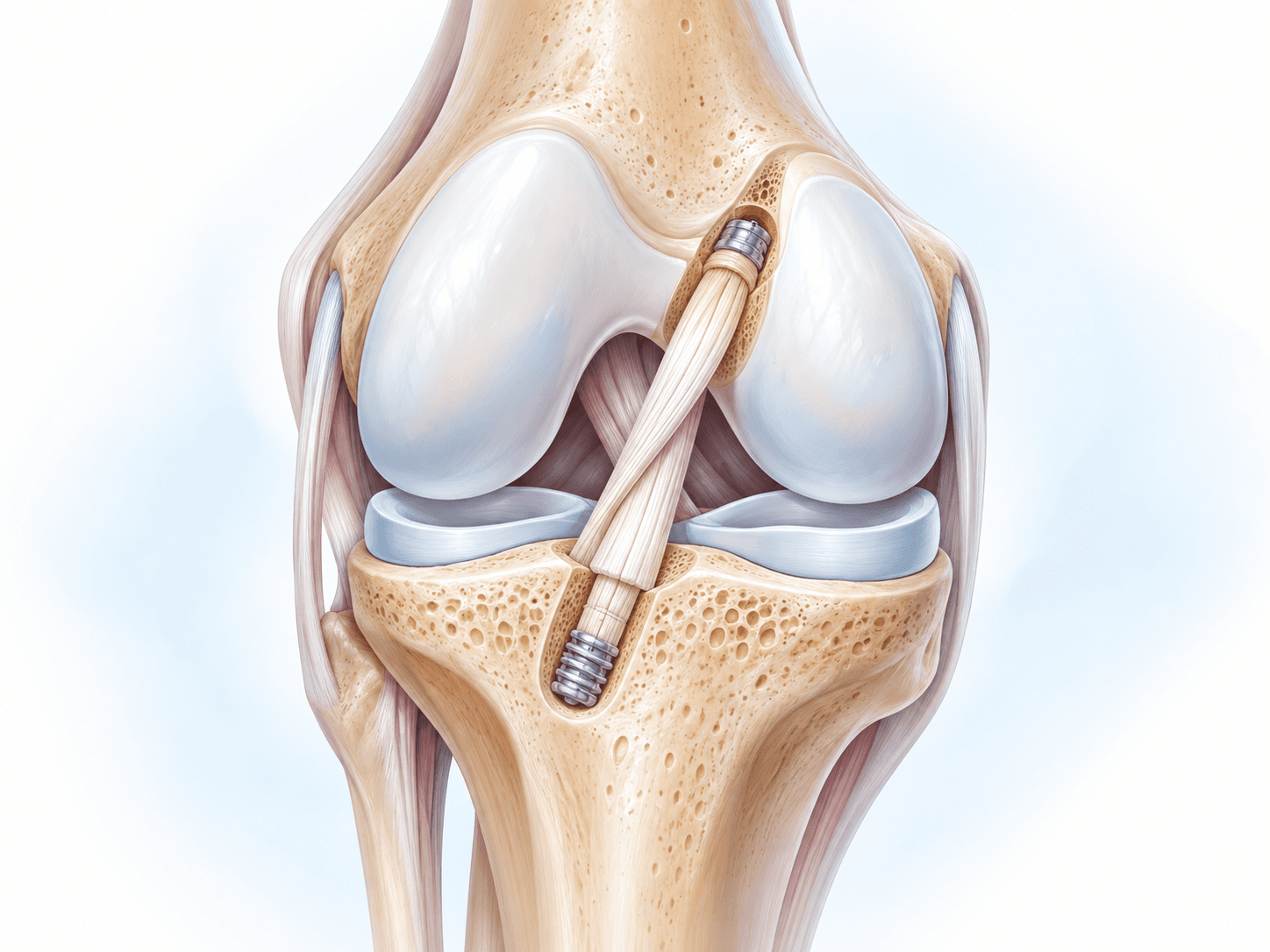
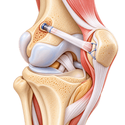
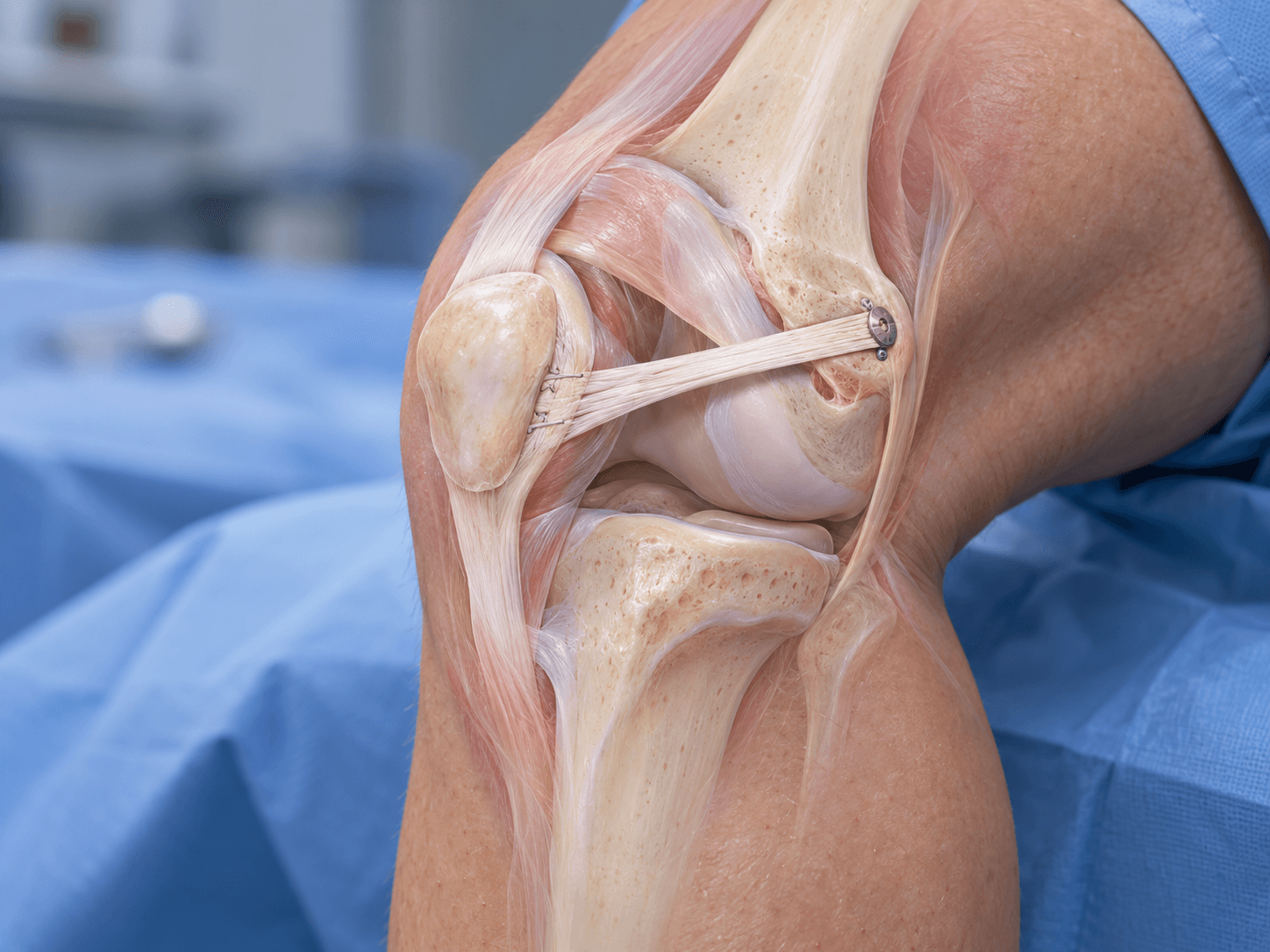
MPFL reconstruction is surgery that replaces the damaged medial patellofemoral ligament with a tendon graft to restore stability to the kneecap. The graft connects the inner side of the patella to the medial femur and acts as a restraint against excessive lateral movement of the patella, particularly during the early part of knee flexion.
The medial patellofemoral ligament, usually abbreviated MPFL, is one of the most important soft-tissue stabilizers preventing the kneecap from moving or dislocating toward the outside of the knee. When a lateral patellar dislocation occurs, the MPFL is frequently stretched or torn. Some patients heal and regain stable function with rehabilitation, while others develop repeated dislocations, apprehension or a persistent sensation that the kneecap is about to slip out.
MPFL reconstruction attempts to recreate this medial restraint using graft tissue. The operation is different from simply tightening the original ligament. A new graft is positioned anatomically so that it supports the patella while still allowing normal movement through the trochlear groove as the knee bends.
A high-quality reconstruction requires more than placing a graft between two bones. The surgeon must understand the patient's patellar anatomy, alignment, trochlear shape, patellar height and rotational profile because recurrent patellar instability is often caused by several factors working together.
What Is the Medial Patellofemoral Ligament?
The MPFL is a broad ligamentous structure extending from the medial side of the patella toward the medial femur. Although it is relatively thin compared with ligaments such as the ACL, it plays a major role in resisting lateral displacement of the kneecap.
The ligament is particularly important during the first part of knee flexion. When the knee is relatively straight, the patella has not yet entered deeply into the trochlear groove, so soft-tissue structures provide much of the stability. As the knee bends, the patella becomes increasingly contained by the bony shape of the trochlea.
This relationship explains why abnormalities of the trochlear groove can be so important. If the trochlea is shallow or dysplastic, the kneecap receives less bony guidance and can depend even more heavily on soft-tissue restraints such as the MPFL.
What Happens During a Patellar Dislocation?
Most patellar dislocations occur toward the outside of the knee. The kneecap moves laterally out of the trochlear groove, stretching or tearing structures on the medial side.
The MPFL is commonly injured during this event. Bone bruising can occur on the medial patella and lateral femoral condyle as the patella dislocates and then returns to its normal position. Cartilage can also be damaged, and occasionally an osteochondral fragment breaks free inside the joint.
After a first dislocation, many patients recover with rehabilitation and do not experience another episode. Others develop recurrent instability because the ligament does not heal with sufficient tension or because underlying anatomy continues to push the patella laterally.
The risk of recurrence is influenced by factors such as young age, trochlear dysplasia, patella alta and other structural abnormalities.
What Is Recurrent Patellar Instability?
Recurrent patellar instability means that the kneecap repeatedly moves abnormally toward the outer side of the knee. This can range from a sensation of subluxation to complete recurrent dislocations requiring reduction.
Patients often describe apprehension when changing direction, descending stairs, running or performing twisting movements. Some stop participating in sport because they no longer trust the knee.
Repeated instability is not merely uncomfortable. Every dislocation can damage the articular cartilage of the patella or trochlea. Over time, recurrent instability can therefore contribute to patellofemoral cartilage injury and degenerative change.
The goal of treatment is to create a stable patella while preserving normal movement.
First-Time Patellar Dislocation Versus Recurrent Instability
A first-time uncomplicated patellar dislocation is often treated without immediate ligament reconstruction. Management can include short-term support, progressive motion, quadriceps strengthening and rehabilitation focused on restoring normal lower-limb control.
Surgery becomes more likely when there is a large osteochondral fracture, a significant loose body or another structural problem requiring operative treatment.
Recurrent instability is different. Once a patient has repeated dislocations or persistent symptomatic subluxation despite rehabilitation, the probability that soft-tissue insufficiency or anatomical risk factors are clinically important becomes much higher.
MPFL reconstruction is therefore primarily associated with recurrent lateral patellar instability, rather than being automatically performed after one uncomplicated first-time event.
MPFL Reconstruction Versus MPFL Repair
MPFL repair attempts to preserve and reattach the patient's existing ligament. Reconstruction uses tendon graft tissue to create a new ligament.
Repair can be considered in selected acute injuries, particularly when there is a clearly defined avulsion from the patella or femoral attachment and the tissue remains suitable. However, chronic instability often involves stretched or scarred ligament tissue that cannot reliably restore normal restraint.
Reconstruction is therefore more commonly used for recurrent instability. It allows the surgeon to establish a new functional ligament using tendon graft rather than depending on previously damaged tissue.
The decision between repair and reconstruction depends on injury timing, tissue quality, associated injuries and the patient's anatomy.
Why MPFL Reconstruction Is Not Always Enough
The MPFL is only one part of patellar stability.
Some patients have a very shallow trochlear groove. Others have a high-riding patella that enters the trochlea later during knee flexion. Some have the tibial tubercle positioned too far laterally, creating a stronger outward pull through the patellar tendon.
Valgus alignment and rotational abnormalities can also increase lateral forces.
If one of these bony abnormalities is severe enough, reconstructing the MPFL alone may not fully correct the underlying mechanics. It can also place excessive tension on the graft.
The surgeon therefore needs to determine whether isolated MPFL reconstruction is appropriate or whether bony correction should accompany it.
What Is Trochlear Dysplasia?
Trochlear dysplasia describes an abnormal shape of the groove in which the patella normally tracks.
A healthy trochlea has a concave groove that helps contain the patella as the knee bends. In trochlear dysplasia, the groove may be shallow, flat or even convex.
The more severe the dysplasia, the less bony stability the patella receives.
Mild dysplasia does not automatically require trochlear surgery. Many patients can be successfully treated with MPFL reconstruction alone.
Severe high-grade dysplasia with persistent instability is more complicated and can lead the surgeon to consider trochleoplasty in carefully selected patients.
What Is Patella Alta?
Patella alta means the kneecap sits higher than normal relative to the femur and tibia.
Because the patella begins from a higher position, it takes longer during knee flexion to engage securely within the trochlear groove.
This increases the period during which the patella depends mainly on soft-tissue restraints.
Patella alta can therefore contribute to recurrent instability.
Mild cases can sometimes be managed with isolated MPFL reconstruction, while more substantial abnormalities may require distalization of the tibial tubercle.
What Is the TT-TG Distance?
The tibial tubercle–trochlear groove distance, commonly abbreviated TT-TG, is an imaging measurement describing the relationship between the tibial tubercle and the center of the trochlear groove.
When the tubercle is positioned farther laterally, the patellar tendon can produce a stronger lateral pull on the kneecap.
A high TT-TG measurement can therefore be one contributor to instability.
The measurement should not be used in isolation. It is influenced by imaging technique, anatomy and knee rotation.
Modern surgical decision-making considers TT-TG alongside patella height, trochlear dysplasia, cartilage status, alignment and the patient's actual instability pattern.
Tibial Tubercle Osteotomy and MPFL Reconstruction
A tibial tubercle osteotomy, or TTO, moves the bony attachment of the patellar tendon.
The surgeon cuts the tibial tubercle and repositions it before fixing it with screws.
Medialization can reduce lateral pull. Distalization can correct substantial patella alta. Anteromedialization can also redistribute patellofemoral contact forces in selected cartilage conditions.
When clinically appropriate, TTO can be combined with MPFL reconstruction.
The reconstruction restores medial ligament restraint, while the osteotomy corrects a bony mechanical factor.
The important point is that TTO should be added because the anatomy justifies it, not routinely because the patient has patellar instability.
Trochleoplasty and MPFL Reconstruction
Trochleoplasty reshapes an abnormally dysplastic trochlear groove.
It is reserved mainly for selected patients with severe trochlear dysplasia and recurrent instability.
The procedure creates or deepens a more functional groove to improve bony containment of the patella.
MPFL reconstruction can be performed at the same time to restore the medial soft-tissue restraint.
Trochleoplasty is considerably more specialized than isolated MPFL reconstruction and should not be treated as standard surgery for all patellar dislocations.
Femoral Osteotomy and Patellar Instability
Some patients have substantial valgus deformity or excessive femoral anteversion contributing to instability.
In these cases, a distal femoral osteotomy can correct coronal or rotational alignment.
The operation changes the bony forces acting across the patellofemoral joint.
MPFL reconstruction may also be required.
This type of combined surgery is most appropriate when careful imaging and clinical evaluation show that deformity is a major driver of instability.
Why Accurate Diagnosis Matters
Patellar instability is not one uniform disease.
Two patients can experience identical lateral dislocations for completely different anatomical reasons. One may have an isolated incompetent MPFL with otherwise normal anatomy, while another has severe trochlear dysplasia, patella alta and rotational malalignment.
If both patients receive exactly the same operation, the outcome may be very different.
The best treatment therefore begins with identifying the complete instability pattern.
MPFL reconstruction works particularly well when used in a knee whose remaining anatomy can support an isolated soft-tissue stabilization.
Conditions treated
Who it's for
- Recurrent lateral patellar dislocation
- Repeated patellar subluxation causing functional symptoms
- Persistent symptomatic patellar instability despite structured physiotherapy
- MPFL insufficiency following previous lateral patellar dislocation
- Patellar apprehension that limits sport, work or normal activity
- Young and active patients with repeated instability
- Athletes unable to return safely because of recurrent patellar instability
- Failed previous MPFL repair
- Selected failed previous patellar stabilization surgery
- MPFL insufficiency associated with mild or moderate trochlear dysplasia
- MPFL insufficiency associated with correctable patella alta
- Patellar instability associated with increased TT-TG distance when combined correction is indicated
- MPFL reconstruction performed with tibial tubercle osteotomy
- MPFL reconstruction performed with trochleoplasty in selected severe trochlear dysplasia
- MPFL reconstruction performed with distal femoral osteotomy in selected valgus or rotational deformity
- Selected cases requiring stabilization during surgery for osteochondral injury
- Persistent lateral instability after failure of appropriate nonsurgical treatment
Good candidates
A good candidate for isolated MPFL reconstruction usually has recurrent lateral patellar instability with evidence that the medial soft-tissue restraint is insufficient, while major bony abnormalities are absent or mild enough that they do not require separate correction.
The surgeon should be able to explain why an isolated graft is expected to control the patella. If substantial patella alta, severe trochlear dysplasia, pronounced tubercle lateralization or major rotational deformity is present, the treatment discussion should address whether those factors also need correction.
A patient should also have realistic expectations about rehabilitation. The operation can provide excellent stability, but normal strength, confidence and sports performance do not return immediately. Physiotherapy is a central part of treatment rather than an optional addition.
Patients With Recurrent Dislocation
Repeated complete dislocation is one of the clearest reasons to consider stabilization surgery.
Each episode can further damage the medial restraints and articular cartilage. Some patients become able to dislocate the kneecap with relatively minor movements once the instability becomes chronic.
The frequency of episodes matters, but frequency is not the only consideration. A patient who has experienced two major dislocations and now avoids normal activities because of persistent apprehension can have a stronger indication than someone with more minor episodes but good function.
The decision should reflect both structural instability and its impact on daily life.
Patients With Recurrent Subluxation
Not every unstable patella fully dislocates.
Some patients experience repeated lateral slipping or subluxation that spontaneously reduces.
These episodes can still produce pain, apprehension and functional limitation.
If the clinical examination and imaging confirm patellar instability, MPFL reconstruction can be considered after appropriate nonsurgical treatment fails.
The diagnosis should distinguish true instability from nonspecific anterior knee pain because reconstruction is not a treatment for every patellofemoral pain syndrome.
Athletes
Sports involving cutting, jumping and rapid directional change can expose an unstable patella to repeated lateral forces.
Athletes may lose confidence even after returning to basic daily activity.
A successful MPFL reconstruction can restore stability and allow many athletes to return to sport.
However, the athlete must rebuild quadriceps strength, hip control, landing mechanics and confidence before unrestricted competition.
The target is stable performance, not simply reaching a postoperative date.
Adolescents
Patellar instability commonly begins in adolescence.
Skeletally immature patients require additional surgical planning because the femoral MPFL attachment lies close to the distal femoral growth plate.
Techniques can be modified to avoid damaging the physis.
The surgeon should therefore have specific experience with pediatric or adolescent patellofemoral instability when the growth plates remain open.
The same anatomical risk factors, including trochlear dysplasia and patella alta, still need evaluation.
Adults
Adults can also develop recurrent instability, either beginning in adolescence or after trauma later in life.
Age itself does not exclude reconstruction.
The more important considerations are cartilage condition, instability pattern, anatomy and functional goals.
An adult with advanced patellofemoral osteoarthritis may have a less predictable response than a younger patient with preserved cartilage.
The condition of the joint therefore matters more than a rigid age cutoff.
Generalized Ligamentous Laxity
Some patients are naturally hypermobile.
Generalized ligamentous laxity can contribute to patellar instability and influence the amount of soft-tissue restraint available.
MPFL reconstruction can still be successful, but graft selection and tensioning require careful consideration.
The surgeon should also evaluate whether instability is isolated to the patella or part of a broader hypermobility pattern.
Patients with syndromic or extreme hypermobility can require more individualized treatment.
When MPFL Reconstruction May Not Be Appropriate
Isolated MPFL reconstruction is less appropriate when a major uncorrected bony abnormality is the dominant driver of instability.
Severe trochlear dysplasia, substantial patella alta, major tubercle malposition or significant rotational deformity may require additional or alternative procedures.
Advanced patellofemoral arthritis can also change the treatment strategy.
Active infection, severe medical instability or inability to complete rehabilitation are additional reasons to delay elective reconstruction.
A patient with anterior knee pain but no true instability should not undergo MPFL reconstruction simply because the patella feels uncomfortable.
Before surgery
Understanding the Instability History
The preoperative assessment begins with a detailed history of every major instability event. The surgeon asks when the first dislocation occurred, how many additional episodes have happened, whether reductions were required and what movements usually trigger instability. Episodes during sport, stairs or simple daily activities can provide information about severity.
The surgeon also asks whether the patient experiences pain between episodes, whether the knee swells and whether the patella feels unstable even when it does not fully dislocate. Previous physiotherapy, braces and other treatments should be documented because surgery is usually considered after an appropriate nonsurgical programme has failed to provide sufficient stability.
Previous operative reports are particularly valuable if the patient has already undergone patellar stabilization, cartilage surgery or tibial tubercle procedures.
Physical Examination
The surgeon evaluates patellar tracking through the entire range of knee movement. Patellar translation is assessed medially and laterally, and the examiner determines whether lateral movement reproduces apprehension.
The patellar apprehension test can demonstrate the patient's sense that the kneecap is about to dislocate. The J-sign can indicate abnormal lateral patellar movement as the knee approaches extension.
Quadriceps strength, especially control of the extensor mechanism, is assessed. Hip strength and lower-limb rotational control can also influence patellar mechanics.
The examination extends beyond the patella because valgus alignment, femoral rotation, foot position and generalized ligamentous laxity may all contribute.
Standard X-Rays
Standing and patellofemoral X-rays are commonly obtained.
A lateral view helps assess patellar height and trochlear morphology. Axial or skyline views demonstrate the relationship between the patella and trochlea.
X-rays also show fractures, degenerative change and previous hardware.
In patients with chronic instability, the surgeon pays particular attention to whether significant patellofemoral arthritis has already developed.
The imaging findings are interpreted together rather than using one measurement to determine the operation.
MRI
MRI provides detailed information about the MPFL, cartilage, menisci and other knee structures. After an acute dislocation, MRI can show the characteristic injury pattern involving the medial patella and lateral femoral condyle.
The study is especially important when an osteochondral fracture or loose body is suspected.
MRI can also help evaluate trochlear dysplasia, patellar height and cartilage damage. The surgeon can assess whether repeated instability has already produced chondral injury on the underside of the patella or within the trochlear groove.
MRI is very useful, but it does not replace physical examination and alignment analysis.
CT and Rotational Assessment
CT is used selectively rather than routinely in every patient.
It becomes particularly useful when rotational deformity is suspected. Excessive femoral anteversion or abnormal tibial torsion can change the direction of forces acting on the patella.
CT can also provide measurements related to the tibial tubercle and trochlear groove.
In complex instability, three-dimensional assessment can help determine whether a derotational femoral or tibial osteotomy is appropriate.
The decision to perform major bony correction should not be based on one imaging value without clinical correlation.
Measuring Patellar Height
Several radiographic indices can assess whether the patella sits abnormally high.
Patella alta is important because a high-riding kneecap engages the trochlea later during flexion.
Measurements such as the Caton-Deschamps or Insall-Salvati indices can contribute to assessment.
The surgeon does not operate on a number alone. The severity of patella alta, recurrent instability pattern and other anatomical findings all matter.
Substantial symptomatic patella alta may lead to consideration of tibial tubercle distalization.
Assessing Trochlear Dysplasia
The trochlear groove is evaluated on lateral radiographs, MRI and sometimes CT.
Different patterns and grades of trochlear dysplasia have been described.
Mild abnormalities are common and do not automatically require trochleoplasty.
High-grade dysplasia associated with recurrent instability is more clinically important.
The surgeon needs to determine whether an MPFL graft alone can provide sufficient stability or whether the abnormal groove remains too dominant a risk factor.
Measuring TT-TG
TT-TG distance can be measured on CT or MRI.
An increased value suggests that the tibial tubercle lies relatively lateral to the trochlear groove.
Historically, threshold values were sometimes treated almost as automatic indications for tibial tubercle osteotomy.
Modern practice is more nuanced.
TT-TG is interpreted alongside patellar height, trochlear anatomy, rotational alignment, symptoms and the patient's overall instability pattern.
Lower-Limb Alignment
Standing alignment should be assessed.
Valgus alignment can increase lateral patellofemoral force.
When deformity is substantial, long-leg standing X-rays may be needed to determine whether the femur or tibia is responsible.
A distal femoral osteotomy can become relevant when clinically important valgus deformity contributes to recurrent instability.
This is especially important in patients who have previously failed isolated soft-tissue stabilization.
Cartilage Assessment
Repeated patellar dislocation can damage cartilage.
The surgeon determines whether focal cartilage injury requires treatment at the time of stabilization.
Small unstable fragments may be removed or treated arthroscopically.
Repairable osteochondral fragments can sometimes be fixed.
Larger focal defects may require cartilage restoration.
The rehabilitation plan then needs to protect both the MPFL graft and the cartilage procedure.
Prehabilitation
Preoperative physiotherapy aims to control swelling, restore full knee extension and improve quadriceps activation. A stronger knee is generally easier to rehabilitate after reconstruction.
Hip abductor and external rotator strength can also be addressed because dynamic valgus and lower-limb control influence patellofemoral mechanics.
The patient learns how to use crutches and, when a brace will be required, how the postoperative brace works.
Prehabilitation cannot correct a mechanically unstable patella, but it can optimize the surrounding muscle system before surgery.
Graft Selection
MPFL reconstruction can use the patient's own tendon or donor tissue.
Common autograft options include gracilis, semitendinosus and quadriceps tendon.
Allograft can also be used.
No single graft has demonstrated universal superiority for every patient.
Graft choice depends on patient age, activity, previous surgery, tissue quality, surgeon preference and whether other reconstructions require the same donor tendons.
Hamstring Autograft
Gracilis tendon is commonly used because it provides sufficient length and strength without requiring a large harvest.
Semitendinosus can also be used.
The tendon is harvested through a small incision, prepared and then fixed between the patella and femur.
Hamstring harvest can produce temporary local discomfort or weakness, although many patients recover well.
In athletes, graft choice can be individualized according to sport and other ligament needs.
Quadriceps Tendon Graft
A strip of quadriceps tendon can be used for MPFL reconstruction.
Some techniques leave the graft attached to the patella, reducing the need for patellar bone tunnels.
This can be attractive when the surgeon wants to minimize the risk of patellar fracture associated with larger or multiple tunnels.
Quadriceps grafts can also be useful in revision cases.
The technique produces a harvest site above the patella and can temporarily affect quadriceps function during rehabilitation.
Allograft
Allograft avoids harvesting the patient's own tendon.
This can reduce donor-site morbidity and preserve autograft options for other procedures.
Modern systematic reviews report good clinical outcomes with both allograft and autograft.
The choice should not be presented as a simple better-versus-worse decision.
Patient age, surgeon experience, graft availability and cost can all influence the final plan.
Planning Femoral Attachment
The femoral attachment is one of the most important technical aspects of MPFL reconstruction.
If the graft is attached too far from the anatomical location, it can become excessively tight in flexion or too loose in extension.
This can produce abnormal patellofemoral pressure, stiffness, pain or recurrent instability.
Preoperative planning and intraoperative imaging therefore focus carefully on femoral attachment placement.
Planning Patellar Fixation
Several methods can secure the graft to the patella.
The surgeon may use small bone tunnels, sockets, suture anchors or soft-tissue techniques.
Each method has advantages and risks.
Large or multiple transverse patellar tunnels can weaken the bone and increase fracture risk.
Modern techniques often aim to achieve reliable fixation while preserving as much patellar bone as possible.
Planning Combined Tibial Tubercle Osteotomy
If TTO is required, the patient needs to understand that the operation and recovery are more extensive than isolated MPFL reconstruction.
A segment of the tibial tubercle is cut, repositioned and fixed with screws.
Bone healing then becomes an additional rehabilitation requirement.
Weight-bearing can be more restrictive, and return to sport can take longer.
Hardware irritation can also lead to later screw removal in some patients.
Medical Assessment
Preoperative testing depends on age, health and hospital protocol.
Young healthy athletes may require relatively limited testing, while older patients or those with medical conditions can need blood tests, ECG or specialist evaluation.
Diabetes and other relevant conditions should be controlled.
Active infection elsewhere in the body should be discussed before elective ligament reconstruction.
Patients should also disclose any history of blood clots or anaesthetic problems.
Medication Review
The team needs a complete medication and supplement list.
Anticoagulants and antiplatelet drugs require individual instructions.
Certain diabetes medications can require temporary adjustment around fasting and anaesthesia.
Patients should not stop important medication independently.
The prescribing clinician and surgical team should coordinate any changes.
How the operation is performed
MPFL reconstruction is usually performed through small incisions around the medial knee. A tendon graft is attached to the medial patella and positioned at the anatomical femoral origin of the MPFL, then tensioned carefully so the kneecap remains stable without becoming overconstrained.
The operation may begin with arthroscopy to inspect the cartilage and remove or treat loose fragments. When additional structural abnormalities require correction, tibial tubercle osteotomy, trochleoplasty or another realignment procedure can be performed during the same surgical episode.
Accurate graft position and tension are central to success. The goal is not to pull the patella firmly inward. The graft should provide restraint only when lateral movement becomes excessive while still allowing normal tracking through flexion.
Anaesthesia and Positioning
The procedure is usually performed under general or spinal/regional anaesthesia.
A nerve block can be added for postoperative pain control.
The patient lies on the operating table with the knee accessible through a full range of motion.
The surgeon needs to be able to assess patellar movement repeatedly during reconstruction.
Fluoroscopy is often available to confirm femoral tunnel positioning.
Arthroscopic Examination
An arthroscope may be inserted through small anterior portals.
The surgeon inspects the patella, trochlea and remaining knee structures.
Cartilage injury is documented.
Loose osteochondral fragments can be assessed and treated.
Arthroscopy also helps confirm whether instability has produced other intra-articular damage.
Not every isolated MPFL reconstruction requires extensive arthroscopic treatment, but visual inspection can be useful when symptoms or imaging suggest associated pathology.
Graft Harvest
When an autograft is used, the selected tendon is harvested.
A gracilis or semitendinosus graft is removed through a small incision and prepared on a sterile table.
The graft is measured and strong sutures are placed in its ends.
For quadriceps tendon reconstruction, a strip of tendon is harvested from the upper patellar region.
The exact harvest technique depends on whether the graft will remain attached to the patella or be completely free.
Preparing the Medial Patella
A small incision is made along the medial border of the patella.
The surgeon identifies the area where the native MPFL attaches.
The patellar surface is prepared according to the fixation method.
Bone sockets, suture anchors or other fixation can be used.
The surgeon avoids unnecessary disruption of the patella.
Preserving bone stock is particularly important in smaller patients and revision cases.
Patellar Tunnel Technique
One method creates one or two controlled bone tunnels or sockets in the patella.
The graft or sutures are fixed within these tunnels.
The technique can provide strong fixation.
However, drilling through the patella creates a stress riser.
Modern techniques therefore emphasize appropriate tunnel size and position to minimize fracture risk.
Large transverse tunnels are generally approached cautiously.
Suture Anchor Fixation
Small anchors can be inserted into the medial patella.
Strong sutures from the anchors secure the graft.
This avoids creating a large complete tunnel through the patella.
Anchor-based fixation has therefore become attractive to many surgeons.
The anchors need to be positioned accurately and securely within sufficient bone.
Soft-Tissue or Quadriceps-Based Fixation
Some quadriceps tendon techniques keep the graft attached near the patella.
This can avoid patellar drilling entirely.
The tendon is redirected toward the femoral attachment.
The technique reduces patellar bone disruption but has different graft-handling considerations.
It can be particularly useful when patellar bone quality or previous tunnels make new bone fixation undesirable.
Identifying the Femoral MPFL Attachment
The femoral attachment lies near the medial epicondylar region.
Small differences in position can substantially alter graft behavior through flexion.
The surgeon uses anatomical landmarks and often fluoroscopy to identify the intended location.
A guidewire is positioned and the knee is moved through flexion to assess how graft length changes.
If the position is incorrect, the guidewire can be adjusted before drilling.
Schöttle Point
A radiographic landmark known as the Schöttle point is commonly used to help locate the femoral MPFL attachment on a lateral fluoroscopic image.
It provides a reproducible reference based on femoral anatomy.
The landmark is a guide rather than a substitute for anatomical understanding.
Accurate lateral imaging is important because rotation of the femur can make the apparent position misleading.
The surgeon therefore combines fluoroscopy with direct anatomy.
Creating the Femoral Socket
Once the position is confirmed, a socket or tunnel is drilled in the femur.
The diameter matches the graft and fixation device.
The direction of drilling is selected to avoid unwanted structures.
In skeletally immature patients, the growth plate must be protected.
Pediatric techniques may use different tunnel orientation or alternative fixation to reduce physeal risk.
Passing the Graft
The graft is passed from the patellar side through the appropriate soft-tissue layer toward the femoral socket.
The graft should travel along the anatomical course of the MPFL rather than lying superficially beneath the skin.
The surgeon ensures that the graft is not twisted.
It is then secured on the femoral side using the selected fixation method.
Graft Tensioning
This is one of the most important steps.
The MPFL graft should not be tightened like a rope pulling the patella inward.
Overtensioning can increase pressure between the patella and trochlea, produce medial patellar tilt, cause stiffness and create pain.
The surgeon positions the knee at an appropriate degree of flexion and centers the patella within the trochlear groove.
The graft is tensioned only enough to prevent excessive lateral translation.
Normal small amounts of patellar mobility should remain.
Checking Patellar Tracking
The knee is moved through flexion and extension.
The surgeon watches the patella enter and travel through the trochlear groove.
The graft should remain appropriately tensioned without becoming excessively tight as the knee bends.
Lateral translation is tested.
The goal is stable but physiological tracking.
If the patella is being pulled too far medially, the tension can be adjusted before final completion.
MPFL Reconstruction With Tibial Tubercle Osteotomy
Why TTO May Be Added
TTO is considered when the patellar tendon attachment contributes significantly to abnormal lateral force or when substantial patella alta needs correction.
The tubercle is cut while preserving an appropriate bony segment.
It is then repositioned according to the desired correction.
Medialization reduces lateral pull.
Distalization lowers a high-riding patella.
Anteromedialization can alter patellofemoral contact pressure.
Fixation
The repositioned tubercle is usually secured with screws.
Compression across the osteotomy encourages bone healing.
The surgeon confirms the new tubercle position using imaging.
Because bone healing is required, postoperative loading is typically more cautious than after isolated MPFL reconstruction.
Combined Stability
The bony procedure and ligament graft perform complementary roles.
The osteotomy corrects the direction of mechanical pull, while the MPFL reconstruction restores medial restraint.
The surgeon should avoid overcorrecting both systems simultaneously because excessive medial force can create new patellofemoral problems.
MPFL Reconstruction With Trochleoplasty
Why Trochleoplasty May Be Added
Severe trochlear dysplasia can leave the patella without a stable bony groove.
Trochleoplasty reshapes the trochlea to improve patellar containment.
It is generally reserved for selected recurrent-instability patients with high-grade dysplasia.
The surgeon elevates an osteochondral flap or otherwise reshapes the trochlear surface depending on technique.
The cartilage-bearing surface is then stabilized in the new configuration.
MPFL reconstruction restores the medial soft-tissue restraint at the same operation.
Why Patient Selection Matters
Trochleoplasty is not a minor addition to MPFL surgery.
It alters the joint surface itself.
The risks and rehabilitation are different from isolated reconstruction.
Patients with mild dysplasia generally do not need trochlear reshaping simply because it appears abnormal on MRI.
The clinical severity of instability and the exact morphology need to justify the procedure.
MPFL Reconstruction With Distal Femoral Osteotomy
Substantial valgus alignment can produce a persistent lateralizing force.
A distal femoral osteotomy corrects that deformity through the femur.
The MPFL graft then provides medial restraint within the corrected alignment.
When excessive femoral anteversion is the problem, the osteotomy can also include rotational correction.
These cases require detailed alignment and rotational planning and are generally performed by surgeons experienced in both patellofemoral instability and deformity correction.
Treating Cartilage Injury
Patellar instability can produce focal cartilage lesions.
Small unstable flaps can be treated arthroscopically.
An osteochondral fragment can occasionally be fixed if it is large, structurally suitable and biologically viable.
Larger defects may require cartilage restoration techniques such as osteochondral transplantation or cell-based repair.
When cartilage restoration accompanies MPFL reconstruction, the rehabilitation may be substantially slower because loading restrictions are dictated by the cartilage procedure.
Lateral Release
Lateral retinacular release should not be considered a routine component of MPFL reconstruction.
Historically, lateral release was used more frequently for patellar disorders.
Modern understanding recognizes that cutting lateral stabilizing tissues indiscriminately can create medial instability or fail to treat the real problem.
A lateral lengthening or release may be considered when there is genuinely abnormal lateral tightness, but it should be based on clinical mechanics rather than performed automatically.
Wound Closure
Once tracking and stability are satisfactory, the surgical field is irrigated.
Incisions are closed.
Sterile dressings are applied.
A hinged knee brace is frequently fitted.
The patient then moves to the recovery area.
The operating report should clearly document graft type, patellar fixation, femoral fixation, any associated osteotomy and the resulting rehabilitation restrictions.
Hospital stay
Immediately After Surgery
The patient is monitored while recovering from anaesthesia. Nurses assess pain, circulation, sensation and the surgical dressings. Mild to moderate swelling is expected because the knee has been operated on and often examined arthroscopically.
Pain is generally managed with several complementary methods. Local anaesthetic, peripheral nerve blocks, paracetamol or acetaminophen and anti-inflammatory medication when medically appropriate can all reduce the need for stronger analgesics. Short courses of stronger pain medication are used when necessary.
The patient begins moving the ankle and activating the quadriceps early. This helps circulation and reduces muscle inhibition.
Day Surgery or Overnight Stay
Isolated MPFL reconstruction is commonly performed as day surgery. Some patients remain overnight depending on hospital policy, pain, mobility or timing of surgery.
Combined procedures can require a longer admission. An MPFL reconstruction with TTO involves a bone osteotomy and usually needs more observation than isolated soft-tissue reconstruction. Trochleoplasty or additional major reconstruction can also justify a longer stay.
The appropriate hospital stay should be determined by safety and surgical complexity rather than by trying to advertise the shortest possible admission.
Weight-Bearing
Many isolated MPFL reconstructions allow early weight-bearing as tolerated while using a brace and crutches. The purpose of crutches is partly comfort and partly control while quadriceps strength returns.
Protocols vary, and not every patient should immediately fully weight-bear. Graft fixation, associated cartilage procedures and surgeon preference can change the plan.
When TTO is performed, weight-bearing is often more restricted because the osteotomy needs to heal. The patient's written discharge protocol should therefore clearly distinguish isolated MPFL reconstruction from combined surgery.
Brace Use
A hinged knee brace is frequently used during the first postoperative weeks.
Some protocols lock the brace in extension during walking and progressively permit greater flexion during exercises.
Other surgeons allow earlier unlocked movement after isolated reconstruction.
There is no universal brace schedule supported by strong evidence for every patient.
The purpose is to protect early healing while allowing enough movement to prevent stiffness and quadriceps shutdown.
Early Range of Motion
Knee flexion usually begins relatively early.
The patient gradually works toward restoring motion without forcing the joint.
Excessive immobilization can contribute to stiffness.
At the same time, aggressive movement that produces substantial swelling can delay recovery.
The physiotherapist therefore progresses motion in a controlled way.
Combined TTO, cartilage restoration or another procedure can impose additional limits.
Physiotherapy Before Discharge
The patient learns how to walk safely with crutches.
Quadriceps sets, straight-leg raises when possible and ankle pumps are commonly introduced.
The physiotherapist also explains how to enter and leave bed, use stairs and manage the brace.
A key early goal is restoring quadriceps activation.
The quadriceps often becomes inhibited after knee surgery, and this can make the leg feel weak even when the reconstructed ligament itself is stable.
Discharge Instructions
The patient should leave with clear instructions about brace settings, weight-bearing, wound care, medication and exercises.
The follow-up date should be arranged.
Warning signs such as fever, significant wound drainage, worsening calf swelling or increasing uncontrolled pain should be explained.
International patients should receive the complete operative report and rehabilitation protocol because a generic note stating only “MPFL reconstruction” does not provide enough detail for the physiotherapist continuing care abroad.
Recovery
Major functional recovery after MPFL reconstruction generally occurs over four to six months, while return to unrestricted pivoting or competitive sport commonly takes approximately six to nine months and can take longer. The exact timeline depends on whether the reconstruction was isolated and whether the patient underwent TTO, trochleoplasty, cartilage repair or another procedure.
Early recovery focuses on swelling control, knee motion and quadriceps activation. Later rehabilitation emphasizes strength, dynamic alignment, jumping, landing and sport-specific control. A patient can regain normal walking relatively early while still being months away from safe return to high-demand sport.
The graft also needs time to integrate with the femur and patella. Rehabilitation therefore aims to restore normal movement without applying unnecessary stress during the early biological healing period.
Why Quadriceps Recovery Matters
The quadriceps controls knee extension and influences patellar movement.
After surgery, pain and swelling can inhibit quadriceps activation quickly.
The leg may therefore feel weak or unstable even though the new MPFL graft is functioning.
Restoring quadriceps strength is one of the central goals of rehabilitation.
The vastus medialis should not be considered in isolation. Modern rehabilitation also emphasizes overall quadriceps strength, hip control, trunk mechanics and whole-limb movement.
Why Hip and Lower-Limb Control Matter
Dynamic valgus during landing or cutting can increase lateral forces at the knee.
Weakness or poor control at the hip can contribute to this pattern.
Rehabilitation therefore includes hip abductors, external rotators and core control in addition to direct knee exercises.
The objective is to improve the entire movement strategy that the athlete uses.
A stable ligament combined with poor landing mechanics is not an ideal return-to-sport outcome.
Weeks 0–2
Main Goal: Control Swelling and Restore Quadriceps Activation
The patient typically uses a brace and crutches. Isolated reconstruction often allows early controlled weight-bearing, while combined procedures can require greater protection.
Swelling management is important because a swollen knee inhibits quadriceps function. Elevation and cold therapy may be used when recommended. Ankle pumps maintain lower-leg circulation.
Knee extension should be restored early. Flexion is increased progressively rather than forced.
Quadriceps contractions begin immediately. Straight-leg raising is introduced when the patient can keep the knee fully controlled without an extension lag.
The surgical wounds are monitored. Increasing drainage, redness or fever requires medical assessment.
Weeks 2–6
Main Goal: Normalize Walking and Increase Knee Motion
The patient gradually increases walking distance. Crutches are reduced as quadriceps control and gait improve, provided weight-bearing is permitted.
Range of motion continues toward normal. Many isolated reconstruction protocols aim for substantial flexion during this phase, although specific limits vary.
Strengthening progresses through controlled closed-chain exercises. Mini-squats, bridges, hip strengthening and carefully selected step work can be introduced.
The patient should avoid twisting or sudden lateral movements.
A reconstructed MPFL can be stable before the neuromuscular system is ready to control those forces safely.
Weeks 6–12
Main Goal: Restore Functional Strength
Most isolated-reconstruction patients are walking more normally by this stage.
The brace can often be discontinued gradually once adequate control is achieved.
Strengthening becomes more demanding.
Stationary cycling, progressive squats, step-ups and leg-press exercises can be used.
Balance work becomes increasingly important.
The patient progresses toward controlled single-leg loading.
The knee should not repeatedly swell significantly after rehabilitation.
Persistent swelling may indicate excessive training load or associated cartilage irritation.
Months 3–4
Main Goal: Develop Single-Leg Control and Prepare for Impact
Daily activities should feel increasingly normal.
The rehabilitation programme focuses on strength symmetry and movement quality.
Single-leg squats, step-downs and balance challenges can expose deficits that are not obvious during ordinary walking.
Running preparation begins when the patient demonstrates sufficient quadriceps strength, minimal swelling and good dynamic knee control.
The presence of an associated TTO or cartilage procedure can delay this stage.
The rehabilitation team should follow the most restrictive surgery performed.
Months 4–6
Main Goal: Begin Running and Controlled Plyometrics
Running can begin when objective criteria are met rather than simply because four months have passed.
The patient should demonstrate adequate strength and tolerate repeated single-leg loading without instability or swelling.
A walk-jog programme usually progresses gradually.
Early plyometric training emphasizes controlled two-leg and then single-leg landing.
Movement quality matters.
The knee should remain aligned without excessive inward collapse, while the patella should remain stable and comfortable.
Months 5–7
Main Goal: Restore Agility and Directional Control
Athletes progress toward acceleration and deceleration.
Lateral movement is introduced progressively.
Cutting and change-of-direction drills become more demanding over time.
This is often the phase where psychological confidence becomes increasingly important.
A patient may have a mechanically stable patella but still hesitate during movements associated with the original dislocation.
Gradual exposure to sports movements helps restore trust in the knee.
Months 6–9+
Main Goal: Return to Sport After Objective Clearance
Many patients return to sport during this period, but timing varies.
The athlete should demonstrate satisfactory strength, range of motion, dynamic balance and sport-specific movement.
There should be no recurrent instability.
Pain and swelling should remain low after demanding training.
Hop tests and strength testing can assist decision-making, although no single test determines readiness.
The patient's sport also matters. Returning to recreational cycling is different from returning to competitive basketball, football or gymnastics.
Return to Running
Running commonly begins several months after isolated MPFL reconstruction.
The patient should have adequate quadriceps strength, good single-leg control and minimal reactive swelling.
Running should begin in a straight line before lateral cutting is introduced.
If the knee develops pain or swelling after each session, training volume may be progressing too quickly.
Combined TTO typically delays running because bone healing needs to be confirmed.
Return to Work
Desk-based work can often resume within one to three weeks after isolated reconstruction, particularly when working remotely.
Commuting, brace use and swelling can influence timing.
Jobs requiring prolonged standing need more recovery.
Heavy work involving climbing, lifting, kneeling or rapid directional movement can take several months.
Combined osteotomy extends the timeline.
Driving
Driving should resume only when the patient has sufficient knee motion and leg control to operate the vehicle safely.
The patient must be able to perform an emergency stop.
A brace can limit safe pedal control.
Strong pain medication also prevents safe driving.
Right-knee surgery usually affects driving more directly.
Individual medical clearance is recommended.
Recovery After MPFL Reconstruction With TTO
The ligament graft and osteotomy heal simultaneously.
Crutches are usually required for longer.
Weight-bearing progresses according to bone healing.
X-rays are used to monitor the tubercle osteotomy.
The patient can regain knee motion while the bone is still healing, but running and impact are delayed until the osteotomy has united sufficiently.
Hardware irritation can become noticeable after activity increases, and some patients later choose screw removal.
Recovery After Trochleoplasty and MPFL Reconstruction
Trochleoplasty changes the trochlear surface and generally requires a more cautious rehabilitation than isolated MPFL reconstruction.
Early motion remains important because stiffness is a recognized concern.
Swelling and patellofemoral discomfort can persist longer.
Return to high-impact sport is individualized.
The patient should not use a standard isolated MPFL protocol after trochlear surgery.
Flying After MPFL Reconstruction
International travel should be planned according to the entire procedure.
An isolated day-case reconstruction and an MPFL plus TTO operation have different mobility and thrombosis considerations.
Long flights involve prolonged sitting and can increase clot risk after lower-limb surgery.
Patients should obtain specific travel clearance.
Hospital discharge should not automatically be interpreted as permission for immediate long-haul flying.
Recovery timeline
- Control swelling, protect the reconstruction and activate the quadriceps.1Control swelling, protect the reconstruction and activate the quadriceps.
Weeks 0–2
Use the brace and crutches according to instructions while gradually restoring knee extension and controlled flexion. Quadriceps activation and safe walking are the main early goals.
- Improve motion and normalize walking.2Improve motion and normalize walking.
Weeks 2–6
Gradually reduce walking aids when permitted. Progress quadriceps, hip and core strengthening while avoiding uncontrolled lateral or twisting movements.
- Restore functional strength.3Restore functional strength.
Weeks 6–12
Progress cycling, squats, step exercises, balance training and controlled single-leg loading. The brace is often discontinued during this period after isolated reconstruction.
- Develop single-leg control and prepare for impact.4Develop single-leg control and prepare for impact.
Months 3–4
Strength training becomes more demanding. The patient works toward symmetrical movement and good dynamic alignment before running is introduced.
- Begin running and controlled plyometric training.5Begin running and controlled plyometric training.
Months 4–6
Introduce progressive straight-line running and landing drills once strength, stability and symptom criteria are satisfied.
- Restore agility and change-of-direction control.6Restore agility and change-of-direction control.
Months 5–7
Progress acceleration, deceleration, lateral movement and increasingly demanding sport-specific drills.
- Return to unrestricted sport after testing and clearance.7Return to unrestricted sport after testing and clearance.
Months 6–9+
Return should depend on strength, patellar stability, movement quality, confidence and sport-specific performance rather than time alone.
Outcomes and success rates
MPFL reconstruction generally produces substantial improvement in recurrent lateral patellar instability when the operation is matched correctly to the patient's anatomy. Most appropriately selected patients experience greater confidence in the knee, improved functional scores and a major reduction in redislocation episodes.
The outcome depends on more than graft strength. Patients with severe trochlear dysplasia, major patella alta, pronounced malalignment or rotational deformity can have less predictable results if those problems are ignored.
A successful MPFL reconstruction therefore begins with appropriate patient selection and complete anatomical assessment.
MPFL Reconstruction Success Rate
There is no single percentage that captures the MPFL reconstruction success rate because studies report different outcomes. Some measure redislocation, others assess return to sport or functional knee scores.
Modern systematic reviews consistently show low recurrent-instability rates after properly performed reconstruction. Reported redislocation rates are often in the low single digits in well-selected cohorts.
This is one reason MPFL reconstruction has become the principal soft-tissue stabilization procedure for recurrent lateral patellar instability.
The important qualification is that a technically successful graft cannot fully compensate for severe untreated bony malalignment.
Recurrent Dislocation After Surgery
Recurrent complete dislocation is uncommon after successful reconstruction.
Some patients can still report subjective instability or apprehension without experiencing a true redislocation.
The cause of recurrent instability should be investigated carefully.
Potential factors include graft failure, traumatic reinjury, inaccurate femoral positioning, severe trochlear dysplasia or an uncorrected alignment problem.
Revision surgery should correct the reason for failure rather than simply repeating the original technique.
Patient-Reported Function
Scores such as the Kujala and IKDC generally improve after MPFL reconstruction.
Patients frequently report better ability to use stairs, run, exercise and participate in sport.
The degree of improvement can be influenced by cartilage damage.
A patient with a stable reconstructed patella can still experience anterior knee pain if repeated previous dislocations have produced significant cartilage injury.
The objective of surgery is therefore stability first, with pain improvement depending partly on the broader condition of the patellofemoral joint.
Return to Sport
Systematic reviews report high rates of return to some level of sport after MPFL reconstruction, commonly above 80%.
Return to the same preinjury level is lower.
One widely cited meta-analysis reported return to sport in more than 90% of athletes, while approximately seven in ten returned to or exceeded their previous level.
Other systematic reviews report overall return closer to the mid-80% range.
The difference illustrates why patients should not be given one guaranteed number.
Sport type, associated procedures, cartilage condition and psychological readiness all influence return.
How Long Does Return to Sport Take?
Across published studies, average return to sport has commonly occurred around six to seven months after isolated reconstruction.
This should not be treated as an automatic clearance point.
Some athletes require nine months or longer before strength and confidence are sufficient.
Combined TTO, trochleoplasty or cartilage restoration generally extends rehabilitation.
Return should therefore be criterion based.
MPFL Reconstruction With TTO Outcomes
Combining MPFL reconstruction with TTO can provide excellent stability when tibial tubercle position or patella height contributes to the problem.
Comparative studies show substantial functional improvements with both isolated MPFL reconstruction and combined surgery.
Patients selected for TTO usually have more significant anatomical abnormalities, which makes simple direct comparison difficult.
The additional osteotomy can lengthen rehabilitation and increases the likelihood of later hardware removal.
TTO should therefore be added because the anatomy requires it rather than because combined surgery is inherently better.
Isolated MPFL Versus MPFL Plus TTO
For patients whose bony alignment is acceptable, isolated MPFL reconstruction can avoid the additional morbidity of bone surgery.
When significant tubercle malposition or patella alta is present, combined correction can better address the mechanical problem.
Systematic reviews have found similar postoperative functional scores between many isolated and combined cohorts, with low instability rates in both.
The important lesson is not that the procedures are interchangeable. They were often performed in different anatomical patient groups.
The operation should follow the anatomy.
Trochlear Dysplasia and Outcomes
Mild and moderate trochlear dysplasia do not necessarily prevent good outcomes after isolated reconstruction.
High-grade dysplasia is more challenging.
When the groove provides very little bony stability, an isolated graft may be asked to perform too much of the stabilizing role.
Selected patients can therefore benefit from combined trochleoplasty.
Current evidence supports careful individualized treatment rather than performing trochleoplasty for every abnormal trochlea.
Graft Choice and Outcomes
Hamstring autograft, quadriceps autograft and allograft can all provide satisfactory stability.
Available reviews do not establish one graft as universally superior.
Some data show excellent recurrence rates with allograft, while autografts remain widely used and avoid donor-tissue considerations.
Technical placement and tensioning likely matter more than small theoretical differences between graft sources.
Femoral Tunnel Accuracy
Incorrect femoral attachment is one of the most important technical causes of poor outcome.
A nonanatomical graft can tighten excessively as the knee bends.
This increases patellofemoral pressure and can cause pain or stiffness.
A graft positioned too loosely can fail to provide adequate restraint.
Accurate placement using anatomy and fluoroscopic landmarks therefore has major clinical importance.
Overconstraint
More tension is not better.
An excessively tight graft can pull the patella medially and increase contact pressure.
The patient may develop pain or loss of flexion.
A successful reconstruction should permit normal physiological patellar movement while preventing abnormal lateral translation.
This is why graft tensioning is as important as graft strength.
Cartilage Damage and Long-Term Outcome
Patients with significant cartilage injury can obtain improved stability while continuing to experience some pain.
The ligament reconstruction does not regenerate lost cartilage.
When a focal defect is clinically important, cartilage restoration may be performed.
Advanced patellofemoral arthritis can limit the benefit of stabilization surgery.
The condition of the cartilage should therefore be included in preoperative expectations.
Long-Term Stability
Mid- and longer-term studies generally show sustained improvements after MPFL reconstruction.
The new ligament is not expected to prevent every future injury.
A sufficiently powerful traumatic event can still dislocate the patella or damage the graft.
The aim is to restore the patella to a stable mechanical environment during normal activity and sport.
Revision MPFL Reconstruction
Revision is considered when instability persists or returns after previous surgery.
The surgeon analyzes femoral tunnel position, patellar fixation, trochlear dysplasia, patella height, TT-TG, limb alignment and rotational anatomy.
CT can be particularly useful when previous tunnels need detailed assessment.
Revision may involve a new graft, different fixation and correction of a previously untreated bony risk factor.
Revision should therefore be planned as a complete instability reconstruction rather than simply exchanging one tendon for another.
Implants and technology
MPFL reconstruction uses biological tendon graft tissue supported by relatively small fixation implants. Unlike knee replacement, no artificial joint surface is implanted.
Common fixation devices include suture anchors, interference screws, cortical buttons and specialized soft-tissue fixation systems.
The specific implant depends on whether the graft is fixed within the patella, on the surface of the patella or using a quadriceps-based technique.
On the femoral side, the graft is commonly secured within a bone socket.
The fixation needs to remain stable while biological healing develops between tendon and bone.
Suture Anchors
Suture anchors can secure the graft along the medial patellar border.
A small anchor is inserted into the bone and strong sutures attach the graft.
The technique avoids drilling a large tunnel completely through the patella.
This can preserve patellar bone and reduce stress-riser concerns.
Anchor size and positioning need to match the patient's anatomy.
Patellar Bone Tunnels
Bone tunnels provide another fixation method.
The graft can be passed through or fixed within sockets drilled into the patella.
This technique provides direct tendon-to-bone healing.
However, excessive tunnel size or multiple large transverse tunnels can weaken the patella.
Modern approaches therefore aim to achieve sufficient fixation while minimizing structural disruption.
Interference Screws
The femoral graft can be fixed inside a socket using an interference screw.
The screw compresses the tendon against the tunnel wall.
Fixation can be secure immediately while biological integration develops.
Interference screws can be metallic or made from biocomposite materials.
The material is less important than accurate tunnel position and appropriate graft tension.
Cortical Buttons
Some techniques use suspensory fixation with a small cortical button.
The button sits against the outer cortex while strong sutures or loops connect it to the graft.
Adjustable-loop systems can allow the surgeon to fine-tune graft position.
These devices are more familiar from cruciate ligament reconstruction but can also be adapted to selected MPFL techniques.
High-Strength Sutures
Modern high-strength synthetic sutures are used throughout MPFL reconstruction.
They secure the graft to anchors or fixation devices.
Suture tape can also be used in selected constructs.
These materials are designed to maintain mechanical stability while healing occurs.
They do not replace the biological graft itself.
Internal Brace Augmentation
Some surgeons use high-strength suture tape alongside the graft as an additional protective construct.
This is sometimes described as internal bracing.
The concept is to protect the biological tissue from excessive elongation during early healing.
The augmentation must not be tensioned excessively because it can overconstrain the patella.
Clinical evidence continues to evolve, so internal brace augmentation should not be presented as necessary for every reconstruction.
Fluoroscopy
Real-time X-ray imaging is commonly used to identify the femoral attachment.
A true lateral image of the femur allows the surgeon to locate recognized radiographic landmarks.
This can reduce the risk of major femoral tunnel malposition.
Fluoroscopy can also confirm hardware position when TTO or another osteotomy is performed.
The technology assists anatomical surgery but does not replace direct understanding of knee anatomy.
Navigation and Computer Assistance
Computer navigation is not routinely required for isolated MPFL reconstruction.
Advanced systems can theoretically help quantify patellar tracking or bony alignment, but standard surgery continues to rely primarily on examination, imaging, fluoroscopy and anatomical landmarks.
Navigation becomes more relevant in complex deformity correction than in straightforward soft-tissue reconstruction.
The most important technological goal remains accurate planning rather than adding complexity for marketing purposes.
3D CT Planning
Three-dimensional CT reconstruction can be valuable in patients with complex rotational or bony abnormalities.
The surgeon can assess femoral anteversion, tibial torsion and the relationship of the patella, trochlea and tibial tubercle.
3D planning can also assist a derotational osteotomy.
Routine isolated MPFL reconstruction does not require three-dimensional CT in every patient.
The technology is most useful when conventional assessment suggests a more complex anatomical problem.
Tibial Tubercle Osteotomy Plates and Screws
TTO is commonly stabilized with screws, although plates can be used in selected techniques.
The screws compress the repositioned bone fragment against the tibia.
They usually remain after healing unless they become symptomatic.
Because the tubercle lies relatively close to the skin, hardware prominence can occasionally be noticeable when kneeling.
This contributes to the secondary hardware-removal rate after combined MPFL and TTO procedures.
Trochleoplasty Fixation
Trochleoplasty techniques can use absorbable sutures, anchors or other low-profile fixation to secure the reshaped osteochondral flap.
The fixation needs to maintain the new groove while bone healing occurs.
Hardware must not protrude into the articular surface.
Because the procedure directly affects cartilage-bearing bone, specialized instrumentation and experience are important.
Pediatric Physeal-Sparing Technology
Skeletally immature patients require femoral fixation that avoids or minimizes injury to the distal femoral growth plate.
The surgeon can use angled sockets, soft-tissue fixation or other physeal-sparing methods.
Fluoroscopy helps identify the growth plate.
Pediatric reconstruction therefore requires different technical planning from adult surgery.
A method appropriate for a fully grown 25-year-old should not automatically be copied in a 12-year-old with substantial growth remaining.
Dynamic Patellar Tracking Assessment
The surgeon assesses patellar movement manually throughout surgery.
Emerging technologies can quantify patellar tracking, but they are not routinely required.
The knee itself remains an important functional test.
After fixation, the surgeon observes how the patella engages the trochlea at different flexion angles.
This dynamic assessment helps detect excessive graft tension.
MRI for Postoperative Assessment
Routine MRI is not required after an uncomplicated successful reconstruction.
It can be useful when recurrent instability, persistent pain or cartilage symptoms develop.
MRI can assess graft continuity, patellofemoral cartilage and other internal structures.
Metal artefact from fixation is generally limited because MPFL implants are relatively small.
CT After Failed Reconstruction
CT is particularly useful in revision planning.
It can identify the location of previous femoral and patellar tunnels.
Rotational anatomy and TT-TG can also be reassessed.
If a previous femoral tunnel is substantially malpositioned, revision may require a new trajectory or occasionally staged bone grafting.
Technology is therefore especially valuable when anatomy has been altered by prior surgery.
Future MPFL Technology
Future developments may include improved dynamic tracking analysis, patient-specific planning and better integration of three-dimensional anatomical data into surgical decision-making.
Biological fixation materials may continue evolving.
However, the major determinants of outcome remain relatively fundamental: correct indication, identification of anatomical risk factors, accurate femoral placement, appropriate graft tension and structured rehabilitation.
A technologically sophisticated operation performed for the wrong instability pattern will not outperform a carefully planned conventional reconstruction.
Risks and how they are managed
All surgery carries risk. Partner hospitals follow enhanced-recovery and infection-prevention protocols, and your surgeon will discuss the risks specific to your case before consent.
- Recurrent patellar instability: The kneecap can remain unstable or dislocate again, particularly after new trauma or when major anatomical risk factors remain untreated.
- Graft failure or stretching: The reconstructed ligament can elongate or rupture, leading to recurrent lateral patellar movement.
- Patellar fracture: Bone tunnels or fixation can weaken the patella. Modern smaller tunnels, anchors and alternative techniques aim to reduce this uncommon but important risk.
- Overconstraint: An excessively tight graft can pull the patella too far medially and increase pressure within the patellofemoral joint.
- Medial patellar instability: Excessive medial restraint can rarely create instability in the opposite direction.
- Knee stiffness: Loss of flexion can occur if scar tissue develops or rehabilitation progresses too slowly.
- Arthrofibrosis: More severe scar formation can occasionally require manipulation or arthroscopic release.
- Persistent anterior knee pain: Stability can improve while pain continues because of pre-existing cartilage damage or abnormal patellofemoral loading.
- Cartilage deterioration: Reconstruction cannot reverse existing cartilage loss, and degeneration can continue over time.
- Incorrect femoral tunnel position: Malposition can cause abnormal graft tension, pain, stiffness or recurrent instability and can require revision surgery.
- Patellar fixation failure: Anchors, sutures or other fixation can rarely loosen or fail before biological healing is complete.
- Infection: Infection is uncommon but can require antibiotics and occasionally surgical washout.
- Blood clots: Deep-vein thrombosis can occur after lower-limb surgery, particularly when mobility is reduced.
- Nerve irritation: Small sensory nerves around the medial knee can be irritated, producing temporary or occasionally persistent numbness.
- Donor-site pain: Hamstring or quadriceps autograft harvest can create local pain, weakness or sensitivity.
- Quadriceps weakness: Temporary quadriceps inhibition is common and can persist when rehabilitation is incomplete.
- Hardware irritation after TTO: Screws used to fix the tibial tubercle can become prominent and sometimes require later removal.
- Delayed union after TTO: The osteotomy can heal more slowly than expected, prolonging weight-bearing restrictions.
- Tibial tubercle nonunion: Rarely, the osteotomy may fail to unite and require additional fixation or bone grafting.
- Tibial tubercle fracture: The bone fragment or surrounding tibia can fracture during surgery or recovery.
- Trochleoplasty-related cartilage complications: Reshaping the trochlea carries additional risks involving the articular surface and postoperative stiffness.
- Persistent apprehension: Some patients remain psychologically cautious even after mechanical stability has been restored.
- Need for revision surgery: Recurrent instability, malposition, stiffness or other complications can require another operation.
- Anaesthetic complications: Nausea, medication reactions, breathing or cardiovascular problems are uncommon but possible with any surgery.
Alternatives
- Structured physiotherapy: First-time dislocations and selected less-severe instability can often be managed with quadriceps strengthening, hip control and movement retraining.
- Patellar stabilization brace: A brace can provide temporary support during rehabilitation or sports progression but does not reconstruct a chronically insufficient ligament.
- Activity modification: Reducing high-risk pivoting or jumping activities can control symptoms in patients who prefer nonsurgical treatment.
- Observation after first-time dislocation: Many uncomplicated first-time dislocations are treated without immediate reconstruction.
- MPFL repair: Selected acute avulsion injuries with good-quality tissue can occasionally be repaired rather than reconstructed.
- Tibial tubercle osteotomy: TTO can address substantial tubercle lateralization or patella alta when bony alignment is a major cause of instability.
- Trochleoplasty: Severe trochlear dysplasia may require reshaping of the trochlear groove rather than relying on isolated soft-tissue stabilization.
- Distal femoral osteotomy: Significant valgus or rotational femoral deformity can require bony correction.
- Derotational osteotomy: Excessive femoral anteversion or tibial torsion can be corrected in carefully selected patients.
- Cartilage restoration: Symptomatic focal cartilage damage caused by repeated dislocation can require an additional joint-surface procedure.
- Osteochondral fragment fixation: A large viable osteochondral fragment from an acute dislocation may be repaired rather than removed.
- Continued conservative management: Patients with manageable symptoms and no recurrent dislocation can continue rehabilitation and monitoring rather than undergo surgery.
What MPFL Reconstruction costs
The contracted Turkey partner package next to approved self-pay benchmarks. Benchmarks are 20th–80th percentile ranges of approved records, normalised to USD.
Turkey package
$4,500 – $7,000
United Kingdom self-pay
$6,500 – $18,050
Germany self-pay
$5,500 – $15,550
Typical self-pay range by country
Surgeons who perform MPFL Reconstruction
All surgeonsSurgeon profiles for this procedure are being added.
Hospitals offering this procedure
Antalya Yaşam Hospital
Private hospital in Muratpaşa, Antalya, with an orthopedics and traumatology department
BHT Clinic Istanbul Tema Hospital
Large private hospital in Atakent, Küçükçekmece, with an orthopedics and traumatology unit
Biruni University Hospital
University hospital in Küçükçekmece, Istanbul, with a published orthopedics and traumatology department
Çankaya Hospital for Orthopedic Care
Çankaya Hospital for Orthopedic Care is an orthopedic hospital in central Ankara with robotic-arm assisted surgery
Sources and references
Peer-reviewed guidance and institutional sources used to write and review this page.
- 01Return-to-sport timing, recurrent instability, complication rates and postoperative functional outcomes. The analysis included 930 patients and reported a high overall rate of return to sport. PubMed
American Journal of Sports Medicine / PubMed
https://pubmed.ncbi.nlm.nih.gov/33720789/
- 02Return-to-play rate, time to return, recurrent instability and functional improvement following reconstruction.
ystematic Review Publisher:
https://pubmed.ncbi.nlm.nih.gov/32866030/
- 03Comparative outcomes of isolated MPFL reconstruction and combined MPFL reconstruction with tibial tubercle osteotomy.
Systematic Review and Meta-analysis / PubMed
https://pubmed.ncbi.nlm.nih.gov/38044955/
- 04Functional results, surgical failure and secondary surgery following combined MPFL and tibial tubercle procedures. PubMed
Systematic Review and Meta-analysis / PubMed
https://pubmed.ncbi.nlm.nih.gov/39465420/
- 05Weight-bearing, range-of-motion rehabilitation and return-to-sport protocols after MPFL reconstruction with and without TTO
Systematic Review / PubMed
https://pubmed.ncbi.nlm.nih.gov/35701590/
- 06Allograft outcomes, recurrent instability, complications and comparison with autograft reconstruction.
Systematic Review / PubMed
https://pubmed.ncbi.nlm.nih.gov/37979146/