Key takeaways
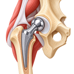
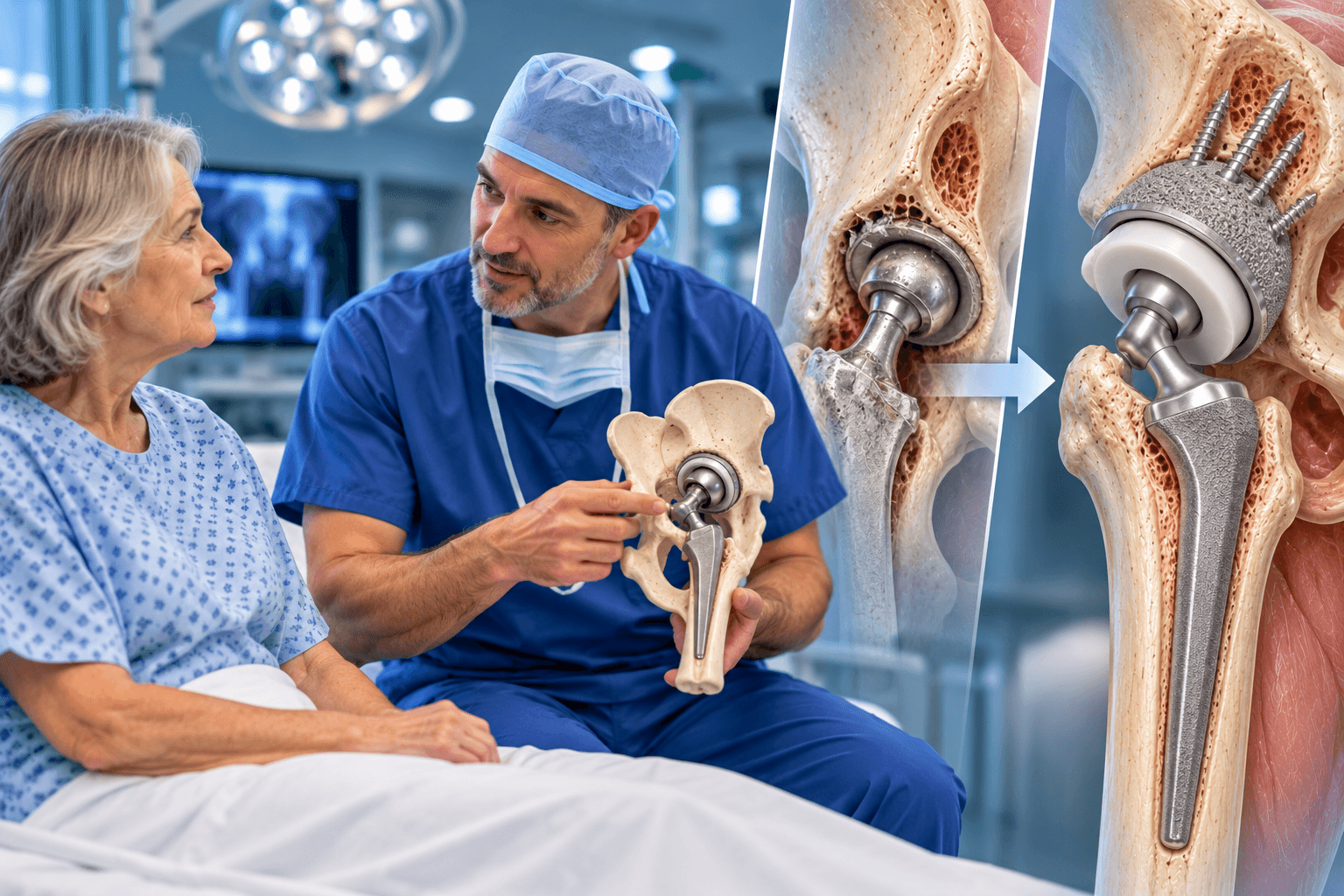
- 1Hip revision surgery is a second operation on a previously replaced hip when part or all of the original prosthesis has failed, become infected, loosened, fractured or become unstable.
- 2A revision can range from a relatively limited liner or femoral-head exchange to complete reconstruction of the acetabulum and femur.
- 3Revision hip replacement is usually more complex than primary hip replacement because scar tissue, bone loss, previous implants and damaged soft tissues can make reconstruction more difficult.
- 4Common reasons include aseptic loosening, recurrent dislocation, periprosthetic joint infection, periprosthetic fracture, implant wear, osteolysis and implant damage.
- 5Not every painful hip replacement requires revision. The cause of pain should be established before another major operation is recommended.
- 6Infection must be actively considered before revision, because the treatment strategy for an infected prosthesis is fundamentally different from the strategy for mechanical loosening.
- 7Some patients require only one revision operation, while chronic infection can require staged treatment with implant removal, an antibiotic spacer and later reimplantation.
- 8Specialized revision implants may include porous metal cups, augments, cages, modular revision stems, long stems, dual-mobility bearings and occasionally constrained liners.
- 9Hip revision surgery recovery time is generally longer than recovery from primary hip replacement, particularly when substantial bone reconstruction or infection treatment is required.
- 10There are alternatives to hip revision surgery in selected situations, but significant mechanical failure, severe bone loss, recurrent instability or chronic prosthetic joint infection frequently requires surgery.
Overview
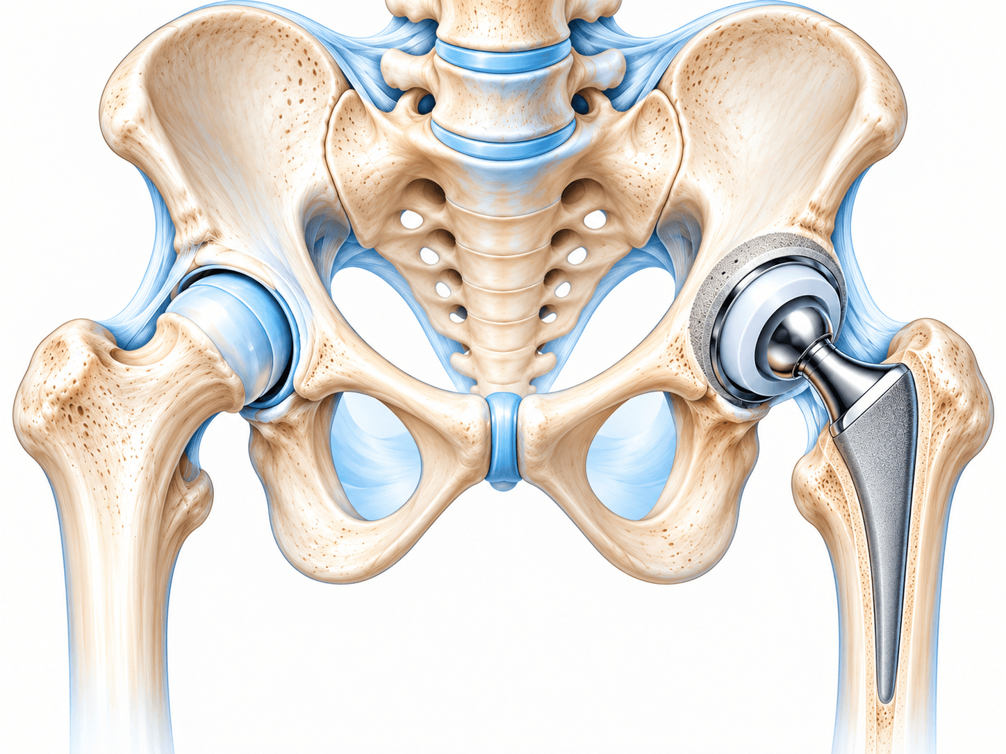
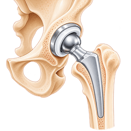
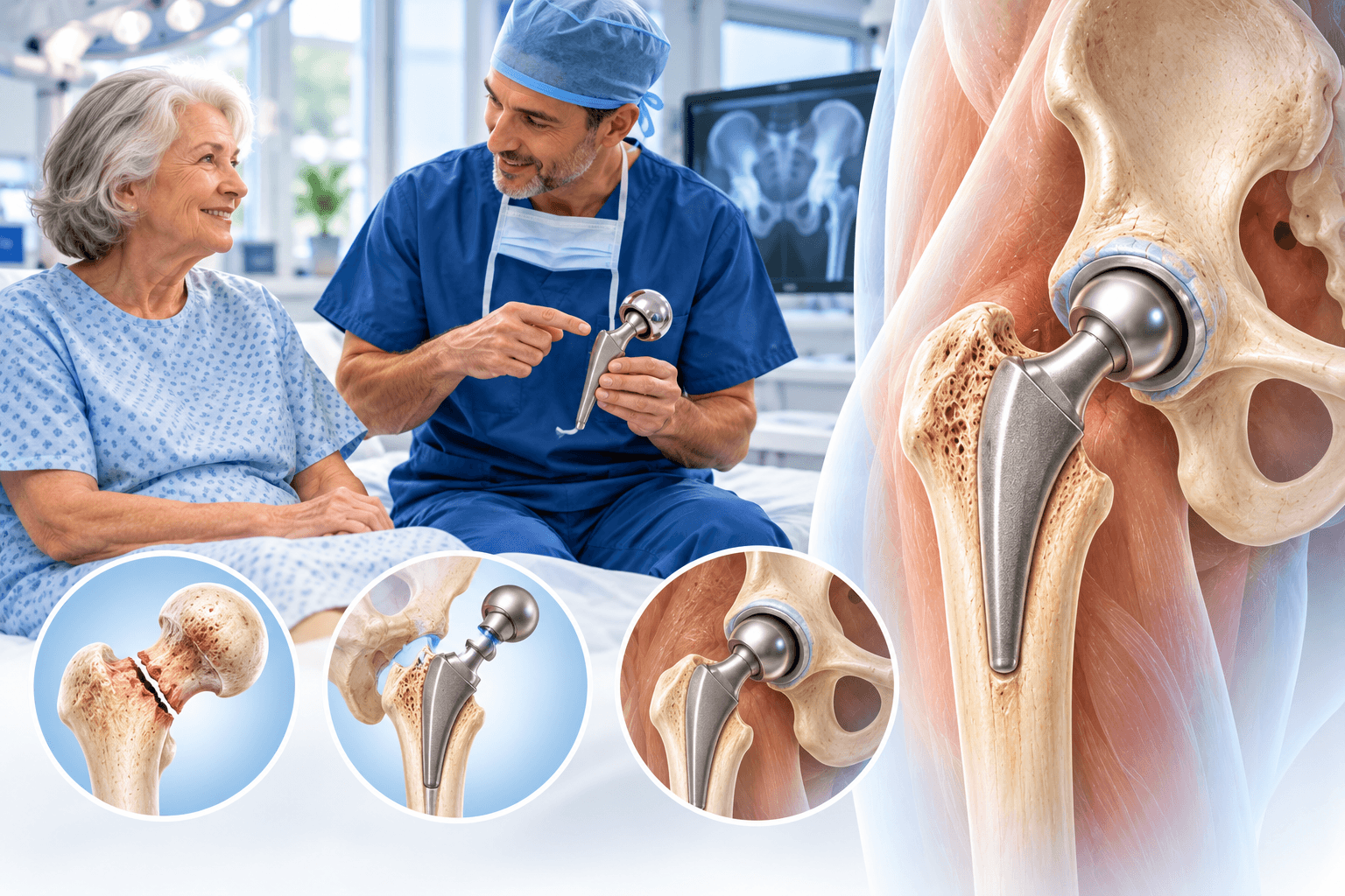
Hip revision surgery is an operation performed when a previous hip replacement needs to be repaired, partially replaced or completely reconstructed. The original artificial hip may have functioned for many years before developing a problem, or revision may become necessary relatively soon after the first operation because of infection, instability, fracture or another complication.
The procedure is also known as revision hip replacement, hip replacement revision, revision total hip arthroplasty or revision THA. Patients sometimes search for the phrase hip replacement revision operation or revised hip replacement surgery. These expressions all refer broadly to surgery performed on a hip that already contains an artificial joint.
Revision is not one single standardized operation. A patient with recurrent dislocation caused by a worn liner may require a relatively limited component exchange, while a patient with severe bone loss and a loose stem may need extensive reconstruction using specialized implants. Infection creates another category entirely because controlling bacteria can become just as important as reconstructing the joint.
How Is Revision Hip Replacement Different From Primary Hip Replacement?
A primary total hip replacement begins with the patient's native hip joint. The surgeon removes the damaged femoral head, prepares the acetabulum and femur and implants the first artificial components.
Revision surgery begins with an artificial joint already present. The surgeon must first determine which components are causing the problem and whether they can safely remain. Existing implants can be tightly bonded to bone, surrounded by scar tissue or associated with areas where bone has been lost over time.
Removing a well-fixed implant without damaging the remaining bone can be one of the most technically demanding parts of the operation. Revision therefore frequently requires specialized extraction instruments, longer operating times and more complex implants than primary replacement.
Does Every Revision Require Removing the Whole Hip Replacement?
No. Some revisions are partial revisions.
If the acetabular shell and femoral stem remain securely fixed and correctly positioned, the surgeon may be able to preserve them. A worn polyethylene liner and femoral head, for example, can sometimes be exchanged while the major metal components remain.
Other patients need only the acetabular component revised because the cup has loosened or is incorrectly positioned. Conversely, a stable cup can sometimes remain while a loose femoral stem is replaced.
A complete revision becomes necessary when both sides are failing, infection requires removal of all components or the existing implant configuration cannot support a stable reconstruction.
Why Do Hip Replacements Need Revision?
Artificial hip replacements can work extremely well for many years, but no implant is immune to biological or mechanical problems.
The fixation between an implant and bone can gradually fail. Polyethylene bearing surfaces can wear. Microscopic wear particles can trigger bone loss known as osteolysis. The artificial hip can dislocate repeatedly. Infection can occur shortly after surgery or years later. A fall can fracture the femur or pelvis around the components.
The reason for revision determines almost every aspect of the operation. A surgeon therefore does not plan “revision hip surgery” in general. The surgeon plans treatment for the specific mechanism of failure.
Aseptic Loosening
Aseptic loosening means an implant has lost stable fixation without an infection being responsible.
The problem can affect the acetabular cup, femoral stem or both. Patients often develop gradually increasing pain after a period when the replacement had previously been comfortable.
A loose acetabular component may migrate or change position. A loose femoral stem can cause thigh or groin pain and progressively damage surrounding bone.
Revision usually involves removing the loose component and creating new fixation using revision implants that obtain stability from healthier remaining bone.
Osteolysis
Osteolysis refers to loss of bone around a joint replacement.
Historically, microscopic polyethylene wear particles were an important cause. The immune system reacts to particles, and the resulting biological response can gradually resorb bone.
Osteolysis can sometimes become extensive before severe symptoms develop. This is one reason long-term follow-up and periodic X-rays remain useful after hip replacement.
When bone loss threatens implant fixation or the structural integrity of the hip, revision may be recommended even before catastrophic failure occurs.
Polyethylene Wear
Modern highly cross-linked polyethylene has significantly improved wear characteristics, but bearing surfaces can still deteriorate over a sufficiently long period.
Progressive liner wear changes the relationship between the femoral head and acetabular component. Debris can contribute to osteolysis.
If the metal shell remains well fixed and appropriately positioned, the surgeon may sometimes exchange only the liner and femoral head.
If bone loss or cup damage is extensive, a more complete acetabular revision can become necessary.
Recurrent Hip Dislocation
Dislocation occurs when the artificial femoral head comes out of the socket.
A single early dislocation does not automatically require revision surgery. Many first-time dislocations can be reduced without reopening the hip.
Repeated instability is different. Recurrent dislocation can result from component malposition, inadequate soft-tissue tension, abductor muscle deficiency, spinal stiffness or neurological problems.
Revision needs to correct the actual cause rather than simply replace components without understanding why instability developed.
Infection
Periprosthetic joint infection is one of the most serious reasons for hip replacement revision.
Bacteria can infect the tissues surrounding the implant shortly after surgery or many years later. Symptoms can include increasing pain, wound drainage, swelling and fever, although chronic infections can present much more subtly.
An infected artificial joint is difficult to treat with antibiotics alone because bacteria can form a protective biofilm on implant surfaces.
Treatment depends on the timing of infection, the organism, implant stability, soft-tissue condition and patient health.
Periprosthetic Fracture
A periprosthetic fracture is a break in the bone surrounding a hip replacement.
The fracture most commonly affects the femur around the stem, although acetabular and pelvic fractures can also occur.
Treatment depends on whether the existing component remains stable. If the femoral stem is still securely fixed, the fracture may be treated with plates, cables or other fixation while retaining the implant.
If the stem is loose, revision to a longer component that achieves fixation beyond the damaged bone is commonly required.
Implant Breakage or Mechanical Failure
Modern hip components are highly durable, but mechanical problems can occur.
Liners can fail or disengage. Certain modular connections can corrode. Rarely, metal components can fracture.
Implant failure should prompt a careful investigation of why it happened. Simply replacing a broken component without correcting abnormal forces can lead to repeated failure.
Revision planning therefore considers component design, alignment, patient activity, bone support and soft-tissue balance.
Adverse Local Tissue Reaction
Some patients with certain metal-containing implant configurations can develop adverse reactions to corrosion products or metal debris.
Local tissue damage can include inflammation, fluid collections and destruction of muscle or bone.
Revision may be needed to remove the problematic bearing or modular interface and reconstruct the hip using another configuration.
The surgeon can use blood tests and advanced imaging when metal-related tissue reaction is suspected.
Painful Hip Replacement
Pain does not automatically mean that revision surgery is necessary.
The surgeon first needs to determine whether the prosthesis is actually responsible.
Pain can arise from the lumbar spine, sacroiliac joint, tendons, bursae, muscle weakness or another nearby condition even when the hip implant remains well fixed.
Revision performed without a clear mechanical or infectious diagnosis can fail to relieve pain. A careful diagnostic work-up is therefore essential before planning surgery.
Leg-Length or Offset Problems
Hip mechanics depend partly on leg length and femoral offset.
A significant problem can affect walking, muscle tension and stability.
Mild perceived leg-length differences are common after hip replacement and do not usually justify revision.
Revision may be considered when the discrepancy is substantial, persistent and clearly related to component position or reconstructive mechanics.
Impingement
Impingement occurs when components, bone or soft tissues make abnormal contact during movement.
Repeated impingement can contribute to pain, liner damage or instability.
The cause can include component orientation, bone anatomy or spinal-pelvic mechanics.
Revision may involve repositioning the cup, changing the femoral component, increasing head size or using a different bearing construct.
Hip Revision for Instability
Revision for instability requires careful analysis.
The surgeon looks at acetabular orientation, femoral version, hip offset, leg length and the condition of the abductor muscles. The relationship between the lumbar spine and pelvis has also become increasingly important because a stiff or fused spine can change functional cup orientation between standing and sitting.
Correcting instability can range from a simple head-and-liner exchange to full component revision.
Dual-mobility components are increasingly used in appropriate revision cases because their design can increase the effective head size and resistance to dislocation.
Hip Revision for Infection
Infection revision is different from routine mechanical revision.
The objective is both to eliminate infection and reconstruct a functional joint.
An early acute infection with stable implants can sometimes be treated using debridement, antibiotics and implant retention. The surgeon opens the joint, removes infected tissue, cleans the area and frequently exchanges modular components such as the femoral head and liner.
More established infection often requires removal of components.
A one-stage or two-stage reconstruction can then be considered depending on the clinical situation.
One-Stage Revision for Infection
In a one-stage revision, the infected prosthesis is removed, infected tissue is thoroughly debrided and new components are implanted during the same operation.
The approach avoids a second major reconstructive procedure.
Patient selection is important. Factors such as identification of the infecting organism, antibiotic sensitivity, soft-tissue condition and overall patient health influence whether a one-stage strategy is suitable.
One-stage revision is well established in experienced centers but is not automatically appropriate for every periprosthetic hip infection.
Two-Stage Revision for Infection
A two-stage revision separates implant removal and definitive reconstruction into two operations.
During the first stage, the surgeon removes infected components and performs extensive debridement. An antibiotic-loaded spacer may be inserted.
The patient then receives antimicrobial treatment while infection is monitored.
If infection control is satisfactory, the second stage removes the spacer and implants the definitive revision hip replacement.
This pathway can involve months of treatment and rehabilitation, which is important for patients to understand before choosing a center abroad.
What Is a Hip Spacer?
An antibiotic spacer temporarily occupies the joint after removal of an infected prosthesis.
Some spacers are designed to allow limited hip movement and partial mobility, while others are more static.
The cement can contain antibiotics selected for the infection.
A spacer is not equivalent to a permanent hip replacement. It is part of an infection-treatment strategy and may have restrictions regarding weight bearing and activity.
Revision for Periprosthetic Fracture
A fracture around the femoral stem must be classified according to its location, bone quality and implant stability.
If the stem remains solidly attached, fracture fixation may be enough.
When the stem has become loose, the surgeon generally needs to bypass the damaged region with a longer revision stem.
Severe fractures can require plates, cables, strut grafts or proximal femoral replacement in exceptional cases.
Revision After Hip Resurfacing
Patients with a previous hip resurfacing can later require conversion to total hip replacement.
Reasons include femoral neck fracture, loosening, adverse metal reaction or persistent pain.
The acetabular component may sometimes be retained, but many cases require revision of both sides.
Metal debris and surrounding tissue quality need particular assessment when the original resurfacing used a metal-on-metal bearing.
Revision After Hemiarthroplasty
A painful hemiarthroplasty can eventually require conversion to total hip replacement.
Common reasons include acetabular erosion, loosening, infection and instability.
The surgeon adds an acetabular component and assesses whether the existing femoral stem can remain.
Conversion surgery generally carries greater complexity than uncomplicated primary total hip replacement because previous surgery and altered anatomy must be managed.
Partial vs Complete Revision Hip Replacement
A partial revision replaces only the problematic portion of the prosthesis.
A complete revision removes and reconstructs both the acetabular and femoral sides.
A smaller revision should not automatically be assumed to be better. Retaining a component that is poorly positioned simply because it remains firmly fixed can leave the original cause of failure unresolved.
The surgeon should preserve components only when they are mechanically and biologically appropriate to keep.
Is Revision Surgery Always More Difficult?
Most revision operations are technically more complex than primary hip replacement, but complexity varies enormously.
A modular head-and-liner exchange can be relatively limited.
Removing a fully ingrown femoral stem from a patient with major bone loss is a very different operation.
The term hip revision surgery therefore covers procedures ranging from modest component exchanges to some of the most demanding reconstructions in adult orthopaedics.
Conditions treated
Who it's for
- Aseptic loosening of the acetabular cup, femoral stem or both
- Periprosthetic joint infection that cannot be controlled with less extensive treatment
- Recurrent or irreducible hip dislocation
- Major component malposition causing instability, pain or impingement
- Periprosthetic fracture associated with a loose hip component
- Severe polyethylene wear with progressive osteolysis
- Mechanical failure of a liner, modular component or other implant part
- Progressive acetabular or femoral bone loss threatening implant stability
- Symptomatic adverse local tissue reaction related to metal debris or corrosion
- Failed hip resurfacing requiring conversion or revision
- Failed hemiarthroplasty requiring conversion to total hip replacement
- Painful implant with a clearly identified mechanical cause
- Substantial leg-length or offset problems caused by implant position in carefully selected patients
- Recurrent instability associated with abductor deficiency
- Fracture nonunion or reconstruction failure around a hip replacement
- Previously revised hip that has developed another mechanical or infectious failure
Good candidates
A good candidate for revision hip replacement has a clearly established problem that is likely to improve through another operation. Revision should not be performed merely because an implant looks old on an X-ray or because a patient has nonspecific discomfort.
The surgeon needs to determine why the existing replacement is failing and whether surgery can meaningfully improve pain, stability or function. This often requires more diagnostic work than before primary hip replacement.
General health is also important. Revision can involve greater blood loss, longer anaesthesia and more extensive reconstruction than primary surgery. Diabetes, cardiovascular disease, kidney disease, nutritional status, smoking, anaemia and other medical conditions should therefore be optimized wherever possible.
Patients With Aseptic Loosening
Patients with a clearly loose component are common candidates.
Symptoms can include progressive groin pain from acetabular loosening or thigh pain associated with femoral stem failure.
X-rays may show migration, radiolucent lines or bone loss.
Revision aims to restore stable implant fixation before progressive bone destruction makes reconstruction even more difficult.
Patients With Recurrent Dislocation
A patient who experiences repeated dislocations despite appropriate conservative management may need revision.
The operation should be based on the cause of instability.
Simply changing the femoral head is unlikely to solve a severely malpositioned acetabular component.
Conversely, removing a well-positioned cup may be unnecessary when instability is primarily related to soft-tissue deficiency.
Patients With Infection
Revision may become necessary when bacteria have established infection around the prosthesis.
The patient should ideally be treated by a multidisciplinary team that can include revision arthroplasty surgeons, infectious-disease specialists and microbiology experts.
The treatment plan depends on how long infection has been present, the organism involved, implant fixation and patient health.
Complex chronic infection should be treated in a center capable of both infection control and difficult reconstruction.
Patients With Bone Loss
Bone loss can affect the acetabulum, femur or both.
The surgeon must determine whether remaining bone can support standard components.
Moderate defects can often be reconstructed using larger porous cups, revision stems or augments.
Massive bone loss can require cages, cup-cage constructs, custom components or major femoral reconstruction.
Patients should understand that the severity of bone loss often determines recovery restrictions.
Older Adults
Age alone does not rule out revision.
A healthy older adult with painful loosening and good functional potential can benefit substantially.
The decision becomes more difficult in a severely frail patient with limited mobility and multiple medical problems.
The expected improvement must justify the physiological burden of surgery.
Younger Patients
Younger patients may undergo revision after implant wear, failure or previous complex reconstructive surgery.
Preserving remaining bone is especially important because another revision may be needed later in life.
Surgeons may therefore use techniques that prioritize biological fixation and bone conservation whenever feasible.
Patients With Multiple Previous Revisions
Each additional operation changes the local anatomy.
Scar tissue increases, bone stock can decrease and muscle function may deteriorate.
Patients who have already undergone several revisions often need highly specialized reconstruction.
Their expected outcome should be discussed realistically because the goal may shift from achieving a nearly normal hip toward creating a stable, pain-controlled and functional limb.
Before surgery
Establishing Why the Hip Replacement Failed
The most important preoperative task is identifying the failure mechanism.
Revision without a clear diagnosis risks solving the wrong problem.
The surgeon begins with the patient's history. The timing and location of pain can provide important clues. Sudden pain after a fall suggests fracture. Progressive thigh pain can occur with stem loosening. Recurrent episodes where the joint comes out of position indicate instability.
The original operative report and implant records are extremely useful. They can identify component brands, sizes and fixation methods, which helps determine whether replacement parts or extraction tools will be needed.
Physical Examination
The surgeon assesses gait, leg length and hip movement.
Muscle strength is evaluated, particularly the abductors that stabilize the pelvis during walking.
The skin and previous surgical scars are examined.
Neurological and vascular status are documented.
The lumbar spine also deserves assessment because spinal stiffness or fusion can influence hip stability and functional component orientation.
Plain X-Rays
Standard pelvic and hip radiographs are fundamental.
The surgeon looks for component migration, fracture, wear, osteolysis and changes around the implants.
Sequential previous X-rays are especially valuable because they reveal whether a component has moved over time.
A single image can sometimes underestimate gradual loosening that becomes obvious when compared with films from earlier years.
CT Scan
CT can provide additional information about bone loss and implant orientation.
Metal-artifact reduction techniques improve visualization around modern components.
Three-dimensional reconstruction can help in complex acetabular defects or pelvic discontinuity.
CT is particularly useful when conventional X-rays do not fully describe the remaining bone available for reconstruction.
Metal Artifact Reduction MRI
MRI can be useful when the surgeon suspects muscle damage, fluid collections or adverse local tissue reaction.
Special imaging sequences reduce distortion caused by the metal implants.
This can help evaluate the abductors and surrounding soft tissues.
MRI is not required for every revision but can provide important information in selected cases.
Blood Tests for Infection
Infection needs to be considered before most revision procedures, even when loosening appears mechanical.
Blood tests commonly include inflammatory markers such as C-reactive protein and erythrocyte sedimentation rate.
Abnormal tests do not prove that a prosthesis is infected, and normal values do not exclude every infection.
They are part of a broader diagnostic assessment.
Joint Aspiration
If infection is suspected, fluid can be aspirated from the hip under sterile conditions.
The laboratory can evaluate cell counts, differential counts and cultures.
Additional synovial tests can be used according to local protocols.
Antibiotics can interfere with culture results, so the diagnostic sequence should be coordinated with the treating team.
Identifying the Organism
When infection is present, identifying the microorganism helps determine antibiotic treatment and revision strategy.
Some infections involve highly sensitive organisms, while others involve resistant bacteria or organisms that are difficult to eradicate.
Culture information can influence whether the surgeon considers implant retention, one-stage revision or staged reconstruction.
Unexpected positive cultures can occasionally emerge during surgery despite an apparently aseptic preoperative work-up.
Implant Identification
Knowing the existing implant system can simplify revision enormously.
Hospital records, implant cards and previous operative notes can identify the manufacturer and component sizes.
Some modular parts are compatible only within specific implant systems.
International patients should bring all available documentation rather than relying on the new hospital to identify an older prosthesis from X-rays alone.
Assessing Bone Loss
Revision planning requires careful evaluation of the remaining acetabular and femoral bone.
Small defects may require little additional reconstruction.
Larger defects can influence the entire strategy.
The surgeon considers where secure fixation can still be obtained and whether bone graft, porous metal augments or custom components may be required.
Acetabular Bone Loss
The socket can lose bone around a loose cup.
Defects can affect the walls, columns or supporting pelvic structure.
Severe loss can lead to pelvic discontinuity, where the upper and lower portions of the pelvis are no longer mechanically continuous.
These situations require specialized revision techniques and should be managed by surgeons familiar with complex acetabular reconstruction.
Femoral Bone Loss
Removing a loose or well-fixed stem can leave defects in the upper femur.
Previous fractures, osteolysis and earlier revisions can further reduce bone stock.
Revision stems are often longer because they need to obtain fixation in healthier bone beyond the damaged region.
Severe proximal femoral deficiency can require modular reconstruction or, in exceptional cases, replacement of a large portion of the proximal femur.
Medical Optimization
The patient's cardiovascular and respiratory health must be assessed.
Anaemia deserves particular attention because revision surgery can involve more blood loss than primary replacement.
Diabetes should be well controlled where possible.
Kidney function influences medication and infection management.
Nutrition is important because malnutrition can impair wound healing and immune function.
Smoking
Smoking is associated with impaired wound healing and infection-related complications.
Stopping nicotine before revision is strongly encouraged.
This is particularly important when surgery requires extensive soft-tissue dissection or bone healing.
Patients undergoing revision already face a higher complication burden than those undergoing routine primary replacement, so modifiable risks deserve serious attention.
Weight and Metabolic Health
Obesity can increase technical difficulty, wound risk and rehabilitation challenges.
Weight optimization should be approached realistically.
Patients with a painful loose implant may not be able to exercise effectively before surgery.
The purpose is to reduce modifiable risk where practical rather than delay necessary reconstruction indefinitely in pursuit of an arbitrary number.
Medication Review
Anticoagulants, antiplatelet drugs, immunosuppressive medication and diabetes treatment require individualized planning.
The patient should provide a complete medication list.
Blood thinners should never be stopped independently.
Revision for infection may also involve complex antibiotic planning, which should be coordinated between the orthopaedic and infectious-disease teams.
Dental and Other Active Infections
Major active infections should be treated before elective aseptic revision whenever feasible.
The presence of a permanent joint prosthesis makes infection prevention particularly important.
Dental management should follow current arthroplasty guidance rather than assuming that every dental procedure automatically requires antibiotics.
The surgeon can advise according to the patient's risk and timing of surgery.
Planning for Blood Loss
Revision hip replacement can involve meaningful blood loss.
Modern blood-conservation strategies can include tranexamic acid, careful surgical technique and correction of preoperative anaemia.
Blood may be cross-matched in complex cases.
The requirement depends on the expected reconstruction and the patient's starting haemoglobin.
Planning Specialized Components
Complex revision implants are not necessarily stocked in the same way as routine primary hip replacements.
The surgeon may need multiple cup sizes, augments, long stems or constrained and dual-mobility options available in the operating room.
Custom-made implants can require weeks of design and manufacturing.
This is another reason detailed preoperative imaging and planning are essential.
Planning for Unexpected Findings
Even excellent preoperative planning cannot predict every finding.
A cup thought to be well fixed can prove loose.
Bone can fracture while removing a stem.
Unexpected infection can be identified.
The surgeon therefore needs backup strategies and access to appropriate revision inventory.
A center performing complex hip revision should be equipped to respond rather than improvise without the correct components.
How the operation is performed
Hip revision surgery begins by exposing the previous hip replacement, identifying the failed components and removing only the implants that need to be exchanged. The surgeon then reconstructs damaged bone and installs new revision components designed to achieve secure fixation and restore hip stability, leg length and biomechanics.
The exact operation is determined by the reason for failure. A liner exchange can preserve most of the original reconstruction. A full revision can require removal of the cup and stem, reconstruction of bone defects and implantation of specialized components.
Because there is no single standardized revision technique, patients should understand exactly which parts of their hip are expected to be revised.
Surgical Approach
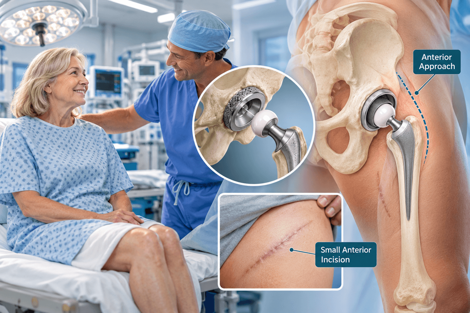
The surgeon usually works through an existing scar when practical, although the previous approach does not always have to be reused.
Revision generally requires broader exposure than primary replacement.
Scar tissue is carefully released.
Important nerves and blood vessels may be less obvious than during the first operation because normal tissue planes have been altered.
The surgeon must gain sufficient exposure without causing unnecessary soft-tissue damage.
Collecting Tissue Samples
When infection is possible, multiple tissue and fluid samples are collected during surgery.
These are sent for microbiological culture and sometimes histological evaluation.
Samples should be obtained before unnecessary contamination occurs.
The results can influence antibiotic treatment after surgery.
Infection testing remains important even when revision was originally believed to be purely mechanical.
Head and Liner Exchange
One of the least extensive revisions is exchange of the modular femoral head and acetabular liner.
The metal shell and femoral stem are preserved if they remain appropriately positioned and securely fixed.
This strategy can be useful for selected cases of liner wear, instability or early infection treated with debridement.
The surgeon must ensure that retaining the existing components does not leave the cause of failure untreated.
Acetabular Cup Removal
A loose cup can often be removed relatively easily.
A well-fixed cup that requires revision is more challenging because bone must be separated from the implant without creating excessive additional damage.
Specialized curved blades and extraction tools can help break the bone-implant interface.
The resulting bone defect is then inspected and classified before reconstruction.
Acetabular Reaming
The remaining socket is prepared to accept the revision component.
The surgeon removes scar tissue and unstable bone while preserving as much viable pelvic bone as possible.
Reaming must create enough contact for fixation without unnecessarily enlarging the defect.
Revision shells are frequently more porous and may accept multiple screws to improve initial stability.
Jumbo Revision Cups
A larger hemispherical cup can sometimes bridge moderate bone defects and achieve fixation in remaining healthy bone.
This strategy is relatively straightforward when sufficient pelvic bone exists.
The larger component should still be positioned according to functional hip mechanics.
Size alone does not solve severe segmental defects or pelvic discontinuity.
Porous Metal Augments
Porous metal augments are modular implants used to fill areas where acetabular bone is missing.
They can support the revision cup and help reconstruct the geometry of the socket.
Modern highly porous metals are designed to encourage biological fixation.
The surgeon selects their shape and position according to the defect.
Multiple augments can sometimes be combined for complex reconstructions.
Bone Graft
Bone graft can be used to restore lost bone.
Morselized graft may fill contained defects.
Structural grafts are used more selectively.
Revision surgery tries to balance immediate mechanical stability with restoration of bone stock.
The exact role of graft varies according to patient age, defect type and implant strategy.
Cup-Cage Reconstruction
Severe acetabular bone loss or pelvic discontinuity can require a cup-cage construct.
A porous cup provides biological fixation while a cage or reinforcing structure provides additional mechanical support.
The technique is more complex than routine acetabular revision.
Weight-bearing restrictions can be more cautious while reconstruction heals.
Custom Triflange Components
Massive pelvic bone loss can exceed the ability of standard implants.
A custom triflange acetabular component can be designed using three-dimensional CT imaging.
The implant is manufactured to match the patient's remaining pelvic anatomy and obtain fixation to stronger regions of bone.
Custom reconstruction can be valuable in highly selected cases but requires substantial planning and specialist expertise.
Removing a Femoral Stem
Femoral component revision begins with determining whether the stem is loose or well fixed.
A loose stem may be relatively straightforward to extract.
Removing a fully ingrown cementless stem can be significantly more difficult.
Special instruments are used to separate the implant from the surrounding femur while trying to preserve bone.
Extended Trochanteric Osteotomy
An extended trochanteric osteotomy, often abbreviated ETO, is a controlled opening of the upper femur that provides access to a well-fixed stem or cement mantle.
Instead of damaging the femur unpredictably while forcing the implant out, the surgeon creates a planned bone flap.
After implant removal and reconstruction, the osteotomy is repaired with cables or wires.
Healing of the osteotomy can influence postoperative weight-bearing and rehabilitation.
Cement Removal
Removing old bone cement from the femoral canal can be technically demanding.
The surgeon uses specialized instruments to break and extract the cement without perforating or fracturing the femur.
Cement can extend far down the canal.
In complex cases, imaging or ultrasonic tools can assist.
The objective is to create a stable bed for the new revision stem while preserving as much bone as possible.
Revision Femoral Stems
Revision stems are frequently longer than primary components.
They can obtain fixation below areas of damaged proximal femur.
Modern modular tapered fluted stems are one important option because the surgeon can establish distal fixation while separately adjusting proximal body size, offset and version.
Other stem designs are available depending on bone-loss pattern and surgeon preference.
Modular Revision Stems
A modular stem is assembled from separate components.
The distal section achieves fixation within the femur.
The proximal body can then be selected to restore leg length, offset and version.
This flexibility is valuable when normal proximal femoral anatomy has been lost.
Modular junctions also create additional engineering interfaces, so component selection must be appropriate to the clinical situation.
Cemented Revision Stems
Cemented revision remains useful in selected circumstances.
Very elderly patients, certain bone-loss patterns or reconstruction strategies can make cemented fixation appropriate.
A cement-in-cement technique can occasionally be used when an existing cement mantle is sound and the revision indication allows it.
Revision fixation should be chosen according to bone and reconstruction rather than a universal rule.
Periprosthetic Fracture Reconstruction
When revision is performed for a fracture, the stem and fracture must be considered together.
A loose stem is commonly replaced with a longer revision component that bypasses the fracture.
The bone can also be stabilized using plates, cables or other fixation devices.
If the stem remains well fixed, the surgeon can sometimes keep it and treat the fracture alone.
Proximal Femoral Replacement
Extremely severe proximal femoral bone loss can occasionally require a proximal femoral replacement.
This type of megaprosthesis replaces a large portion of the upper femur.
It can be used in salvage situations where conventional revision fixation is impossible.
The operation carries substantial risks and should be considered a specialized reconstruction rather than routine revision hip surgery.
Trial Components
Trial components allow the surgeon to assess the reconstructed hip before definitive implantation.
Leg length, offset, stability and range of motion are evaluated.
Revision cases can be particularly challenging because previous bone loss and soft-tissue damage distort normal landmarks.
The surgeon may need to balance perfect anatomical restoration against what is mechanically achievable with the remaining tissue.
Dual-Mobility Components
Dual-mobility cups contain a small femoral head locked within a larger polyethylene component that also articulates with the acetabular shell.
The design increases the effective head size and jump distance.
This can help reduce instability in selected high-risk revision patients.
Recent comparative evidence continues to support an important role for dual-mobility components in revision THA where dislocation risk is a major concern. PubMed
Constrained Liners
A constrained liner mechanically captures the femoral head to resist dislocation.
These implants can be useful when soft-tissue deficiency makes stability difficult to achieve.
Constraint also transfers greater forces to the implant-bone interface.
For this reason, constrained liners are usually reserved for selected instability situations rather than being used routinely.
Correcting Component Position
A revision for recurrent instability often requires correction of component orientation.
The acetabular cup can be removed and repositioned.
Femoral version may also need adjustment.
The surgeon considers standing and sitting pelvic mechanics where relevant.
A technically successful revision addresses the cause of instability instead of merely using a larger head to compensate for poorly positioned components.
Abductor Deficiency
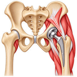
The abductor muscles stabilize the pelvis and contribute to hip stability.
Previous surgery, trochanteric injury or tissue damage can leave the abductors deficient.
These patients have a higher risk of limp and instability.
Revision planning may include soft-tissue repair, trochanteric fixation or a stability-enhancing bearing such as dual mobility or, in selected circumstances, a constrained liner.
One-Stage Infection Revision Technique
In a one-stage infection revision, all removable infected components are extracted during one procedure.
The surgeon performs extensive debridement and removes contaminated tissue.
The surgical field is thoroughly irrigated.
New revision components are then implanted and targeted antimicrobial treatment continues after surgery.
This strategy requires careful patient and infection selection.
First Stage of Two-Stage Revision
The first stage involves removal of the infected prosthesis and aggressive debridement.
Multiple samples are collected.
An antibiotic-loaded spacer is frequently inserted.
The patient's antibiotic programme is then guided by microbiology and infectious-disease assessment.
The interval before reimplantation varies according to clinical progress and institutional protocol.
Second Stage of Two-Stage Revision
The second stage occurs when the treating team believes definitive reconstruction is appropriate.
The spacer is removed and the joint is reassessed.
Additional tissue samples can be collected.
Revision implants are then used to reconstruct the hip.
Patients should understand that infection management remains complex even when the second-stage operation proceeds successfully.
Wound Closure
Revision surgery can create a larger surgical wound than primary replacement.
The surgeon closes deep tissues and skin carefully.
Drains are used selectively.
High-risk patients can receive specialized dressings or closed-incision negative-pressure therapy according to local protocol.
Wound monitoring is particularly important because revision itself carries a higher infection burden than first-time arthroplasty.
Hospital stay
Immediately After Revision Surgery
After surgery, the patient is monitored in the recovery area while anaesthesia wears off.
Blood pressure, oxygen levels, pain and neurological status are assessed.
The surgical team checks the operated leg and wound.
Complex revisions can require closer haemodynamic monitoring because the operation may involve greater fluid shifts and blood loss than primary total hip replacement.
Some medically fragile patients or extremely complex reconstructions may require short-term higher-level monitoring.
Pain Management
Multimodal pain control is generally used.
This can combine paracetamol or acetaminophen, regional anaesthetic techniques and carefully selected additional medication.
The aim is to reduce pain enough that the patient can breathe comfortably, sleep and begin mobilizing.
Pain treatment also needs to avoid excessive sedation, particularly in older adults who may be susceptible to confusion.
How Painful Is Hip Revision Surgery?
Patients frequently ask how painful hip revision surgery is compared with their first hip replacement.
There is no single answer because revision ranges from a relatively limited liner exchange to extensive bone reconstruction.
A complex full revision can produce more postoperative soreness because scar tissue must be released and bone may need to be reconstructed. Femoral osteotomy, fracture fixation or major acetabular reconstruction can add discomfort.
Modern anaesthesia and multimodal analgesia make this pain treatable. The most intense postoperative pain generally improves progressively over the first days, while muscular soreness and deep aching can continue for several weeks.
Blood Tests After Surgery
Haemoglobin and other laboratory values may be monitored after complex revision.
Blood transfusion is not automatically required merely because haemoglobin decreases.
The decision is based on the patient's symptoms, cardiovascular condition and degree of anaemia.
Kidney function and electrolytes may also be monitored, particularly after long operations or in patients receiving antibiotics for infection.
Early Walking
Mobilization usually begins as soon as the reconstruction and medical condition permit.
Some patients can stand the same day or the morning after surgery.
Other patients require temporary restrictions because bone graft, an osteotomy or a complex implant needs protection.
Revision rehabilitation should therefore never copy a generic primary-hip protocol without considering what was reconstructed.
Weight-Bearing Restrictions
Weight bearing can range from full weight bearing as tolerated to substantial protection for several weeks.
A straightforward component exchange can allow rapid progression.
Major acetabular reconstruction or femoral osteotomy can require partial or limited weight bearing.
The operative report should clearly state the permitted amount.
International patients should have this instruction in writing for their physiotherapist after returning home.
Physiotherapy
The physiotherapist begins with transfers, standing and safe walking.
Rehabilitation aims to protect the reconstruction while preventing unnecessary deconditioning.
Patients may need a walker initially.
Progression to crutches or a cane depends on balance, strength and weight-bearing restrictions.
Abductor weakness can make gait recovery considerably slower than after a primary replacement.
Length of Hospital Stay
A relatively uncomplicated revision can sometimes require only a few nights.
More complex surgery frequently requires longer hospitalization.
Infection, major blood loss, medical complications or the need for inpatient rehabilitation can extend the stay.
The decision should reflect actual medical and functional readiness rather than a fixed package duration.
Discharge Planning
Before discharge, the patient needs a clear medication plan, wound-care instructions and rehabilitation protocol.
Blood-clot prevention should be explained.
The patient should know their exact weight-bearing status and any hip precautions.
Follow-up imaging should already be planned.
International patients additionally need operative documentation and implant records before leaving the country.
Recovery
Recovery after revision hip replacement is usually slower and more variable than after primary hip replacement.
Patients should not compare themselves automatically with someone who had a routine first-time replacement.
The tissues have already undergone at least one previous operation. Scar tissue must heal again, muscles can be weaker, and the new components may rely on damaged bone that requires time to integrate.
The early goal is safe mobility while protecting the reconstruction. Long-term rehabilitation focuses on restoring strength, gait and endurance.
First 24–72 Hours
The first days focus on pain control, medical stability and safe transfers.
The patient begins ankle exercises and other simple movements.
Walking starts with professional assistance when safe.
If full weight bearing is allowed, confidence can increase relatively quickly.
If weight bearing is restricted, physiotherapists teach the patient how to protect the operated limb while using a walker or crutches.
Week 1
The patient commonly remains dependent on a walking aid.
Swelling and bruising are expected.
Fatigue can be significant because revision surgery places a substantial physiological demand on the body.
The patient should focus on short, safe walking sessions and prescribed exercises rather than testing the hip with long distances.
A larger amount of early activity does not necessarily produce faster healing.
Weeks 2–6
Pain and swelling generally decrease.
Wound healing progresses.
Patients begin rebuilding confidence and endurance.
Walking distance can increase gradually within any weight-bearing restrictions.
Muscle recovery is often slower than after primary replacement, especially when abductors were damaged before surgery or needed additional reconstruction.
The patient may continue using a walker or crutches throughout this phase.
Weeks 6–12
Many patients experience a meaningful improvement in mobility.
X-rays can help determine whether the reconstruction is stable and whether weight-bearing restrictions can be advanced.
Patients can transition to lighter walking aids when appropriate.
The emphasis shifts increasingly toward strength, gait quality and functional independence.
A limp can persist because the muscles surrounding the hip require prolonged rehabilitation.
Months 3–6
This period often represents the transition from basic recovery toward broader function.
Walking endurance improves.
Patients return gradually to household, occupational and recreational activities.
Those who underwent complex bone reconstruction may only begin unrestricted rehabilitation during this period.
The hip often still feels different from a primary replacement because revision components can be larger and the surrounding tissues may have sustained more damage.
Months 6–12
Improvement can continue for a full year.
Strength, balance and confidence can increase gradually.
Some residual weakness or stiffness can remain after difficult revisions.
The final goal is not always a completely normal-feeling hip.
For a patient with massive bone loss or multiple previous operations, a stable joint with substantially reduced pain and reliable walking can represent an excellent outcome.
Hip Revision Surgery Recovery Time
The phrase hip revision surgery recovery time does not have one universal numerical answer.
A relatively limited revision can recover in a timeframe approaching primary total hip replacement.
A major acetabular or femoral reconstruction can require protected weight bearing and rehabilitation for several months.
For general patient education, 12–24 weeks for major functional recovery is a reasonable broad range. Complete improvement can continue for 6–12 months.
Walking After Revision Hip Replacement
Most patients are encouraged to start walking early.
The crucial issue is how much weight the reconstruction can tolerate.
Patients with strong immediate fixation may be allowed to bear weight as tolerated.
Others need protected weight bearing because grafted bone, an osteotomy or pelvic reconstruction needs time to heal.
The surgeon's written instructions are more important than generic advice found online.
Walker, Crutches and Cane
A walker commonly provides the safest initial support.
Crutches can be suitable for stronger patients.
A cane is introduced later when the patient can place sufficient weight through the operated side and maintain good balance.
Walking aids should not be removed according to a predetermined date.
The patient should progress when gait quality and reconstruction stability support it.
Physiotherapy After Revision
Rehabilitation is individualized.
Exercises typically focus on hip and leg strength, safe range of motion, balance and walking mechanics.
Patients with abductor deficiency can need prolonged strengthening.
Some complex reconstructions require therapists to avoid specific loading patterns initially.
The physiotherapist therefore needs to understand exactly what the surgeon repaired.
Returning to Work
Desk-based work can sometimes resume within several weeks after an uncomplicated revision.
Complex surgery can require considerably longer.
Physical jobs involving lifting, climbing or prolonged standing may require three months or more.
The surgeon should understand the actual occupational tasks before providing clearance.
A patient who can walk comfortably at home may still be far from ready for a physically demanding job.
Driving
Driving requires safe control of the leg, adequate reaction time and freedom from impairing pain medication.
Right-sided revision deserves particular caution because the operated leg controls braking and acceleration in most vehicles.
Complex reconstruction or prolonged weight-bearing restrictions can delay driving.
The decision should be individualized rather than based on a universal postoperative week.
Sleeping
Comfortable sleep often remains difficult during the first weeks.
Patients can use pillows for support.
Sleeping on the operated side may remain uncomfortable longer than after primary replacement because the scar and soft tissues have been operated on more than once.
Any approach-specific movement precautions should be respected until the surgeon relaxes them.
Stairs
Patients can often learn stair technique before discharge.
A railing and walking aid may be necessary.
The ability to climb stairs does not mean repeated stair exercise is appropriate.
Patients with partial-weight-bearing restrictions need specific instruction on how to protect the reconstruction while negotiating steps.
Swelling
Swelling around the hip and thigh is common.
It can extend toward the knee and lower leg.
The amount should gradually decrease.
New severe swelling, calf pain, shortness of breath or chest discomfort requires urgent assessment because venous thromboembolism is a possible complication.
Persistent Limp
A limp can remain for several reasons.
Abductor muscles may have been damaged by previous surgery, implant failure or repeated dislocation.
Leg-length changes and general weakness also affect gait.
Some patients regain nearly normal walking.
Others continue using a cane because permanent muscle deficiency remains even though the revision implant itself is stable.
Recovery After Infection Revision
Infection revision deserves its own recovery expectations.
A one-stage procedure can resemble a complex mechanical revision but includes extended antimicrobial treatment.
A two-stage pathway is much longer because the patient undergoes removal of the infected implant, a period with a spacer and later reimplantation.
Recovery should therefore be measured across the entire treatment episode rather than from the date of the final operation alone.
Recovery After Extended Trochanteric Osteotomy
An ETO requires bone healing.
The surgeon may restrict weight bearing or active abductor loading while the osteotomy unites.
Cables or wires hold the bone segment in position.
Rehabilitation can therefore be slower than after a revision where the femur did not need to be opened.
Recovery After Acetabular Reconstruction
A large revision cup with excellent initial fixation may allow relatively early loading.
More extensive reconstructions involving augments, cages or pelvic discontinuity can require greater protection.
The surgeon uses serial imaging to assess stability.
Patients should not increase weight bearing simply because pain has improved.
Mechanical healing can lag behind subjective comfort.
Flying After Hip Revision Surgery
Long-distance flights should be planned carefully.
Revision surgery temporarily increases blood-clot risk.
The patient may also be using a walker and require assistance through an airport.
Complex wounds or infection treatment make early international travel even less desirable.
Medical-travel patients should remain locally long enough for initial wound and radiographic assessment rather than scheduling departure according to the shortest possible package.
Recovery for International Patients
International patients should leave with the operative report, implant information, discharge summary, medication plan and rehabilitation protocol.
If cultures were taken, the mechanism for communicating final microbiology results must be clear.
Patients undergoing infection revision may need weeks of antimicrobial monitoring.
Remote follow-up is helpful, but it cannot replace emergency local care if a serious complication develops after the patient returns home.
Recovery timeline
- Achieve safe transfers, pain control and initial protected walking.1Achieve safe transfers, pain control and initial protected walking.
Days 0–7
The patient begins mobilizing according to the surgeon's weight-bearing instructions. Wound monitoring, blood-clot prevention and management of postoperative anaemia are priorities. Complex reconstruction can require a walker and strict loading limitations during this stage.
- Improve household mobility while protecting the revision construct.2Improve household mobility while protecting the revision construct.
Weeks 2–6
Walking distance increases gradually. Swelling and surgical pain usually decrease. Patients continue using an appropriate walking aid and avoid advancing activity beyond the limits created by bone graft, osteotomy or reconstructive fixation.
- Progress strength and weight bearing when imaging and healing permit.3Progress strength and weight bearing when imaging and healing permit.
Weeks 6–12
Follow-up X-rays help guide rehabilitation. Some patients transition from a walker to crutches or a cane. Others remain protected because their reconstruction needs additional healing time.
- Regain broader daily function and endurance.4Regain broader daily function and endurance.
Months 3–6
Strengthening becomes increasingly important. Many patients return to a wider range of household, social and occupational activities. Persistent abductor weakness or complex bone reconstruction can continue to influence gait.
- Approach mature functional recovery.5Approach mature functional recovery.
Months 6–12
The hip continues adapting to the revision construct. Muscle strength, balance and walking confidence can improve for many months. Long-term follow-up focuses on implant stability, infection surveillance where relevant and preservation of remaining bone.
Outcomes and success rates
What Is a Successful Hip Revision?
The definition of success depends on why revision was necessary.
For aseptic loosening, success means creating stable new fixation and reducing pain.
For recurrent dislocation, success requires a hip that remains stable during normal activity.
For infection, control of infection becomes a central outcome alongside reconstruction.
For a patient with major bone loss, preserving enough structure to allow walking may represent an excellent outcome even if function does not equal that of an uncomplicated primary replacement.
Pain Relief
Many patients obtain substantial pain relief when the cause of failure has been accurately identified and corrected.
Pain from a loose stem or cup can improve significantly after stable reconstruction.
Infection-related pain can improve when infection is eradicated.
However, revision should not be promised as a cure for every painful hip because muscle damage, nerve problems and spine disease can continue causing symptoms even after the implant is mechanically stable.
Functional Improvement
Revision can improve walking, stability and everyday independence.
The amount of improvement depends partly on how much tissue remains functional.
A patient with intact abductor muscles and good bone stock has a different prognosis from someone who has undergone five previous surgeries and lost substantial proximal femoral bone.
Revision outcomes should therefore be interpreted according to the starting condition.
Revision Is Not the Same as Primary Hip Replacement
A primary hip replacement usually has more favorable predictability because the surgeon begins with better bone and fewer scars.
Revision patients already have a failed reconstruction.
The operation often has greater technical complexity and complication risk.
The fact that revision outcomes may be less predictable does not mean the surgery is unsuccessful. It reflects the more difficult problem being treated.
Implant Survivorship
Modern revision components can provide durable reconstruction for many years.
Survival depends on the reason for revision, amount of bone loss, implant fixation and patient characteristics.
A revision performed for simple liner wear has a very different long-term risk profile from a reconstruction for pelvic discontinuity or chronic infection.
It is therefore misleading to publish one universal revision success percentage.
Outcomes for Instability
Recurrent instability remains an important challenge after revision THA.
Modern dual-mobility constructs are widely used in patients at elevated dislocation risk.
Recent systematic reviews have reported lower dislocation-related failure with dual-mobility components compared with conventional fixed-bearing constructs in many revision settings, while also emphasizing that much of the available evidence remains observational rather than high-level randomized evidence.
Outcomes for Infection
Periprosthetic joint infection is among the most difficult revision indications.
Both one-stage and two-stage strategies can achieve infection control in appropriately selected patients.
Contemporary evidence does not support a simplistic statement that one strategy is universally superior for every infected hip.
Patient selection, microorganism, soft-tissue condition and the experience of the treating center are critical to interpreting outcomes.
Outcomes With Severe Acetabular Bone Loss
Major acetabular defects require more sophisticated reconstruction.
Modern porous metals, augments, cup-cage constructs and custom implants have expanded the surgeon's options.
The key objective is obtaining secure fixation to remaining healthy pelvic bone while restoring the hip center and stability.
Current revision literature continues to emphasize defect-specific reconstruction rather than a single implant solution for all acetabular bone loss.
Risk of Rerevision
A revision hip can itself eventually require another revision.
The risk is influenced by the original indication.
Infection and instability are particularly challenging because they can recur.
Bone loss can also make any future operation more difficult.
Patients should therefore think of revision not merely as replacing old parts but as preserving as much biological and mechanical foundation as possible for the future.
Patient Satisfaction
Satisfaction is generally highest when expectations match the complexity of the reconstruction.
A patient with a relatively straightforward loose cup may reasonably expect excellent pain relief and strong function.
A person with chronic infection, severe muscle deficiency and several previous operations may define success differently.
Clear preoperative counseling is therefore an important part of good revision surgery.
Implants and technology
Revision Acetabular Cups
Revision cups are designed to achieve secure fixation despite previous bone loss.
They are commonly made with highly porous surfaces that encourage bone integration.
Multiple screw holes provide additional fixation options.
Some shells are substantially larger than primary cups because they need to engage healthier remaining pelvic bone.
Porous Metal Technology
Highly porous titanium and tantalum-based structures have become important in revision arthroplasty.
Their architecture provides high friction for initial stability and a surface that supports biological fixation.
Porous metal augments can fill irregular defects that previously required large structural grafts.
These materials have expanded reconstruction options for severe acetabular bone loss.
Acetabular Augments
Augments are modular pieces positioned against deficient pelvic bone.
They create support for the revision cup.
The surgeon can use different shapes and sizes to rebuild the socket.
Augments should not be considered separate cosmetic additions. They are structural parts of a mechanical reconstruction.
Revision Femoral Stems
Revision stems can be longer and more robust than primary stems.
Their purpose is often to bypass damaged proximal bone and obtain fixation farther down the femur.
Tapered fluted stems are widely used when suitable.
Other designs may be preferred for specific defect patterns.
The correct implant depends on the location and quality of remaining bone.
Modular Stems
Modular revision stems allow independent control of distal fixation and proximal hip mechanics.
The distal section secures the implant.
A separate proximal body can then adjust version, offset and leg length.
This flexibility is valuable in distorted anatomy.
The surgeon must also understand the mechanical demands placed on modular junctions.
Dual-Mobility Cups
Dual-mobility technology has become particularly relevant to revision hip replacement because instability is a common reason for failure.
The design creates two articulations and increases the effective head size.
Modern evidence supports its usefulness in lowering dislocation risk in many revision populations compared with standard fixed-bearing constructs.
Dual mobility does not eliminate instability. Component position, muscle function and spinal-pelvic mechanics remain important.
Constrained Liners
Constrained liners physically retain the femoral head within the socket.
They can provide stability when the soft tissues are severely deficient.
Because constraint transfers greater forces to the implant and bone, these liners are not automatically the best solution for every unstable revision.
Recent comparative literature continues to evaluate constrained liners against dual-mobility constructs for complex instability, with patient selection remaining essential.
Large Femoral Heads
Larger heads can increase jump distance and range of motion before impingement.
They are one strategy for reducing instability.
The optimal choice between a large fixed head and dual mobility depends on the patient's risk and component configuration.
Recent comparative analyses suggest dual mobility can provide an advantage for dislocation prevention in some revision populations, but reconstruction should still be individualized.
Bone Graft
Bone graft can help restore deficient bone stock.
It can be taken from the patient or obtained from donor bone banks depending on the reconstruction.
Morselized graft is useful for selected contained defects.
Larger structural grafts are less commonly required than in the past because modern porous metal components provide additional alternatives.
Custom Implants
Custom implants are designed from the patient's CT scan.
They are particularly useful when standard components cannot achieve reliable fixation because of massive pelvic bone loss.
Custom triflange components can bridge complex defects and obtain fixation through multiple pelvic regions.
Manufacturing requires time, so these implants are used for planned complex reconstruction rather than urgent routine revision.
Navigation
Computer navigation can help measure component orientation.
Revision anatomy can be difficult because normal landmarks have been altered.
Navigation can provide additional information but does not solve the problem of poor bone stock or soft-tissue deficiency.
Its value depends on the reconstruction and the surgeon's workflow.
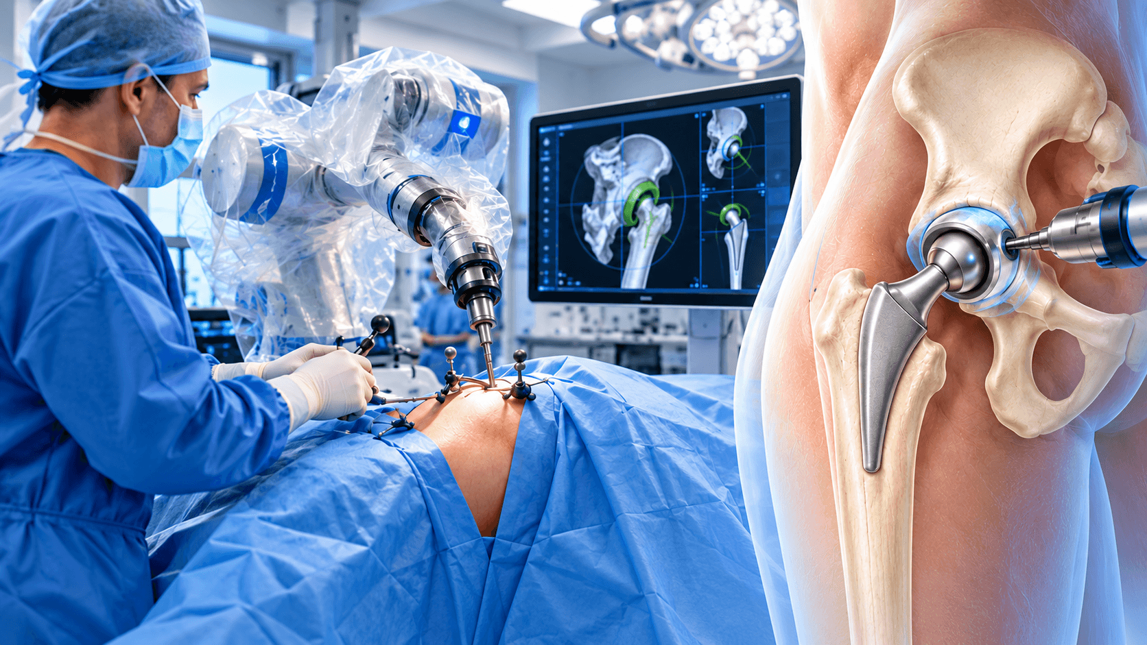
Robotic Assistance
Robotic systems can be used in selected revision cases, particularly when an acetabular component is being reconstructed.
However, revision surgery often involves unexpected findings and highly variable bone defects.
Robotics remains an adjunct rather than the defining technology of complex revision.
Experience in implant extraction and bone reconstruction is usually more important than the presence of a robotic platform.
Three-Dimensional Planning
CT-based three-dimensional planning can be highly valuable for severe bone loss.
The surgeon can visualize defects before surgery and anticipate the location of remaining supportive bone.
Virtual reconstruction can also assist in designing custom implants.
Three-dimensional printed models may be useful in exceptional complex cases for surgical planning and education.
Intraoperative Imaging
Fluoroscopy can help assess component position, leg length and fracture fixation.
Its use varies according to surgical approach and case complexity.
Imaging does not replace direct assessment of implant stability.
It provides an additional source of information to support the surgeon's decision-making.
Risks and how they are managed
All surgery carries risk. Partner hospitals follow enhanced-recovery and infection-prevention protocols, and your surgeon will discuss the risks specific to your case before consent.
- Periprosthetic joint infection: Revision surgery carries a greater infection burden than routine primary replacement. Infection can require prolonged antibiotics, debridement or another staged revision.
- Recurrent infection: Patients revised because of infection can experience recurrence even after extensive treatment. Further surgery may be necessary.
- Dislocation: Revision hips have a higher instability risk because muscles, capsule and normal anatomy may already be compromised.
- Recurrent dislocation: Some patients remain unstable despite revision and require additional reconstruction using different component positions or stability-enhancing implants.
- Periprosthetic fracture: Bone can fracture while old components are removed, during implantation of revision components or after a later fall.
- Aseptic loosening: A revision cup or stem can eventually lose fixation and require rerevision.
- Bone loss: Removing old implants can sacrifice additional bone. Surgeons use specialized extraction techniques to preserve as much bone as possible.
- Failure of bone graft or reconstruction: Grafted or reconstructed areas may fail to incorporate or may lose mechanical support.
- Pelvic discontinuity complications: Severe acetabular reconstruction can fail mechanically or biologically and occasionally requires further complex surgery.
- Nonunion of an extended trochanteric osteotomy: An osteotomy used to remove a femoral stem may heal slowly or fail to unite.
- Trochanteric migration: A repaired trochanteric fragment can move, potentially affecting abductor function and stability.
- Abductor weakness: Previous surgery and soft-tissue damage can leave permanent weakness, limping or instability.
- Nerve injury: The sciatic, femoral or other nerves can be stretched or injured during extensive exposure or reconstruction.
- Blood-vessel injury: Major vascular injury is uncommon but potentially serious, particularly in complex pelvic revisions.
- Leg-length difference: Restoring stability and fixation can sometimes require accepting a small leg-length difference.
- Blood clots: Deep-vein thrombosis and pulmonary embolism remain recognized risks after major hip surgery.
- Bleeding and anaemia: Revision can involve more blood loss than a primary replacement, and some patients require transfusion.
- Wound-healing problems: Multiple previous operations can compromise skin and soft tissues, increasing the risk of drainage and delayed healing.
- Hematoma: Blood can collect around the surgical site and occasionally require drainage.
- Implant breakage: Revision components can fail mechanically, particularly when bone support is poor or abnormal stresses remain.
- Persistent pain: Not every patient becomes completely pain-free. Muscle damage, nerve injury, spine disease or extensive reconstruction can cause residual symptoms.
- Stiffness: Scar tissue and previous surgery can limit motion even after successful implant revision.
- Heterotopic ossification: Bone can develop in surrounding soft tissue and, in severe cases, restrict movement.
- Medical complications: Cardiovascular, respiratory, kidney and neurological complications can occur, particularly in older or medically complex patients.
- Need for further revision: Every revision hip carries a possibility of another operation in the future because of infection, instability, loosening, fracture or another mechanical problem.
Alternatives
- Observation and regular imaging: A stable implant with mild symptoms or limited osteolysis may sometimes be monitored rather than revised immediately.
- Activity modification: Reducing activities that provoke symptoms can be appropriate when the implant remains mechanically stable and surgery is not yet justified.
- Pain medication: Medication can manage symptoms when revision risks exceed expected benefit, although it does not correct mechanical loosening.
- Physiotherapy: Strengthening can improve symptoms caused by weakness or soft-tissue dysfunction when the prosthesis itself is stable.
- Treatment of spine or tendon disorders: Pain caused by lumbar disease, trochanteric pain or iliopsoas irritation may improve without replacing hip components.
- Closed reduction for a first dislocation: Many isolated hip dislocations can be reduced under sedation or anaesthesia without immediate revision surgery.
- Brace after dislocation: Selected patients can temporarily use a brace after reduction, although bracing does not correct major component malposition.
- Antibiotic suppression: Long-term suppressive antibiotics may be considered in selected patients with chronic infection who cannot reasonably undergo major revision. This is infection management rather than definitive mechanical reconstruction.
- Debridement with implant retention: Selected acute periprosthetic infections can sometimes be treated with surgical debridement, exchange of modular components and antibiotics without removing well-fixed major components.
- Fracture fixation without stem revision: A periprosthetic femoral fracture can sometimes be stabilized while retaining the stem if the existing implant remains securely fixed.
- Nonoperative or palliative management: Frail patients with very high operative risk may choose symptom-focused care when the burdens of complex reconstruction outweigh likely benefit.
- Resection arthroplasty: In exceptional salvage situations involving severe infection or failed reconstruction, components can be removed without immediate replacement. Function is significantly compromised, so this is not a routine alternative.
What hip revision surgery costs
The contracted Turkey partner package next to approved self-pay benchmarks. Benchmarks are 20th–80th percentile ranges of approved records, normalised to USD.
Turkey package
$13,000 – $22,000
United States self-pay
$59,350 – $107,950
United Kingdom self-pay
$22,250 – $48,200
Germany self-pay
$20,900 – $45,950
Typical self-pay range by country
Surgeons who perform hip revision surgery
All surgeonsHospitals offering this procedure
Antalya Yaşam Hospital
Private hospital in Muratpaşa, Antalya, with an orthopedics and traumatology department
BHT Clinic Istanbul Tema Hospital
Large private hospital in Atakent, Küçükçekmece, with an orthopedics and traumatology unit
Biruni University Hospital
University hospital in Küçükçekmece, Istanbul, with a published orthopedics and traumatology department
Çankaya Hospital for Orthopedic Care
Çankaya Hospital for Orthopedic Care is an orthopedic hospital in central Ankara with robotic-arm assisted surgery
Sources and references
Peer-reviewed guidance and institutional sources used to write and review this page.
- 01Patient-focused overview of revision THA, including loosening, instability, infection, fracture, partial versus complete revision and why revision surgery is generally more technically demanding than primary hip replacement. AAHKS
American Association of Hip and Knee Surgeons
https://www.hipkneeinfo.org/es/hip-care/revision-total-hip-arthroplasty/
- 02Revision indications, differences between primary and revision THA and registry-based information regarding common causes of revision.
American Academy of Orthopaedic Surgeons / American Joint Replacement Registry
https://orthoinfo.aaos.org/globalassets/pdfs/aaos_ajrr_2018-patient-supplement.pdf
- 03Diagnostic evaluation of suspected prosthetic joint infection, including inflammatory markers and principles surrounding infection assessment before revision arthroplasty.
merican Academy of Orthopaedic Surgeons, 2017
https://www.aaos.org/quality/quality-programs/diagnosis-and-prevention-of-periprosthetic-joint-infections/
- 04Modern strategies for acetabular bone loss, porous revision shells, augments and reconstruction of severe defects including pelvic discontinuity. PubMed
Journal of the American Academy of Orthopaedic Surgeons / PubMed, 2024
https://pubmed.ncbi.nlm.nih.gov/38412446/
- 05Comparative evidence on dual-mobility and conventional fixed-bearing components in revision THA, particularly dislocation-related rerevision and overall complications.
Journal of Arthroplasty / PubMed, 2026
https://pubmed.ncbi.nlm.nih.gov/41611102/
- 06Updated systematic review evaluating instability after revision THA and the role of dual-mobility constructs in reducing dislocation risk.
Journal of Arthroplasty / PubMed, 2026
https://pubmed.ncbi.nlm.nih.gov/41611102
- 07Comparison of dual-mobility and large-head constructs with emphasis on dislocation prevention in primary and revision arthroplasty.
PubMed-indexed systematic review, 2026
https://pubmed.ncbi.nlm.nih.gov/41379986/?utm_source=chatgpt.com
- 08Contemporary comparison of one-stage and two-stage revision pathways for periprosthetic hip infection. PubMed
Journal of the American Academy of Orthopaedic Surgeons / PubMed, 2025
https://pubmed.ncbi.nlm.nih.gov/39303283/