

Key takeaways
- 1Avascular necrosis of the shoulder is the death of a segment of bone in the humeral head, the ball of the joint, after its blood supply fails.
- 2The commonest triggers are long courses of corticosteroid tablets, heavy alcohol use, sickle cell disease and a previous fracture of the upper arm.
- 3Early disease can be silent or cause only a dull ache, so the problem often goes unnoticed until late, after the dead bone has begun to flatten.
- 4MRI is the most sensitive test and can reveal the problem before it shows on a plain X-ray.
- 5Staging, based on how much bone the disease involves and whether the surface has collapsed, decides whether joint-preserving care or replacement is realistic.
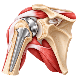
- 6When the humeral head has collapsed and the socket has worn, shoulder replacement is the dependable way to restore comfort and movement.
- 7Treatment of avascular necrosis of the shoulder in turkey can suit planned surgery once a specialist has reviewed imaging and the underlying causes, such as steroid use, are clear.
Overview
What is avascular necrosis of the shoulder?
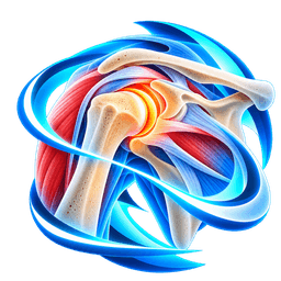
Avascular necrosis of the shoulder is a condition in which the blood supply to the head of the humerus fails and an area of bone dies. It is sometimes called osteonecrosis. Without treatment, the weakened bone can cave in and the joint can wear out. This page explains the causes, stages, tests and treatment, including options in turkey.
What is avascular necrosis of the shoulder?
"Avascular" means without blood vessels and "necrosis" means tissue death. Living bone constantly renews itself, and it needs a steady flow of blood to do that. When flow to a patch of the humeral head stops, the cells in that patch die and the area can no longer repair normal daily stress.
The cartilage on top is not yet harmed at first. As the underlying bone softens, the surface may buckle like the shell of a boiled egg that someone has pressed. Doctors call this humeral head collapse. After that, the socket wears and arthritis follows.
Who gets it?
It is more often seen in people between 30 and 60 than classic wear-and-tear arthritis, and it is less common than the same problem in the hip. Many patients have a clear risk factor, such as months of high-dose steroids for asthma, lupus or an organ transplant, or sickle cell disease. Some have no known cause.
How serious is it?
The condition is not life-threatening, yet it can be disabling. Small, early areas sometimes stay stable for years. Larger areas near the weight-bearing centre of the ball tend to progress. Speed of progression is hard to predict, which is why regular review and imaging matter after a diagnosis.
How is this page organised?
We start with anatomy and blood supply, then staging, tests and look-alike problems. After that we cover joint-saving treatments, replacement surgery and travel planning. Our shoulder hub links to related pages, and the free case review is available when you want an opinion on your scans.
Anatomy
What happens in the body with avascular necrosis of the shoulder
To understand why bone in the shoulder can die, it helps to know how fragile the blood supply of the humeral head is. Unlike muscles, which have rich blood flow, the ball of the joint depends on a small number of vessels that can fail after injury, drugs or disease.
How does the humeral head get its blood?
Two main arteries feed the upper humerus: the anterior and posterior circumflex humeral arteries. They send branches into the bone, mostly through the front groove and around the neck of the humerus. Because the head is covered by cartilage on most of its surface, blood must arrive through narrow channels near its base.
The ball itself is a rounded cap, about 4 to 5 cm across in adults, that sits against the shallow socket of the shoulder blade (the glenoid). A thick layer of cartilage covers both surfaces. Muscles of the rotator cuff surround the joint and keep the ball centred while the arm moves.
What goes wrong in the bone?
Scan reports often call this pattern humeral head osteonecrosis, and you may see that phrase on your own paperwork. It means the same thing as the title of this page: a patch of the ball has lost its blood supply, and the bone inside it is no longer alive or able to renew itself.
When blood flow stops, the bone marrow and the bone cells within the affected zone die over days to weeks. The body then tries to repair the area by growing new vessels and removing dead bone. This repair process temporarily makes the area weaker, since the body clears old bone faster than it lays down new bone.
If the dead area is small, the repair may succeed and the head keeps its shape. If it is large, the supporting layer beneath the cartilage can crack, which lets the surface sink. A crescent-shaped gap on imaging, called a subchondral fracture line, is an early sign of this.
Why does the socket suffer next?
Once the head loses its smooth round shape, it grinds against the glenoid with each movement. The glenoid cartilage wears, and secondary arthritis develops. This is why late-stage avascular necrosis of the shoulder may look like shoulder osteoarthritis on an X-ray, although its cause and typical age are different.
Symptoms & causes
Avascular necrosis of the shoulder symptoms and causes
Common symptoms
- A deep, dull ache in the shoulder or upper arm that is felt even at rest in the early phase, often leading people to suspect a muscle strain.
- Pain that gradually becomes sharper with overhead movement, lifting or reaching, as the dead bone loses strength under load.
- Night pain that interferes with sleep, particularly when lying on the affected side, a symptom that often brings people to the clinic.
- Clicking, catching or grinding when the arm rotates, which suggests the surface of the ball has become irregular after collapse.
- Stiffness, especially a loss of outward rotation and lifting, that develops over months as the capsule tightens and the joint surface changes.
- Reduced range of motion that makes dressing, washing hair or reaching a high shelf slow and uncomfortable.
- Weakness or heaviness in the arm that is mostly due to pain inhibiting the muscles instead of true nerve damage.
- Pain in both shoulders in some people, since risk factors such as steroids or sickle cell disease affect the whole body.
- Absence of symptoms in early disease, so the first sign may be an abnormal scan taken for another reason, for example when doctors assess the hip for osteonecrosis.
Causes and risk factors
- Long-term or high-dose corticosteroids, such as prednisolone taken for months for lupus, asthma or after a transplant, which are among the most frequent triggers.
- Heavy and sustained alcohol intake, which may change fat metabolism and clog the small vessels of the bone.
- Sickle cell disease, where abnormally shaped red cells block small bone vessels and cause repeated bone damage.
- Previous fracture of the upper arm, especially a displaced break near the ball, which can tear the blood vessels that feed the bone.
- Previous shoulder dislocation with injury to the circulation, or surgery near the humeral neck.
- Radiation treatment or chemotherapy to the chest or shoulder region, which can harm bone blood supply over time.
- Deep sea diving and compressed air work, where nitrogen bubbles form in small vessels (decompression-related osteonecrosis).
- Other illnesses and conditions, such as Gaucher disease, clotting disorders, HIV infection and some autoimmune diseases, and cases with no identifiable cause.
Types
Types and stages of avascular necrosis of the shoulder
Doctors describe this condition in stages, because the stage reflects how much damage has occurred and therefore which treatments still make sense. Different staging systems exist, but all rely on X-ray and MRI findings.
How are the stages described?
Knowing the avascular necrosis of the shoulder stages explains why advice can change between two scans a year apart. Disease can move from one stage to the next over 6 to 24 months, or stay put for a long time, so each stage describes a moment in time rather than a fixed sentence.
The Cruess system, a modification of an older hip classification, is the one most surgeons use for the shoulder. The table below summarises it in plain words.
| Stage | Imaging findings | Typical approach |
|---|---|---|
| Stage I | Normal X-ray, abnormal MRI showing dead bone and fluid | Protection, medicines, close monitoring, sometimes core decompression |
| Stage II | Patches of sclerosis or lucency on X-ray; head still round | Joint-preserving procedures may suit |
| Stage III | Crescent sign: a subchondral fracture line under the cartilage | Joint-preserving surgery less predictable; discuss replacement timing |
| Stage IV | Collapse of the humeral head with a flattened or irregular surface | Hemiarthroplasty is sometimes used for a healthy socket |
| Stage V | Collapse with damage to the socket (secondary arthritis) | Total shoulder replacement is usually recommended |
Why does the size of the dead area matter?
Small lesions, affecting less than about 15% of the head surface, often remain stable. Medium and large lesions have a higher risk of collapse, mainly if they sit in the area that bears most load when you raise the arm. MRI measures the size and position of the lesion.
What do primary and secondary forms mean?
Doctors say "primary" when they find no cause. "Secondary" describes cases linked to a known factor, for example steroids or trauma. Secondary cases often involve other joints, and doctors may screen the hips and knees, since osteonecrosis can occur in several places at once.
Is it related to a fracture?
Post-traumatic cases follow a break of the upper arm, particularly displaced four-part fractures. Our page on proximal humerus fracture explains why some breaks threaten the blood supply, and why surgeons sometimes choose replacement at the time of injury.
Diagnosis
How is avascular necrosis of the shoulder diagnosed?
This condition is diagnosed from a medical history, a physical examination and imaging, with MRI giving the earliest and clearest answer. Because early disease may be silent, doctors often search for it when a risk factor is present or when shoulder pain does not match a plain X-ray.
What does the history reveal?
Expect questions about steroid courses in recent years, alcohol intake, sickle cell disease, past injuries, radiation, diving and other joint pain. Your doctor wants to know when the shoulder pain began, whether it is present at rest, and whether the other shoulder or a hip is also sore.
What will the physical examination show?
Typical findings include tenderness around the joint, limited and painful movement, and sometimes a grating sensation as the arm rotates. Muscle bulk may shrink if the problem has lasted a long time. The doctor will also check the neck and the rotator cuff to exclude other sources of pain.
Which imaging gives the answer?
Plain X-rays in two views are the starting point. In early disease they may look normal, which is why the diagnosis is sometimes missed for months. In later disease they show sclerosis, a crescent line or flattening of the ball.
An MRI can show dead bone and the surrounding repair zone long before any change on an X-ray. A CT scan describes the bone surface and the socket in detail and guides surgical planning. Bone scans are rarely needed now.
Should doctors check other joints?
Often, yes. When doctors find avascular necrosis in one shoulder, an MRI of the other shoulder and the hips may be advised, particularly if the cause is steroids or sickle cell disease. Finding silent disease early gives more treatment choices and prevents surprises later.
What should you bring to a remote review?
Gather the images from every scan in digital form, as well as reports, a list of past and current medicines with doses and dates (especially steroids), and a short timeline of symptoms. Read about medical record review to see how a specialist uses these records before suggesting a plan.
Tests you may have
- Plain X-rays in at least two views to look for sclerosis, a crescent line, flattening of the ball and joint space loss.
- MRI of the shoulder to detect dead bone and marrow changes at stage I, and to measure the size and position of the lesion.
- CT scan to define bone collapse and the shape of the glenoid, which is essential when you are planning replacement.
- MRI or X-rays of the opposite shoulder and the hips, because osteonecrosis frequently affects more than one joint.
- Blood tests for sickle cell status, cholesterol and fats, clotting problems and inflammation, depending on the suspected cause.
- Review of medicine records, including steroid doses and duration, since stopping or adjusting treatment may be part of care.
- Ultrasound or MRI of the rotator cuff when you are weighing replacement, so the surgeon can choose the implant type.
Look-alikes
Conditions that can feel like avascular necrosis of the shoulder
Pain deep in the shoulder with a normal early X-ray has many possible causes, and this condition is one that is easily missed. The comparison below shows how specialists separate it from common alternatives.
Which conditions mimic it?
| Look-alike condition | How it differs | How doctors tell them apart |
|---|---|---|
| Shoulder osteoarthritis | Gradual wear across both ball and socket, usually after 60 | X-ray shows joint space loss and spurs rather than a localised dead area |
| Rotator cuff tear | Pain and weakness lifting the arm, a tendon problem | MRI or ultrasound shows a tendon defect with a normal humeral head |
| Frozen shoulder | Stiffness in all directions, normal bone | Normal MRI of bone; capsule thickening in the joint |
| Shoulder impingement | Painful arc during lifting, linked to tendon irritation | Bone signal on MRI is normal |
| Bone bruise or stress fracture | Follows an injury or overuse; fluid-like signal on MRI | Pattern is diffuse and settles within months, without a dead-bone margin |
| Bone tumour or infection | Pain at rest, systemic features, sometimes fever | Blood tests and MRI features; biopsy when uncertain |
| Neck nerve irritation | Arm pain with tingling, neck movement worsens it | Neck examination, nerve tests, neck MRI |
Why is it often found late?
Early disease does not show on X-rays, so people may receive treatment for months for a "strained muscle" or impingement. A history of steroid use should trigger a lower threshold for MRI. If your shoulder pain is worse than your X-ray suggests, ask whether an MRI would help.
Can it look like transient bone marrow oedema?
Occasionally. Some people develop temporary fluid in the bone that causes pain and resolves in 6 to 12 months without dead bone. The MRI pattern makes the difference and by following changes over time. This is a reason for repeat imaging, rather than acting on a single scan.
What about problems that coexist?
Cuff tears, arthritis in the opposite joint and stiffness from guarding the arm can all coexist. A careful surgeon decides which problem is causing your pain and tells you what each treatment can and cannot fix. Ask for this reasoning in your consultation.
Non-surgical
Non-surgical treatment for avascular necrosis of the shoulder
Non-surgical care for avascular necrosis of the shoulder aims to reduce pain, protect the weakened bone and address the cause. It cannot bring back dead bone, but it can help small early lesions stay stable and make life comfortable while you make other decisions.
What is the first step?
The first step is to deal with the trigger. If steroids are involved, the prescribing doctor reviews whether the dose can be lowered or changed, but you must never stop them suddenly yourself, since this can be dangerous. Reducing alcohol, treating high blood fats and stopping smoking support bone health.
How is activity adjusted?
Doctors advise avoiding heavy lifting, pushing, or loaded overhead work while the bone is vulnerable. This is often described as protected use, since the shoulder carries no body weight but still bears the load of the arm and anything it holds. A sling may help briefly in a painful flare, not for weeks.
Which exercises are safe?
Doctors encourage gentle range-of-motion exercise, because immobility leads to stiffness. A physiotherapist can teach pendulum movements and assisted lifting in a pain-free range. Strengthening starts cautiously, usually after the pain settles. Expect programmes of 6 to 12 weeks, with review scans if symptoms change.
What medicines can help?
Doctors use simple pain relief such as paracetamol first, with non-steroidal anti-inflammatory drugs (NSAIDs) added if safe for you. Some doctors also prescribe bisphosphonates, which slow bone breakdown, for early disease. Evidence for this use in the shoulder is limited, so treat it as one option to discuss, not a standard cure.
Are there other non-surgical options?
Extracorporeal shockwave therapy, pulsed electromagnetic fields and hyperbaric oxygen have all had trials in osteonecrosis. Studies disagree and are mostly small, and they come largely from the hip. Do not expect these approaches to replace surgery for advanced disease, and be cautious about expensive courses that promise a cure.
How is progress followed?
A typical schedule is a clinic visit and X-ray at 6 weeks, then every 3 months for the first year. Doctors repeat the MRI if symptoms change, or after 6 to 12 months to check whether the dead area has grown. Keep copies of every scan so you can compare each new image with the last.
Repeat X-rays or MRI every 3 to 6 months show whether the lesion is stable. If pain continues, if the ball starts to flatten, or if function drops, the plan changes. This monitoring is a key part of care and prevents missing the moment when surgery would be most helpful.
Self-care
Exercises and self-care for avascular necrosis of the shoulder
Daily habits can ease symptoms and protect the shoulder while you and your doctor follow the lesion. Self-care does not replace medical treatment, so check with your doctor or physiotherapist before starting any exercise, particularly if you are taking steroids or have a collapsed bone surface.
How can you manage pain at home?
Use a heat pack for 15 to 20 minutes to relax stiff muscles, and a cold pack after a busy day if the joint feels hot and sore. Take simple painkillers as advised, and try not to wait until pain is severe. Rest the arm in a supported position, with a small pillow under the elbow.
How should you sleep?
If lying flat is uncomfortable, try 2 or 3 pillows to raise the upper body by about 30 degrees. Place a rolled towel between the arm and the chest to take tension off the joint. Avoid sleeping on the sore side for the first weeks, and switch positions slowly to prevent sudden jolts.
Sleep on your back or on the opposite side with a pillow hugging the sore arm. A recliner or a wedge pillow reduces pressure at night. Poor sleep makes pain feel worse, so settling the nights is one of the most useful goals of early care.
Which movements are suitable?
- Gentle pendulums: let the arm hang and swing slowly for 1 minute, 2 to 3 times a day.
- Elbow and wrist bending: keep the lower arm mobile to avoid swelling and stiffness.
- Assisted raising with the good arm: clasp both hands and lift to a comfortable height, hold 5 seconds, repeat 10 times.
- Shoulder blade squeezes: draw the blades gently together for 5 seconds, 10 repetitions.
What lifestyle changes matter?
Small habits add up over 3 to 6 months. Aim for 150 minutes of gentle aerobic activity a week, such as brisk walking, which supports circulation without loading the arm. Keep a symptom diary noting pain at rest, at night and with activity, and bring it to each appointment, since patterns over weeks are more useful than a single snapshot.
Limit alcohol, since heavy intake is a recognised risk factor. Keep to a balanced diet with enough calcium and vitamin D, unless your doctor advises otherwise. Stay physically active with walking or stationary cycling, and discuss smoking cessation support, which improves bone and wound healing.
What should you avoid?
Avoid heavy lifting, throwing, push-ups and contact sport, which place load on the weakened ball. Do not stop prescribed medicines on your own. Be careful with repeated steroid injections into the joint, as they may be unhelpful in this condition, and ask your doctor before accepting any.
Check with your doctor or physiotherapist before starting new exercises, and stop any movement that causes sharp pain.
Treatment
Avascular necrosis of the shoulder treatment options
Surgery for this condition ranges from procedures that try to save the joint to replacement of the damaged surfaces. The right choice depends on the stage, the size of the lesion, your age and whether the socket has worn.
What is core decompression?
Core decompression is a small operation in which the surgeon drills one or more channels into the humeral head to lower pressure and encourage new blood vessels. It may be combined with bone graft or bone marrow cells. It is used mostly in stages I and II, and results are best when done before collapse.
Can surgeons graft the bone?
Some surgeons place a bone graft under the cartilage or a strut of bone to support the weakened area. Others use a vascularised graft. These operations are specialised, are not widely available and have limited published data in the shoulder. Ask how often your surgeon performs them.
When is arthroscopy used?
Keyhole surgery (shoulder arthroscopy) can remove loose cartilage or release a tight capsule in selected cases, which may ease pain for a time. It does not fix the bone itself and does not prevent collapse, so surgeons treat it as a stopgap in specific patients.
What is partial shoulder replacement?
In a partial shoulder replacement (hemiarthroplasty), the collapsed ball is replaced by a metal head and the socket is retained. It is appropriate when the socket cartilage is still healthy, which is more common in younger patients. Pain relief is generally good, but it can fade if the socket wears. See the turkey page for partial replacement.
What is total shoulder replacement?
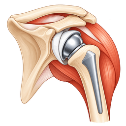
In total shoulder replacement, the surgeon replaces both the ball and the socket. It is the usual choice once the glenoid has worn and the rotator cuff is healthy. Most patients report significant pain relief, and the implant may last many years. Details of care are on the turkey page for total replacement.
How do the options compare?
| Option | Best suited to | Main trade-off |
|---|---|---|
| Core decompression | Stage I to II, round head | Unpredictable once collapse starts |
| Bone graft procedures | Selected young patients with a crescent line | Specialised; limited shoulder evidence |
| Partial replacement | Collapsed head, healthy socket | Socket may wear later |
| Total replacement | Collapse plus socket arthritis | Needs a working cuff; implant wear over decades |
You can compare costs through the total shoulder replacement cost guide and the partial shoulder replacement cost guide.
When surgery is considered
Surgery is worth considering for avascular necrosis of the shoulder once the bone has collapsed or pain stays uncontrolled, and joint-saving procedures are no longer sensible. Earlier operations are for selected people only, so a specialist opinion at the first sign of a worsening scan is valuable.
What signals that it is time?
- Pain that disturbs sleep or daily tasks despite medicines and activity changes for 3 months.
- Progression on repeat imaging, such as a crescent sign or flattening of the ball.
- Loss of motion that stops you from working, dressing or caring for yourself.
- Evidence of socket wear, which makes joint preservation unlikely to succeed.
- A general health review that shows you are fit for anaesthesia and rehabilitation.
Why do age and activity matter?
A person aged 35 who plays sport or does manual work places very different demands on an implant from a person aged 70 who is retired. Some surgeons therefore delay replacement as long as pain is tolerable, while others act sooner to avoid further bone loss. Both approaches can be reasonable, so ask your surgeon to explain the thinking.
Because this condition often strikes people in their 30s to 50s, they may outlive an implant. Surgeons weigh the benefit of relief now against the chance of revision later. Younger patients are usually told about activity limits and the possibility of a second operation, which is part of an honest conversation.
Which questions should you ask?
- At what stage am I, and what is the size of the dead area?
- Do I need treatment for the cause, such as steroid review or sickle cell care?
- Which operation do you recommend now, and what is the plan if it fails?
- How many shoulder replacements for osteonecrosis do you do each year?
- What activities can I return to, and what should I avoid?
What about the other joints?
If the hips or knees are also affected, the order of operations needs planning. Some patients prioritise the joint that limits walking, while others prioritise the arm used for work. Share all your scans, since this broader picture changes surgical advice.
Procedures
Procedures that may treat avascular necrosis of the shoulder
Costs
Avascular necrosis of the shoulder treatment cost in Turkey
| Procedure | Turkey package | US self-pay | Saving |
|---|---|---|---|
| Total Shoulder Replacement | $9,000 – $14,000 | $40,017 | ~71% |
| Partial Shoulder Replacement | $8,000 – $12,500 | $36,050 | ~72% |
Packages are contracted partner prices with validity dates; benchmarks are source-backed estimates. Open a cost guide for the full country comparison.
In Turkey
Treating avascular necrosis of the shoulder in Turkey
Treatment of this condition in turkey is a sensible option for planned, non-urgent surgery when your scans can be reviewed remotely and you are fit to travel. Many private hospitals in turkey perform shoulder replacement regularly and are experienced with international patients.
Why consider turkey?
People often choose it for faster access to a shoulder specialist, bundled care that includes hospital stay and physiotherapy planning, and the option of staying nearby for early recovery. Our why turkey guide gives the reasons, while orthopedics in turkey shows how services are organised.
What is the pathway?
- Remote review: upload scans and a short history through the free case review.
- Plan: the team confirms the stage, advises whether replacement is needed and which type.
- Cause check: any steroid, alcohol or sickle cell issue is reviewed with your own doctor.
- Admission: you arrive a few days early for tests and anaesthesia review.
- Surgery: hospital stay is typically 2 to 3 nights.
- Early recovery: 10 to 14 days in turkey for wound checks and first physiotherapy.
- Return home: continued rehabilitation and scheduled follow-up reviews.
What records are important?
Send the images themselves as well as the reports, since a surgeon will want to measure the lesion personally. Include any older scans, even if they looked normal, because comparing them shows how fast the bone has changed. Records from 2 or 3 years back are often very informative.
For shoulder replacement for avascular necrosis in turkey, surgeons usually want a recent MRI and CT scan, X-rays, a list of medicines including steroid dates, and blood results if you have sickle cell disease. The guide to treatment planning shows how the plan is built and what happens at each step.
How can you check quality?
Ask about hospital accreditation, the surgeon's experience with shoulder replacement and with osteonecrosis, the implant brand and whether you will receive implant stickers or records. Ask how emergencies are handled and who will answer your questions after you fly home. See also questions to ask before surgery abroad.
What logistics should you plan?
Plan 2 to 3 weeks away, arrange a companion, and book a flexible return flight. Review flying after surgery and rehabilitation before you travel. For costs, see the total shoulder replacement cost guide.
When should you not travel?
Avoid travel if you have an active infection, a sickle cell crisis, unstable heart or lung disease, or an uncontrolled blood sugar level. If steroids are essential, your own doctor should agree on a safe plan first. Shoulder osteonecrosis surgery in turkey is planned, so you can wait until you are stable.
Complications
Complications of avascular necrosis of the shoulder
If this condition progresses, the main consequence is joint destruction with persistent pain and stiffness, and surgical treatment has its own risks. Knowing both helps you weigh timing and choose a realistic plan.
What happens without treatment?
Small lesions may remain quiet, but larger ones often progress to collapse within months to a few years. The flattened head damages the socket, producing arthritis and a stiff, painful shoulder. Sleep and independence suffer, and the rotator cuff may weaken through disuse.
Delay can also narrow your options. Joint-saving procedures only work before collapse, and severe bone loss in the socket can complicate later replacement. This is why a scan and specialist review soon after diagnosis are valuable, even if you choose to wait.
What are the risks of surgery?
Rates differ between studies and between hospitals, so ask your surgeon for figures from their own practice. As a rough guide, most patients have no major complication, and problems that do occur are often treatable when caught early.
Core decompression is a small procedure, but the bone can still collapse later, and fracture through the drill tract is rare. Replacement carries the usual surgical risks, which are uncommon but important.
- Infection: occurs in a small proportion of patients and may need antibiotics or further surgery, with a higher risk in people on steroids.
- Stiffness: reduced range of motion if rehabilitation is not followed.
- Implant loosening or wear: more likely in young and active patients over time.
- Nerve injury or fracture: uncommon but possible during surgery.
- Blood clots: a low risk, higher with prolonged travel or reduced mobility.
Does the underlying cause change the risk?
Yes. Long-term steroids can slow healing and raise infection risk, and sickle cell disease needs special anaesthesia planning, with attention to hydration, oxygen and temperature. Tell the surgical team everything about your medical history so they can prepare.
Can the disease recur or affect other joints?
Osteonecrosis may involve other bones, so follow-up of the hips, knees and the opposite shoulder is sensible when risk factors persist. Replacing one joint does not protect another. Early detection of problems elsewhere gives more treatment options.
Urgent care
When to seek urgent care for avascular necrosis of the shoulder
- Fever, chills or a hot, red, swollen shoulder, especially after a joint injection or surgery: seek same-day medical care for possible infection.
- Sudden severe shoulder pain after a minor movement, with an arm that will not lift: attend an emergency department, since the weakened bone may have collapsed or broken.
- New numbness, tingling, pallor or coldness in the arm or hand: seek urgent assessment for nerve or blood-flow problems.
- Severe pain in the shoulder, chest or back with a sickle cell crisis: contact emergency services or your haematology team immediately.
- A surgical wound that leaks, opens or smells: contact your surgical team the same day.
- Calf swelling or sudden shortness of breath after surgery or a flight: call emergency services, as this may be a blood clot.
Prevention
How to lower your risk of avascular necrosis of the shoulder
Not every case of this condition can be prevented, but many risk factors are modifiable, and early detection gives the best chance of protecting the joint. Prevention is mostly about being aware of your own risk and acting early.
How can you lower the risk?
- Use steroids only when needed and at the lowest effective dose and duration, and never stop them abruptly without medical advice.
- Limit alcohol to within recommended limits, and seek help if drinking is heavy.
- Manage sickle cell disease and clotting problems with your specialist team.
- Treat high cholesterol and blood fats, and avoid smoking.
- Follow safe diving practice, including decompression stops and certified training.
Should high-risk people be screened?
There is no routine screening for the general public. People on prolonged high-dose steroids who develop new shoulder or hip pain should mention it early and ask whether an MRI is appropriate. A normal X-ray does not exclude early disease, which is the main reason to speak up.
How can you protect the joint once diagnosed?
Plan daily tasks so that heavy items sit between waist and chest height, ask for help with luggage and groceries, and use the stronger arm for pulling and pushing. Wearing a supportive bag strap across the body rather than on the sore shoulder also reduces strain during long days.
Avoid heavy loading, keep the shoulder moving gently, take medicines as advised and attend follow-up scans. These steps cannot promise that collapse will be avoided, but they give small lesions the best chance and reduce unnecessary strain.
What cannot be prevented?
Sometimes no cause is found, and a displaced fracture that cuts the blood supply cannot be undone by lifestyle. If your trauma was severe, discuss monitoring with your surgeon. The aim is to catch the problem early, not to blame yourself, since many causes are beyond personal control.
Outlook
Living with avascular necrosis of the shoulder: outlook and recovery
The outlook for avascular necrosis of the shoulder depends on the stage at diagnosis, the size of the lesion and the underlying cause. Early small lesions may remain stable, while advanced disease usually needs replacement, which gives good pain relief for most people.
What is the natural course?
The course varies widely. Some people have a slow, mild course for years, while others have rapid collapse within months. Large lesions in the central load-bearing part of the head are more likely to collapse. Regular imaging is the best way to know where you are on this spectrum.
What is recovery after replacement like?
| Time after surgery | What to expect |
|---|---|
| Weeks 0 to 6 | Sling support, gentle assisted movement, hand and elbow exercises |
| Weeks 6 to 12 | Active movement starts; sling is discontinued; light daily tasks |
| 3 to 6 months | Strengthening; return to most normal activities and driving |
| 6 to 12 months | Continued gains in comfort and strength; final result emerges |
What can you do afterwards?
Most people return to desk work in 4 to 8 weeks and to lighter physical work after 3 to 6 months. Swimming, cycling, golf and light gym work are usually possible. Heavy lifting, contact sport and repeated overhead loading are generally discouraged, because younger patients put more strain on implants.
How long do results last?
Many shoulder implants work well for 10 to 15 years or more, though results for younger and active patients are less predictable than for older ones, and revision may be needed in some. Regular check-ups with X-rays allow wear to be caught early. See also rotator cuff tear arthropathy for what failed cuffs mean for implant choice.
What is the long-term picture?
Satisfaction is high after replacement when expectations are realistic. Patients commonly tell us that sleeping through the night was the first big change, usually within 2 to 3 weeks, and that reaching and lifting returned more slowly over the following months. Keep attending review visits at 1 year and every 2 to 5 years afterwards.
Most people report much less pain, better sleep and a shoulder that allows ordinary life. Movement is usually good for daily tasks but may not return to full range. Realistic goals, steady rehabilitation and management of the underlying cause give the best chance of a satisfying result.
Surgeons
Specialists who treat avascular necrosis of the shoulder
FAQ
Avascular necrosis of the shoulder: frequently asked questions
What is avascular necrosis of the shoulder?
What are the symptoms of avascular necrosis of the shoulder?
What causes avascular necrosis of the shoulder?
Can avascular necrosis of the shoulder heal on its own?
Is the pain worse at night?
Does avascular necrosis of the shoulder always need surgery?
What is the best treatment for humeral head collapse?
How long is recovery after shoulder replacement for avascular necrosis?
Is avascular necrosis of the shoulder treatment in turkey safe?
Can I have a remote review before travelling to turkey?
How long should I stay in turkey after surgery?
Should I be worried about my hips if I have this in my shoulder?
Sources
Sources for this avascular necrosis of the shoulder guide
Peer-reviewed guidance and institutional sources used to write and review this page.
- 01Osteonecrosis
National Institute of Arthritis and Musculoskeletal and Skin Diseases, 2023
https://www.niams.nih.gov/health-topics/osteonecrosis
- 02Osteonecrosis
Mayo Clinic, 2023
https://www.mayoclinic.org/diseases-conditions/avascular-necrosis/symptoms-causes/syc-20369859
- 03
- 04Shoulder Joint Replacement
American Academy of Orthopaedic Surgeons (OrthoInfo), 2023
https://orthoinfo.aaos.org/en/treatment/shoulder-joint-replacement/
- 05Shoulder Injuries and Disorders
MedlinePlus, 2023
https://medlineplus.gov/shoulderinjuriesanddisorders.html
- 06