


Key takeaways
- 1Laminectomy removes some or all of the lamina, the bony roof over the spinal canal, to relieve pressure on the spinal cord or nerve roots.
- 2It is most commonly used for lumbar spinal stenosis causing leg pain, numbness, weakness or difficulty walking.
- 3A cervical laminectomy can decompress the spinal cord in patients with multilevel cervical stenosis or degenerative cervical myelopathy.
- 4A laminectomy does not automatically require spinal fusion.
- 5Fusion is considered when there is pre-existing instability, deformity, significant spondylolisthesis or when decompression requires removing enough supporting structures to make the spine unstable.
- 6High-quality contemporary research supports decompression without fusion for many carefully selected lumbar stenosis patients, including some with low-grade degenerative spondylolisthesis.
- 7Laminotomy vs laminectomy: a laminotomy removes only a portion of the lamina, while a laminectomy removes a larger portion or the entire lamina at the treated level.
- 8Minimally invasive decompression can achieve the same goal through smaller corridors in selected patients.
- 9Most patients begin walking soon after surgery.
- 10Laminectomy recovery commonly takes about six to twelve weeks for routine lumbar decompression, although improvement can continue for several months.
- 11The operation is usually more reliable for relieving leg symptoms caused by nerve compression than for treating nonspecific lower-back pain.
- 12Postoperative pain is usually most noticeable during the first several days and should gradually decrease.
- 13A laminectomy can provide long-lasting decompression, but later degeneration, recurrent stenosis or instability can occasionally produce new symptoms.
- 14Post laminectomy syndrome is an older umbrella term for persistent or recurrent pain after spinal surgery. It does not identify one single cause.
- 15Modern clinicians increasingly use terms such as persistent spinal pain syndrome because persistent pain after surgery can result from nerve injury, scar tissue, adjacent disease, recurrent stenosis, instability, hip or sacroiliac pathology and several other mechanisms.
- 16Serious complications such as neurological injury, infection, epidural hematoma or major dural complications are uncommon but important.
- 17The quality of patient selection is as important as the technical operation: stenosis seen on MRI alone is not a reason for surgery unless it correlates with meaningful symptoms.
Overview
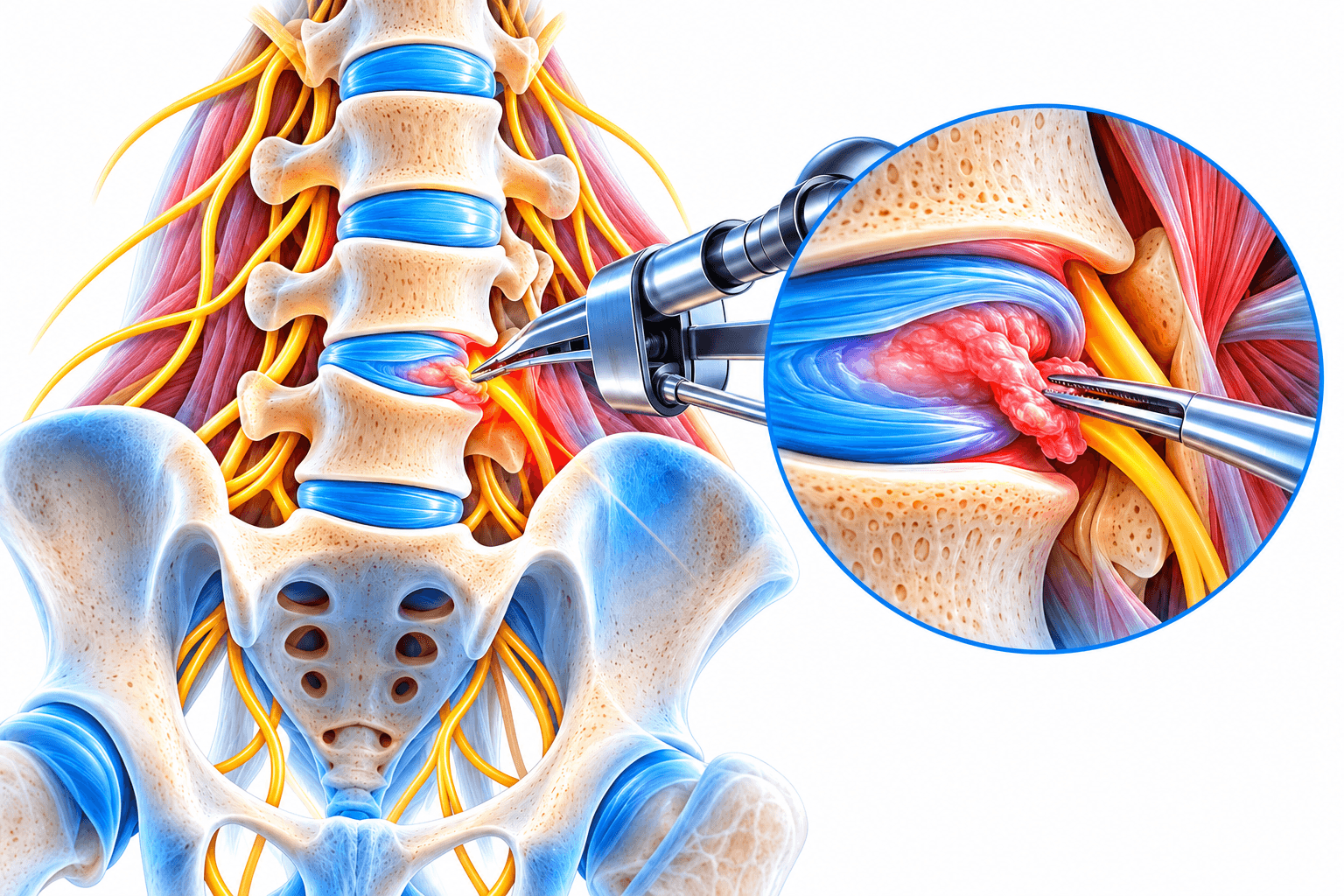
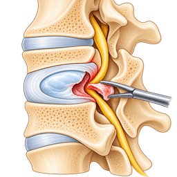
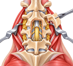
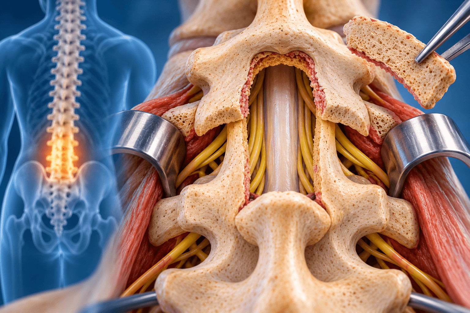
A laminectomy is a spinal decompression operation in which a surgeon removes part or all of the lamina to create additional space for the spinal cord, cauda equina or nerve roots.
The lamina form the posterior roof of the spinal canal. When degenerative changes cause the canal to narrow, the nerves can become compressed between enlarged joints, thickened ligaments, bulging discs and surrounding bone.
Removing the lamina increases the available space.
The surgeon can also remove other structures contributing to compression, particularly thickened ligamentum flavum and selected portions of enlarged facet joints.
What Is the Lamina?
Each vertebra contains a vertebral body at the front and a bony arch at the back.
The lamina are portions of this posterior arch.
The two lamina meet toward the midline near the spinous process and create part of the protective roof over the spinal canal.
Removing the lamina does not mean removing the vertebral body.
The vertebra can remain structurally stable if enough of the supporting joints, ligaments and other elements are preserved.
Why Does Removing Bone Help?
Spinal stenosis is fundamentally a space problem.
Nerves require enough room within the spinal canal and neural foramina.
Age-related degeneration can enlarge facet joints, thicken ligaments and reduce the available space.
A laminectomy enlarges the canal mechanically.
Unlike medication or injection, which can reduce inflammation around compressed nerves, decompression actually changes the available anatomical space.
What Is Spinal Stenosis?
Spinal stenosis means narrowing around neurological structures.
In the lumbar region, it can compress the cauda equina and nerve roots.
In the cervical region, it can compress the spinal cord as well as exiting nerve roots.
Stenosis can result from bone spurs, thickened ligament, disc degeneration, facet-joint enlargement, spondylolisthesis or combinations of these problems.
Lumbar Spinal Stenosis
Lumbar stenosis is one of the most common reasons for laminectomy.
Typical symptoms include pain, heaviness, numbness or weakness in the buttocks and legs while standing or walking.
Many patients feel better when sitting or bending slightly forward.
This pattern is known as neurogenic claudication.
Neurogenic Claudication
Neurogenic claudication occurs when narrowing of the lumbar spinal canal produces symptoms in the legs during standing or walking.
Patients can describe aching, burning, numbness, heaviness or weakness.
Sitting or leaning forward commonly brings relief.
A classic example is someone who can walk farther while leaning over a shopping cart than while walking upright.
Lumbar Laminectomy
Lumbar laminectomy decompresses the nerves in the lower back.
The surgeon reaches the posterior spine, removes the necessary lamina and thickened ligament, and enlarges narrowed regions around the nerve roots.
The operation can involve one level or several levels.
The objective is to relieve neural compression while preserving as much mechanical stability as possible.
Is Lumbar Laminectomy the Same as Lumbar Decompression?
Laminectomy is one type of lumbar decompression.
A decompression operation can combine laminectomy, laminotomy, medial facetectomy and foraminotomy.
Discectomy can also be added when disc material compresses a nerve.
For this reason, hospital documents can use “lumbar decompression” even when a formal laminectomy forms part of the operation.
Cervical Laminectomy
A cervical laminectomy removes the posterior bony roof of selected neck vertebrae to create additional space for the spinal cord.
It is most commonly used for multilevel cervical spinal-cord compression.
Because removing the posterior structures can affect neck stability and alignment, cervical laminectomy is frequently combined with posterior fusion when instability or kyphosis is a concern.
In selected well-aligned patients, laminoplasty may be considered as a motion-preserving alternative.
Cervical Myelopathy
Cervical myelopathy means dysfunction of the spinal cord caused by compression in the neck.
Symptoms can include hand clumsiness, difficulty buttoning clothes, dropping objects, numbness, weakness, poor balance and walking difficulty.
Some patients develop urinary urgency or more advanced neurological dysfunction.
The goal of decompression is often to prevent further neurological deterioration, although improvement can also occur.
Cervical Laminectomy for Myelopathy
When compression affects multiple cervical levels, removing the lamina can create room for the spinal cord.
The effectiveness of posterior decompression depends partly on cervical alignment.
A spine with reasonable lordosis allows the spinal cord to move away from anterior compression after posterior decompression.
Severe kyphosis changes those mechanics and can require a different approach.
Thoracic Laminectomy
Thoracic laminectomy is less common.
It can be used for tumors, cysts, selected disc pathology, ossification or other lesions compressing the thoracic spinal cord.
The thoracic spinal cord has less tolerance for manipulation than lumbar nerve roots.
Thoracic decompression therefore requires particularly careful planning.
Laminectomy for a Spinal Tumor
Tumors inside or around the spinal canal sometimes require a laminectomy to create access.
The procedure allows the surgeon to visualize the spinal cord, nerve roots or tumor.
The amount of lamina removed depends on the lesion.
Fusion can be necessary when the operation involves substantial structural removal or an unstable tumor-related vertebral segment.
Laminectomy for Infection
An epidural abscess or other infection can occasionally require decompression.
The objective is to drain infection and relieve dangerous neurological compression.
This is different from elective degenerative-stenosis surgery.
Antibiotic therapy and the patient's systemic condition become central parts of treatment.
Laminectomy for a Cyst
Synovial or facet cysts can compress lumbar nerves.
The surgeon can remove the cyst and decompress the nerve.
Some cysts arise in a mechanically degenerated segment.
Whether fusion should accompany cyst removal depends on instability and the amount of facet joint that must be removed.
Laminectomy for a Herniated Disc
A large disc herniation does not usually require a full laminectomy.
Microdiscectomy can often be performed through a much smaller opening.
A limited laminotomy may provide enough access.
More extensive laminectomy is used only when the anatomy or accompanying stenosis requires broader decompression.
What Is a Decompressive Laminectomy?
The term emphasizes that the purpose is decompression.
The surgeon is not removing bone because the lamina itself is diseased.
Bone is removed because the neural structures need more space.
The procedure can therefore include several complementary decompression steps.
What Is a Laminotomy?
A laminotomy removes only part of the lamina.
The surgeon creates a window through the bone.
This may be sufficient to remove thickened ligament or reach a compressed nerve.
Compared with a full laminectomy, more posterior bone is preserved.
Laminotomy vs Laminectomy
Laminotomy removes a smaller portion of the lamina, whereas laminectomy removes a broader portion or essentially the entire lamina at the treated level.
The choice is not about which operation is universally better.
The surgeon should remove only as much bone as necessary to achieve safe and complete decompression.
A narrow focal stenosis may be treated through a laminotomy, while severe central multilevel stenosis can require broader laminectomy.
Hemilaminectomy
A hemilaminectomy removes lamina from only one side.
It is useful for unilateral compression, tumors and selected decompression procedures.
The opposite side remains intact.
This preserves additional posterior anatomy.
Unilateral Laminotomy for Bilateral Decompression
A minimally invasive technique can approach from one side and decompress both sides of the canal.
The surgeon undercuts the lamina and ligament on the opposite side.
This preserves more midline structures.
It can be effective for selected lumbar stenosis patients.
What Is Laminoplasty?
Laminoplasty is used mainly in the cervical spine.
Instead of removing the lamina completely, the surgeon reshapes and opens it like a door to enlarge the spinal canal.
Small plates or other devices can hold the expanded lamina open.
The objective is to preserve more posterior structure and cervical motion than laminectomy with fusion.
Laminoplasty vs Cervical Laminectomy
Both can decompress the cervical spinal cord.
Laminoplasty is generally best suited to selected patients with preserved cervical alignment and no major instability.
Laminectomy with fusion can be preferable when instability, substantial axial pain, kyphosis risk or deformity makes stabilization desirable.
Recent systematic reviews continue to show that both can produce meaningful neurological improvement, with different trade-offs in mobility, blood loss and complications.
Laminectomy Without Fusion
Many lumbar laminectomies are appropriately performed without fusion.
This is particularly true when the spine is mechanically stable and adequate decompression can be achieved while preserving stabilizing facet structures.
Avoiding unnecessary fusion preserves motion and reduces implant-related surgical burden.
Modern evidence increasingly supports selective rather than automatic use of fusion.
Laminectomy With Fusion
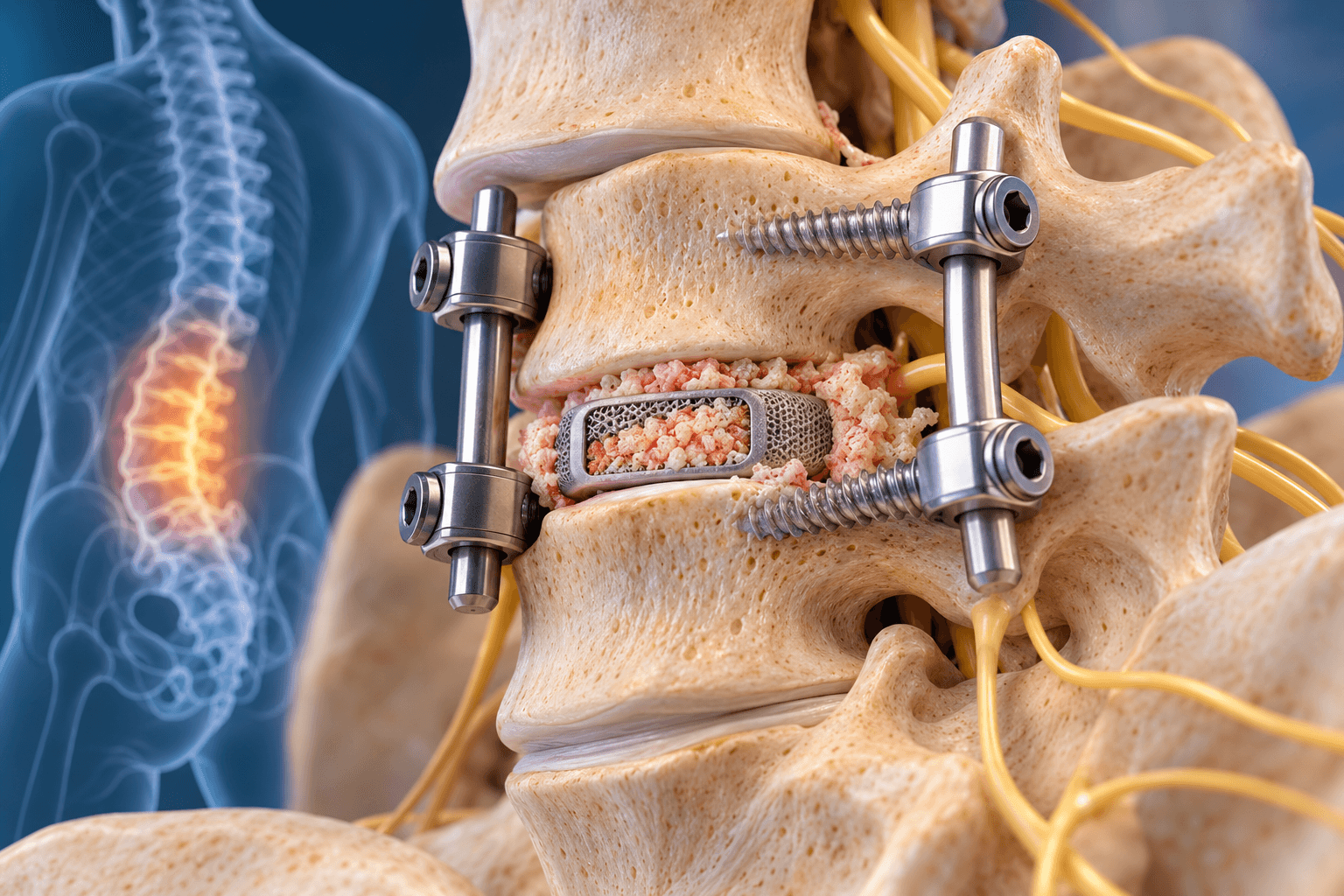
Laminectomy with fusion combines decompression with permanent stabilization of the treated spinal level.
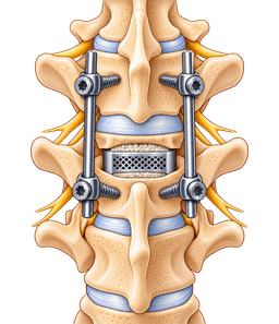
After neural compression is relieved, screws and rods can stabilize the vertebrae.
Bone graft is placed so that the vertebrae heal together.
The recovery is significantly different from decompression alone because biological bone fusion takes months.
Why Would Fusion Be Added?
The strongest reasons include established instability, substantial spondylolisthesis, deformity or expected iatrogenic instability after decompression.
Removing a large proportion of both facet joints can destabilize a segment.
Prior surgery can also affect stability.
Fusion should have a clear mechanical rationale rather than being added simply because implants are available.
Does Spondylolisthesis Always Require Fusion?
No.
This is an important point for patient education.
Several randomized trials and longer-term studies have shown that selected patients with lumbar stenosis and low-grade degenerative spondylolisthesis can do well with decompression alone.
Fusion remains appropriate when the slip is unstable, mechanical symptoms are substantial, foraminal collapse is significant or decompression would destabilize the segment.
Decompression Alone vs Fusion
Fusion adds operating time, implants, bone-healing requirements and additional potential complications.
Its benefit must therefore justify those costs.
A stable stenotic segment does not necessarily become better simply because it has been fused.
The decision should be individualized using symptoms, standing imaging, dynamic instability, alignment and planned bone removal.
Is a Laminectomy a Major Surgery?
Yes, a standard laminectomy is considered spinal surgery and should be taken seriously, although the magnitude varies enormously.
A one-level minimally invasive lumbar decompression can be a relatively limited outpatient procedure.
A multilevel cervical laminectomy with fusion is major surgery requiring implants, several hours in the operating room and a much longer recovery.
The term “laminectomy” alone therefore does not define surgical severity.
How Long Is Laminectomy Surgery?
A routine one- or two-level lumbar laminectomy often takes approximately one to three hours.
Broader multilevel decompression can take longer.
The NHS describes lumbar decompression operations as commonly lasting around two to four hours when the entire decompression procedure is considered. nhs.uk
Cervical laminectomy with fusion and complex revision procedures can take several hours.
Is Laminectomy Minimally Invasive?
It can be.
Minimally invasive techniques use tubular retractors or smaller muscle-splitting exposures.
The surgeon can perform laminotomy or partial laminectomy through these corridors.
The goal is to reduce muscle injury without compromising decompression.
Not every stenosis pattern is best treated through the smallest possible incision.
Open vs Minimally Invasive Laminectomy
Open surgery provides broad visualization and can be efficient for multilevel disease.
Minimally invasive surgery preserves more paraspinal muscle attachment and midline structures.
Studies generally suggest advantages such as reduced blood loss and shorter early recovery in selected patients, while long-term clinical success depends primarily on adequate decompression and correct indication.
A tiny incision is not valuable if stenosis remains inadequately treated.
How Does a Laminectomy Differ From Microdiscectomy?
Microdiscectomy primarily treats a herniated disc compressing a nerve.
The surgeon makes a small opening and removes the offending disc fragment.
Laminectomy is generally used for broader bony and ligamentous stenosis.
A patient can undergo both when stenosis and disc herniation coexist.
How Does It Differ From Spinal Fusion?
Laminectomy creates space.
Fusion eliminates movement.
These are different objectives.
Some patients require only decompression.
Others need decompression plus stabilization.
Confusing the two can lead patients to believe that every stenosis operation permanently stiffens the spine, which is not true.
Conditions treated
Who it's for
- Lumbar spinal stenosis causing neurogenic claudication that significantly limits walking or standing
- Persistent lumbar radiculopathy caused by central or lateral recess stenosis
- Progressive neurological weakness caused by nerve compression
- Multilevel lumbar stenosis that cannot be adequately decompressed through a smaller approach
- Degenerative lumbar stenosis that remains disabling despite appropriate nonsurgical treatment
- Cervical spinal stenosis causing degenerative cervical myelopathy
- Progressive hand dysfunction, balance problems or weakness caused by cervical spinal-cord compression
- Selected cervical radiculopathy requiring posterior decompression
- Thoracic spinal-cord compression in selected structural disorders
- Spinal tumors requiring posterior surgical access
- Epidural abscess or other compressive infection requiring urgent decompression
- Symptomatic facet or synovial cyst causing nerve compression
- Revision stenosis after previous decompression
- Ossification or hypertrophy of posterior spinal ligaments causing neurological compression
- Selected congenital spinal stenosis with disabling symptoms
- Cauda equina compression requiring urgent surgical decompression
- Laminectomy with fusion when instability, deformity or necessary facet removal makes decompression alone mechanically unsafe
Good candidates
A good candidate has symptoms that correspond to anatomically significant compression.
Someone with severe central lumbar stenosis, short walking tolerance and classic neurogenic claudication can be an excellent candidate.
Someone with only nonspecific back aching and stenosis that does not correlate with symptoms is much less predictable.
The purpose is to treat neural compression, not simply an abnormal MRI.
Patients With Neurogenic Claudication
These are among the classic candidates.
They can often stand or walk only limited distances before developing leg symptoms.
Sitting rapidly improves symptoms.
When conservative care fails and imaging demonstrates appropriate stenosis, decompression can substantially improve walking capacity.
Patients With Lumbar Radiculopathy
Stenosis can compress one or several nerve roots.
Patients can experience shooting pain, numbness or weakness along a specific nerve distribution.
Foraminotomy and lateral-recess decompression can be added to laminectomy.
The surgeon targets the exact anatomical areas causing compression.
Patients With Predominantly Back Pain
Results are less predictable.
Laminectomy principally treats nerve compression.
It does not rebuild degenerated discs or eliminate every source of mechanical back pain.
Patients whose main complaint is axial back pain should receive careful counseling before decompression.
Patients With Cervical Myelopathy
These patients can benefit from timely decompression because spinal-cord dysfunction can progressively impair walking and hand function.
The goal is often to stop deterioration.
The degree of neurological recovery varies.
Long-standing severe spinal-cord damage may not fully reverse even after adequate decompression.
Older Adults
Spinal stenosis becomes more common with age.
Older adults can achieve substantial improvement after decompression.
Chronological age alone is not a contraindication.
Frailty, cardiovascular health, cognitive function and ability to rehabilitate are more important than age by itself.
Patients With Medical Comorbidities
A limited decompression can sometimes be less physiologically demanding than fusion.
This makes avoidance of unnecessary fusion particularly meaningful in older or medically complex patients.
However, anaesthesia and surgery still carry risk.
Optimization is essential.
Patients With Spondylolisthesis
The surgeon must decide whether the slip is stable.
Standing and dynamic imaging can help.
Stable low-grade degenerative slips can sometimes undergo decompression alone.
Mobile or mechanically unstable slips more often require fusion.
Patients With Scoliosis
Mild stable degenerative scoliosis with focal stenosis can sometimes be decompressed without major reconstruction.
Larger curves, significant imbalance or unstable deformity require a more comprehensive strategy.
Removing posterior structures from a deformed spine without considering balance can accelerate instability.
Revision Patients
Previous surgery creates scar tissue around nerves.
Revision decompression can still be effective when recurrent compression is clearly demonstrated.
The risks of dural tear and nerve injury are generally higher.
The surgeon should review previous operative reports and imaging carefully.
Poor Candidates
Poor candidates include patients whose symptoms do not correlate with stenosis, people with predominantly non-neurological back pain, uncontrolled medical conditions and those who could obtain similar benefit from a less invasive treatment.
Severe instability is not necessarily a contraindication to decompression, but it changes the operation because fusion may need to be added.
Before surgery
History
The surgeon asks where the pain occurs, how far the patient can walk and which activities trigger symptoms.
Leg heaviness while standing suggests stenosis differently from pain that appears only while sitting.
Numbness, weakness and bowel or bladder symptoms are documented.
The duration and progression of neurological deficits are particularly important.
Neurological Examination
Strength, sensation and reflexes are tested.
Gait is assessed.
Cervical patients undergo examination for myelopathy, including hand dexterity and upper motor-neuron signs.
Lumbar patients are evaluated for individual nerve-root deficits.
The neurological examination determines urgency as well as prognosis.
Red Flags
Sudden bladder or bowel dysfunction, saddle anesthesia and rapidly progressive bilateral leg weakness can indicate cauda equina syndrome.
Progressive spinal-cord dysfunction in the neck can also require timely treatment.
These conditions should not be managed as routine elective tourism cases while neurological function deteriorates.
X-Rays
Standing X-rays show alignment and degenerative changes.
They can identify spondylolisthesis and scoliosis.
This matters because MRI is usually performed lying down and may not reveal how the spine behaves under body weight.
Flexion-Extension X-Rays
Dynamic films can evaluate instability.
The patient bends forward and backward under controlled conditions.
Abnormal translation can make fusion more appropriate.
Normal dynamic studies support decompression alone when other mechanical factors are favorable.
MRI
MRI is the principal imaging study for spinal stenosis.
It shows the spinal canal, discs, ligaments, spinal cord and nerve roots.
The surgeon identifies the exact levels and regions of compression.
MRI abnormalities must still be interpreted in relation to symptoms because stenosis can exist in patients who function well.
CT
CT gives more detailed information about bone.
It can be particularly useful after previous surgery, in congenital narrowing or when ossified structures contribute to stenosis.
CT can also clarify facet anatomy.
CT Myelography
When MRI cannot be performed or provides inadequate information, CT myelography can outline neural compression after contrast is introduced into the spinal fluid.
It is more invasive than MRI.
It remains valuable in selected cases, especially around substantial metal artifact.
Electromyography
EMG and nerve-conduction studies are not required for every stenosis patient.
They can help when symptoms could result from peripheral neuropathy, plexus disease or another neurological condition.
They can also help clarify multi-level symptoms.
A normal or abnormal EMG alone does not decide whether laminectomy is appropriate.
Conservative Treatment
Many lumbar stenosis patients initially receive exercise-based rehabilitation, activity modification and symptom-directed medication.
Some benefit from epidural injection.
Surgery becomes more attractive when neurological compression continues to substantially limit function despite appropriate treatment.
Progressive weakness can justify earlier surgery.
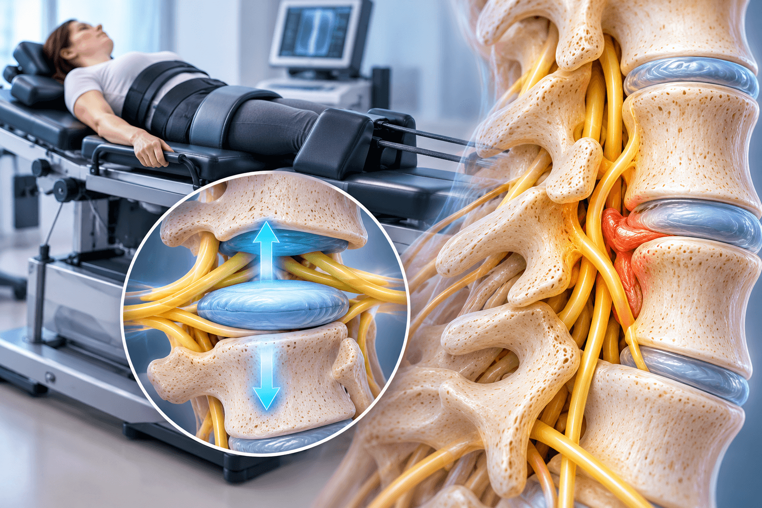
Physical Therapy
Therapy aims to maintain strength and cardiovascular conditioning.
Flexion-tolerant exercises can sometimes reduce stenosis symptoms.
Therapy cannot physically remove bone or thickened ligament.
Patients with severe fixed stenosis can therefore improve only partially.
Epidural Steroid Injection
Injection can reduce inflammation around a compressed nerve.
Relief is often temporary.
It does not enlarge the spinal canal.
Repeated injections should not postpone necessary surgery when neurological weakness is progressing.
Medical Optimization
Blood pressure, diabetes, heart disease and pulmonary disease are assessed.
Anemia is corrected when possible.
Nutrition is reviewed.
A routine one-level decompression and a multilevel cervical procedure carry different physiological demands, so preoperative testing is individualized.
Smoking
Smoking increases wound and pulmonary complications.
Its effect on bone fusion is particularly important when fusion is added.
Even without fusion, cessation can improve general surgical recovery.
Patients should be encouraged to stop before surgery rather than simply reducing cigarettes for a few days.
Anticoagulants
Spinal surgery occurs near neurological structures where postoperative bleeding can be dangerous.
Blood-thinning medication therefore requires careful planning.
Patients should never stop prescribed anticoagulants independently.
The surgeon, anaesthetist and prescribing clinician coordinate the safest schedule.
Infection Screening
Active systemic infection can increase the risk of spinal surgical infection.
Patients with fever, urinary infection or infected skin lesions should notify the surgical team.
Elective surgery can need postponement.
Preparing the Home
Frequently used items should be positioned at comfortable height.
A stable chair with arms can simplify standing.
Loose clothing helps.
Patients should arrange help for shopping, heavy cleaning and lifting during the early recovery period.
Preparing for Walking
Walking is usually encouraged soon after surgery.
Comfortable shoes are useful.
Some older patients temporarily use a walker.
The goal is not permanent dependence but safe early mobility.
Work Planning
Desk workers can often return earlier than manual workers.
The patient should clarify whether the job involves lifting, prolonged driving or climbing.
A realistic plan is preferable to promising every patient a return at exactly two or four weeks.
Medical Travel Planning
International patients should send MRI images rather than only the radiology report.
Standing X-rays and dynamic studies can be important when instability is uncertain.
The surgeon should determine in advance whether decompression alone or laminectomy with fusion is anticipated.
That distinction changes the operation, implants, price and recovery substantially.
How the operation is performed
Laminectomy surgery is performed by exposing the posterior spine, identifying the correct vertebral levels, removing the required lamina and thickened ligament, and enlarging the spinal canal until the compressed neurological structures have adequate space.
The surgeon may also perform foraminotomy, medial facetectomy or discectomy.
The amount removed is tailored to the stenosis.
A well-planned decompression aims to release nerves without removing unnecessary stabilizing anatomy.
Anaesthesia
General anaesthesia is most common.
The patient is fully asleep.
The anaesthesia team monitors heart rate, blood pressure, oxygenation and other physiological parameters.
Complex cervical or thoracic surgery can use additional monitoring.
Positioning for Lumbar Laminectomy
The patient is commonly positioned face-down.
Special pads support the chest and pelvis while reducing abdominal pressure.
This helps decrease venous congestion and bleeding.
Pressure points and peripheral nerves are carefully protected.
Positioning for Cervical Laminectomy
Posterior cervical procedures are usually performed face-down or in another controlled prone configuration.
The head is stabilized.
The neck is positioned to maintain safe spinal-cord dimensions.
Intraoperative imaging confirms the surgical levels.
Identifying the Level
Wrong-level spine surgery is a major preventable error.
Fluoroscopy or another imaging method confirms the appropriate vertebrae.
The surgeon correlates imaging with anatomical landmarks before removing bone.
Incision
Traditional open laminectomy uses a midline incision.
The length depends on the number of levels.
Minimally invasive decompression uses smaller incisions and muscle-splitting corridors.
Scar size does not necessarily indicate the amount of internal decompression achieved.
Muscle Exposure
In open surgery, paraspinal muscles are gently separated from the posterior vertebrae.
Excessive muscle damage is avoided.
Modern techniques preserve attachments when possible.
Minimally invasive surgery achieves access through a narrower corridor.
Removing the Lamina
A high-speed burr and surgical instruments can thin and remove bone.
The surgeon works carefully near the ligament protecting the underlying nerves.
The exact amount varies.
A complete laminectomy removes more bone than a laminotomy.
Ligamentum Flavum Removal
The ligamentum flavum lies beneath the lamina.
It commonly becomes thickened in degenerative stenosis.
Once the protective bony work is complete, the surgeon removes the thickened ligament.
This often produces a major increase in available canal space.
Medial Facetectomy
The facet joints can enlarge and compress the lateral recess.
The surgeon may remove the medial portion of a facet.
Preserving enough of the joint is important for stability.
Excessive bilateral facetectomy can create instability and potentially require fusion.
Lateral Recess Decompression
The lateral recess is where a nerve root travels before entering its foramen.
Bone and ligament can narrow this zone.
The surgeon undercuts the facet and removes compressive tissue.
The nerve root should then lie freely without excessive manipulation.
Foraminotomy
The neural foramen is the opening through which a spinal nerve exits.
Foraminotomy enlarges this opening.
It can be performed during laminectomy when foraminal stenosis contributes to symptoms.
The surgeon balances adequate decompression with preservation of facet stability.
Discectomy
If a disc fragment compresses the nerve, the surgeon can remove it.
Not every laminectomy requires disc surgery.
The disc is left alone when it is not part of the compression.
Unnecessary disc removal can alter spinal mechanics.
Confirming Decompression
The surgeon visually and mechanically confirms that neural structures have adequate space.
The dura should no longer be constricted by thick ligament or bone.
Nerves are handled gently.
“More bone removal” is not automatically better once adequate decompression has been achieved.
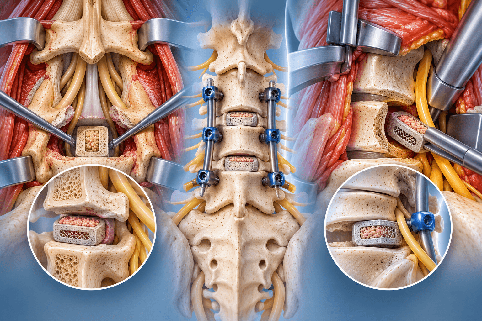
Laminectomy With Fusion Technique
When fusion is planned, pedicle screws or cervical fixation are placed.
Rods connect the screws.
Bone graft is added across prepared surfaces.
The neural structures are decompressed while the instrumentation stabilizes the vertebrae.
Recovery then follows both decompression and fusion principles.
Lumbar Laminectomy With Fusion
This is commonly performed when stenosis coexists with mechanical instability.
The surgeon may add TLIF, PLIF or posterolateral fusion.
An interbody cage can restore disc height.
However, fusion should not be considered a required component of every lumbar decompression.
Cervical Laminectomy With Fusion
Posterior cervical laminectomy often includes fusion when several levels are decompressed and maintenance of alignment is important.
Lateral-mass or pedicle screws can stabilize the neck.
Rods connect the construct.
Fusion reduces the risk of progressive postoperative instability or kyphotic change in appropriately selected patients.
Cervical Laminectomy Without Fusion
This can still be appropriate in selected patients with stable alignment and carefully preserved posterior structures.
However, postoperative kyphotic change is an important consideration.
Modern practice more often compares laminectomy with fusion against laminoplasty for multilevel cervical myelopathy.
Patient selection depends heavily on preoperative lordosis, instability and neck pain.
Minimally Invasive Lumbar Laminectomy
Tubular retractors allow access through a small muscle corridor.
The surgeon removes part of the lamina and undercuts the opposite side if necessary.
This can preserve midline ligaments and muscle attachments.
The procedure is particularly attractive for selected one- or two-level stenosis.
Unilateral Approach for Bilateral Decompression
The surgeon enters from one side.
Bone beneath the spinous process is undercut.
The opposite ligament and lateral recess are decompressed.
This allows bilateral neural decompression while preserving more posterior anatomy.
It requires technical experience and appropriate anatomy.
Endoscopic Decompression
Endoscopic systems use a camera through a narrow working channel.
They can treat selected stenosis patterns.
The incision is extremely small.
However, endoscopic expertise has a learning curve, and broad multilevel stenosis is not automatically better treated endoscopically.
Adequate neural decompression remains more important than incision size.
Intraoperative Neuromonitoring
Neuromonitoring is not essential for every routine lumbar laminectomy.
It is more commonly used during cervical, thoracic or complex revision surgery.
Electrical signals can detect changes in spinal-cord or nerve function.
This provides an additional safety tool but cannot eliminate neurological risk.
Dural Tear
The dura is the membrane containing spinal fluid and neural structures.
It can be torn accidentally, especially when scar tissue is present from previous surgery.
Small tears can usually be repaired during the operation.
A repaired dural tear does not automatically mean long-term problems will develop.
Wound Closure
After decompression, bleeding is controlled.
A drain is used selectively.
Muscle and fascia are closed.
The skin is closed with sutures, staples or absorbable material.
A sterile dressing is applied.
How Long Is Laminectomy Surgery?
An isolated one-level operation can sometimes be completed in around an hour.
A common multi-component lumbar decompression takes approximately two to four hours, according to NHS patient guidance. nhs.uk
Multilevel cervical surgery, revision decompression and laminectomy with fusion can take considerably longer.
Hospital stay
Recovery Room
The patient wakes under close observation.
Strength and sensation in the arms or legs are checked.
Pain and nausea are treated.
Blood pressure and breathing are monitored.
A catheter can remain temporarily after longer procedures.
Getting Out of Bed
Early mobilization is encouraged.
Patients commonly stand and walk on the day of surgery or the next morning.
A physiotherapist or nurse initially assists.
Walking reduces deconditioning and helps prevent blood clots.
Hospital Stay After Lumbar Laminectomy
Many modern patients leave within one or two days, and selected limited decompressions can be outpatient procedures.
Broader lumbar decompression can require a longer stay.
NHS guidance for lumbar decompression notes that some patients stay approximately two to five days depending on the operation and clinical circumstances. nhs.uk
Cervical Laminectomy Hospital Stay
Hospitalization is often longer when fusion is added.
Neurological function, swallowing where relevant, walking and pain control are monitored.
A multilevel posterior cervical reconstruction can require several nights.
Drain
A wound drain can be placed when the surgeon expects significant fluid or blood accumulation.
It is commonly removed within the first postoperative days.
Many routine decompressions do not require one.
Pain Medication
Multimodal pain treatment reduces reliance on high-dose opioids.
Medication can include acetaminophen, short-term opioids, muscle-relaxant medication and other drugs according to patient health.
NSAID policy depends partly on whether fusion is also performed.
Bowel Function
Constipation is common after anaesthesia and opioid treatment.
Walking, fluids and prescribed bowel medication help.
This is usually temporary.
Patients should not become alarmed by a short delay in normal bowel function unless other concerning symptoms are present.
Urination
Temporary urinary retention can occur.
A catheter is sometimes used overnight.
Patients should be able to urinate safely before routine discharge.
New bladder dysfunction accompanied by neurological changes requires urgent assessment.
Discharge
Patients typically leave when they can walk safely, pain is controlled with oral medication, neurological status is stable and home support is adequate.
They receive instructions about wound care, activity, medication and warning signs.
International patients should also receive the operative report and postoperative plan before traveling.
Recovery
Laminectomy Recovery
Laminectomy recovery commonly takes about six to twelve weeks after routine lumbar decompression, although many patients begin walking and performing basic daily activities much earlier.
Recovery differs from fusion because there is no requirement to wait for two vertebrae to grow together when decompression is performed alone.
Muscles and surgical tissues still need time to heal.
Neural recovery can continue for many months.
First 24–72 Hours
Incisional and muscle pain are expected.
Walking begins early.
Some patients notice immediate improvement in preoperative leg pain.
Others experience temporary nerve irritation or numbness.
The surgical region feels stiff.
Pain medication makes movement more manageable.
How Bad Is the Pain After a Laminectomy?
Pain after a laminectomy is usually moderate during the first several postoperative days and should progressively improve.
Patients commonly describe incisional soreness, tightness or aching in the spinal muscles.
Posterior cervical surgery can cause significant neck and shoulder muscle soreness.
A limited minimally invasive lumbar decompression is often less painful than open multilevel surgery.
The severity is therefore determined by surgical extent rather than the word “laminectomy” alone.
Is Nerve Pain Normal After Surgery?
Some residual or fluctuating nerve pain can occur.
A chronically compressed nerve does not necessarily become normal immediately after decompression.
Inflammation can persist.
Tingling can change as the nerve recovers.
However, new progressive weakness, severe neurological deterioration or loss of bladder/bowel control is not routine recovery.
First Week
Walking is encouraged several times a day.
The incision remains tender.
Patients become increasingly independent with bathing and dressing.
Fatigue is common.
Long periods of sitting can feel uncomfortable.
The objective is gentle regular activity rather than staying in bed.
Weeks 2–4
Walking distance increases.
Most patients reduce stronger pain medication.
Household activity expands.
Many patients with sedentary employment begin considering return to work.
Heavy lifting and repetitive spinal twisting remain limited.
Weeks 4–6
A substantial proportion of routine lumbar decompression patients feel considerably more functional.
NHS guidance notes that return to work at around four to six weeks can be possible depending on the occupation and individual recovery. nhs.uk
Physical therapy can be introduced or progressed.
Manual workers generally require additional time.
Weeks 6–12
Strength and endurance continue improving.
Patients return to more normal exercise.
Residual numbness can continue even when pain has improved.
Older patients who were severely deconditioned before surgery can need longer to rebuild walking capacity.
Three Months
Many uncomplicated lumbar laminectomy patients have returned to most normal daily activity by three months.
The surgical wound is mature.
Muscular stamina continues improving.
Persistent severe symptoms at this stage deserve reassessment but do not automatically mean the operation failed.
Nerves can recover more slowly than muscles.
Six Months
Most routine decompression patients have reached a stable level of functional recovery.
Neurological improvement can continue.
Patients with longstanding weakness or numbness can have incomplete recovery because chronic compression can permanently injure nerves.
How Long Does It Take to Recover From a Laminectomy?
For an uncomplicated lumbar decompression without fusion, major recovery often occurs over six to twelve weeks.
Light daily activity returns sooner.
Heavy manual work can take two to three months or more.
A laminectomy with fusion follows a substantially longer recovery because bone healing commonly continues for six to twelve months.
Lumbar Laminectomy Recovery
Lumbar recovery focuses on restoring walking, trunk endurance and lower-limb strength.
Many patients are surprised that they can walk soon after surgery.
The limiting factor initially is usually wound soreness rather than instability.
The surgeon provides lifting restrictions based on how much anatomy was removed.
Cervical Laminectomy Recovery
Recovery depends strongly on whether fusion is included.
Posterior neck muscles can remain sore for several weeks.
Patients undergoing surgery for myelopathy can require neurological rehabilitation.
Balance and hand function may improve gradually.
Fusion cases require additional precautions while bone heals.
Walking
Walking is one of the best early activities.
Patients start with short frequent distances.
The goal increases gradually.
A walker is sometimes used temporarily for balance.
This does not mean surgery has made the patient permanently dependent on walking aids.
Sitting
Sitting can initially worsen lumbar soreness.
The patient should change position periodically.
A firm supportive chair is often easier than a deep sofa.
There is no need to maintain an unnaturally rigid posture all day.
Bending
Early repeated bending is limited.
The restriction protects healing muscles and helps avoid sudden strain.
Patients progressively return to normal movement.
Unlike fusion, a successful decompression alone does not intentionally eliminate motion at the surgical level.
Lifting
Light objects are introduced first.
Patients keep loads close to the body.
Heavy lifting returns gradually.
The surgeon may impose stricter limits after multilevel or revision surgery.
Twisting
Forceful repetitive twisting is avoided early.
Normal gentle turning returns as comfort improves.
Golf and racquet sports require later rotational loading than ordinary daily movement.
Physical Therapy
Not every patient requires prolonged formal therapy.
Older or deconditioned patients can benefit significantly.
Therapy addresses walking, leg strength, trunk endurance and safe return to activity.
If fusion has been added, the programme follows the fusion protocol.
Core Strengthening
Gentle abdominal activation progresses to broader stabilization exercises.
Aggressive sit-ups are not necessary.
Modern rehabilitation encourages functional movement rather than teaching the patient to keep the spine rigid forever.
Return to Desk Work
Many uncomplicated patients return within two to six weeks.
Working from home can permit an earlier staged return.
The ability to change positions regularly helps.
Long commuting can be more difficult than the work itself.
Return to Manual Work
Manual employment can require six to twelve weeks or more.
Heavy construction, warehouse work and repetitive lifting place greater demand on the spine.
A laminectomy with fusion can delay unrestricted heavy work for several months.
Driving
Driving resumes when the patient is no longer taking sedating medication, can sit comfortably and can respond quickly.
Neck movement is particularly important after cervical surgery.
Patients should not drive simply because a calendar says two weeks have passed.
Sleeping
Patients can sleep in a comfortable position.
A pillow under the knees can help while lying on the back.
A pillow between the knees can make side sleeping more comfortable.
There is generally no requirement to sleep in one rigid posture after uncomplicated lumbar decompression.
Showering
The surgical team provides wound-specific instructions.
Many incisions can get briefly wet once appropriately sealed.
Soaking in pools or baths is delayed until the wound is fully healed.
A shower chair can help patients with poor balance.
Stairs
Stairs are generally allowed once the patient can use them safely.
A physiotherapist can teach technique before discharge.
There is usually no biological reason to avoid all stairs after an uncomplicated laminectomy.
The main concern is fall risk.
Sex After Laminectomy
Sexual activity can resume when pain is controlled and the patient can avoid uncomfortable spinal loading.
Early positions should minimize bending and twisting.
No universal waiting period applies to all patients.
Fusion and extensive cervical reconstruction create different considerations.
Gym After Laminectomy
Walking and low-impact cardiovascular exercise return first.
Stationary cycling can be introduced when comfortable.
Resistance training follows.
The patient should rebuild movement quality before returning to heavy squats or deadlifts.
Running
Impact exercise returns after sufficient healing and conditioning.
Some patients are able to resume recreational running.
Severe arthritis or other musculoskeletal conditions can remain limiting even when the spine is well decompressed.
Swimming
Swimming can be a useful low-impact activity once the incision has fully healed.
Patients begin with shorter sessions.
Cervical patients should consider neck comfort with different strokes.
Golf
Putting and short swings return before full swings.
Rotation is gradually reintroduced.
Many patients return to recreational golf after successful lumbar decompression.
Permanent Restrictions After Laminectomy
An uncomplicated laminectomy without fusion usually does not create universal permanent restrictions.
Many patients eventually return to unrestricted normal activity.
Long-term limitations depend on residual degeneration, instability, neurological deficits and other health conditions.
If fusion is added, restrictions become more procedure-specific.
Scar
A traditional lumbar laminectomy creates a midline back scar.
The length reflects the number of levels and exposure.
Minimally invasive approaches produce smaller scars.
Scar appearance generally fades over time.
The size of the scar does not reliably predict the quality of nerve decompression.
How Many Years Does a Laminectomy Last?
Is a Laminectomy Permanent?
The bone that has been removed does not normally regenerate into the original stenotic lamina.
In that sense, the anatomical decompression is durable.
However, the remaining spine continues aging.
Facet joints can enlarge further, discs can degenerate and adjacent levels can develop stenosis.
How Many Years Does a Laminectomy Last?
There is no fixed expiration date for a laminectomy. Many patients remain improved for many years, and some never require another operation.
The durability depends on the original diagnosis, number of levels treated, spinal alignment, stability and ongoing degeneration.
Recurrence of symptoms does not necessarily mean the removed lamina “grew back.”
A different level or another degenerative structure can become responsible.
Recurrent Stenosis
Scar tissue and continued degenerative changes can narrow neural spaces again.
Recurrent stenosis can appear at the same or adjacent levels.
Some patients remain asymptomatic despite radiographic narrowing.
Repeat surgery is considered only when clinically significant compression returns.
Instability After Laminectomy
Removing too much supporting facet or ligament can contribute to instability.
This is one reason modern decompression aims to preserve stabilizing anatomy.
Patients with pre-existing spondylolisthesis or deformity can also progress independently of the surgery.
Fusion is considered if symptomatic instability develops.
Adjacent-Level Disease
The spine continues aging above and below the operated levels.
Because decompression without fusion preserves motion, adjacent-level mechanical effects are generally different from those following fusion.
Nevertheless, new stenosis can develop anywhere in a degenerative spine.
Revision Laminectomy
Revision decompression can be successful when recurrent compression is clearly identified.
Scar tissue makes surgery more technically demanding.
The dural tear risk is higher.
The surgeon carefully evaluates whether instability now makes fusion necessary.
Post Laminectomy Syndrome
What Is Post Laminectomy Syndrome?
Post laminectomy syndrome is an older umbrella term used to describe persistent or recurrent pain after spinal surgery.
Despite the name, it does not mean the laminectomy itself necessarily caused the pain.
It can occur after many different spinal procedures, including fusion, discectomy and decompression.
Modern pain specialists increasingly prefer more descriptive terminology such as persistent spinal pain syndrome type 2.
Is Post Laminectomy Syndrome a Single Disease?
No.
It is a clinical description.
One patient can have recurrent disc herniation.
Another can have nerve damage.
Another can develop adjacent stenosis, sacroiliac pain, facet pain or pseudarthrosis after a fusion.
Combining all of these under one label can prevent accurate diagnosis.
Why Can Pain Persist After Decompression?
A nerve compressed for years can sustain permanent injury.
Decompression removes ongoing pressure but cannot always reverse established neurological damage.
The patient can therefore have persistent numbness, burning or weakness even when postoperative imaging shows excellent decompression.
Wrong Pain Generator
Another possibility is that stenosis was not the main cause of preoperative symptoms.
Hip arthritis, peripheral neuropathy, vascular claudication and sacroiliac pain can mimic spinal symptoms.
This highlights why accurate preoperative diagnosis is so important.
Scar Tissue
Epidural scar forms naturally after surgery.
Most scar tissue is harmless.
Occasionally it can be associated with persistent nerve symptoms.
Seeing scar tissue on MRI does not automatically prove that it is responsible for pain.
Recurrent Disc Herniation
A new disc fragment can compress a nerve after previous discectomy or decompression.
Treatment depends on symptom severity and neurological findings.
Not every recurrent herniation requires another operation.
Recurrent or Progressive Stenosis
Degenerative bone and ligament can continue changing.
A level that was adequately decompressed years earlier can become narrowed in another region.
Adjacent levels can also become stenotic.
New MRI findings are correlated with new symptoms.
Spinal Instability
Symptoms can arise if a decompressed segment becomes unstable.
Standing and dynamic X-rays can reveal abnormal movement.
Fusion can be considered when instability clearly correlates with pain or neural compression.
Persistent Spinal Pain Syndrome
The newer terminology recognizes that persistent pain after spinal surgery is complex.
Treatment begins with identifying potentially correctable structural causes.
When no surgically correctable lesion exists, pain-management strategies can include rehabilitation, medication, psychological support and selected neuromodulation techniques.
Spinal Cord Stimulation
Spinal cord stimulation can be considered for selected patients with chronic neuropathic pain after previous surgery when additional decompression or reconstruction is not indicated.
A trial is generally performed before permanent implantation.
It is not a treatment for untreated severe mechanical compression.
Recovery timeline
- Control postoperative pain, protect the incision and establish safe independent walking.1Control postoperative pain, protect the incision and establish safe independent walking.
Days 0–14
The patient takes frequent short walks and avoids heavy lifting. Incisional soreness and muscle stiffness are expected. Neurological status is monitored. New weakness, bladder dysfunction or progressive numbness requires urgent assessment rather than waiting for a routine appointment.
- Increase walking tolerance and return to light daily activity.2Increase walking tolerance and return to light daily activity.
Weeks 2–6
Pain medication usually decreases. Household tasks and light office work return progressively. Bending and lifting restrictions are relaxed according to the operation. Many patients notice clear improvement in neurogenic claudication by this stage.
- Restore strength, endurance and broader functional independence.3Restore strength, endurance and broader functional independence.
Weeks 6–12
Formal physiotherapy can emphasize trunk and lower-limb conditioning. Most uncomplicated decompression patients are returning toward normal life. Manual workers and patients with multilevel surgery can remain restricted.
- Return to demanding work and recreational exercise where appropriate.4Return to demanding work and recreational exercise where appropriate.
Months 3–6
Strengthening becomes less restricted. Persistent nerve symptoms continue to be monitored because neurological recovery can be slow. A patient with worsening symptoms should be reassessed rather than simply told to exercise harder.
- Reach mature neurological and functional recovery.5Reach mature neurological and functional recovery.
Months 6–12
Most soft-tissue healing is complete. Residual numbness or weakness is increasingly likely to reflect chronic nerve damage rather than ordinary surgical inflammation, although some neurological improvement can still continue.
Outcomes and success rates
How Successful Is Laminectomy?
Laminectomy is an established treatment for symptomatic spinal stenosis when patient selection is appropriate.
The most reliable improvements generally involve leg pain, neurogenic claudication and walking tolerance.
Back pain can improve as well, but results are less predictable because axial pain has many causes.
Lumbar Laminectomy Outcomes
Patients with classic lumbar stenosis commonly report improved walking capacity and reduced leg symptoms.
The operation removes mechanical compression rather than treating every degenerative abnormality visible on MRI.
Long-term satisfaction is therefore strongest when symptoms and imaging clearly correspond before surgery.
Neurological Recovery
Pain often improves before numbness.
Weakness can require months.
A severely damaged nerve may never regain completely normal function.
The surgeon should therefore avoid promising that decompression will erase every neurological symptom.
Walking Improvement
Improved walking is one of the central goals of lumbar stenosis surgery.
Patients who previously had to stop every few minutes can often increase their distance substantially.
Other causes of limited walking, including hip arthritis, vascular disease and heart or lung disease, can remain.
Back Pain Improvement
Some patients experience meaningful improvement because standing and walking mechanics normalize.
Others retain degenerative back pain.
Laminectomy should therefore not be marketed primarily as an operation to cure nonspecific axial pain.
Cervical Laminectomy Outcomes
The principal goal in cervical myelopathy is protection of the spinal cord.
Hand function, balance and gait can improve.
The amount of recovery depends on preoperative severity and duration.
Advanced spinal-cord injury can leave residual deficits.
Cervical Laminectomy vs Laminoplasty
Recent evidence indicates that both posterior laminectomy with fusion and laminoplasty can produce meaningful neurological improvement in appropriately selected multilevel cervical myelopathy patients. Comparative studies show trade-offs in operative time, blood loss, range-of-motion preservation and complications rather than one universally superior operation. PubMed
Laminectomy Alone vs Laminectomy With Fusion
Fusion is not automatically associated with better results.
For many lumbar stenosis patients, decompression alone avoids additional surgical burden without compromising long-term clinical outcomes.
Five-year randomized evidence has continued to support decompression alone as a valid treatment for selected lumbar stenosis patients, including some with degenerative spondylolisthesis. PubMed
Risk of Later Fusion
A minority of patients who undergo decompression alone later develop symptomatic instability or recurrent stenosis requiring fusion.
The possibility should be discussed preoperatively.
However, the fact that some patients eventually need fusion is not a reason to fuse every stable spine during the first operation.
Reoperation
Reasons for reoperation include recurrent stenosis, disc herniation, instability, infection and adjacent-level disease.
Revision risk increases over years simply because the spine continues aging.
Long-term success should therefore be measured by function and symptom control, not by expecting the spine never to change again.
Durability
Many patients remain improved for years after a single decompression.
The removed lamina itself is not expected to recreate the original stenosis.
New degenerative disease can appear elsewhere.
Maintaining healthy body weight, strength and general conditioning supports long-term spinal function but cannot completely prevent age-related degeneration.
Implants and technology
Does Laminectomy Require Implants?
A standard laminectomy without fusion usually requires no permanent spinal implant.
The purpose is decompression.
Temporary surgical tools are removed before closure.
Implants become relevant when fusion or stabilization is added.
Pedicle Screws
Lumbar or thoracic laminectomy with fusion can use pedicle screws.
These screws anchor into the vertebrae.
Rods connect them.
They maintain mechanical stability while bone graft forms the fusion.
Cervical Lateral-Mass Screws
Posterior cervical fusion commonly uses screws placed into the lateral masses.
Pedicle screws can also be used in selected anatomy.
Rods connect the construct.
The surgeon chooses fixation based on bone quality and levels involved.
Navigation
Three-dimensional navigation can assist with level identification, decompression planning and screw placement when fusion is added.
Routine limited laminectomy does not necessarily require advanced navigation.
Technology should have a purpose rather than being added primarily for marketing.
Intraoperative CT
Three-dimensional imaging can be useful during complex revision or instrumented surgery.
It allows immediate verification of screw position.
For a straightforward decompression without instrumentation, routine intraoperative CT may offer less additional value.
Robotics
Robotics primarily assists with planned spinal instrumentation trajectories.
It does not independently perform the laminectomy.
A decompression without screws generally derives little direct benefit from robotic pedicle-screw guidance.
If fusion is required, robotic assistance can be incorporated into the fixation step.
High-Speed Burr
A high-speed surgical burr allows controlled thinning of the lamina and facet bone.
Different burr sizes are selected according to anatomy.
The surgeon works progressively rather than aggressively removing large blocks near neural tissue.
Kerrison Rongeur
The Kerrison is a specialized spinal instrument used to remove thin pieces of bone and ligament.
It is widely used during decompression.
The instrument allows precise enlargement of the canal and foramina.
Microscope
An operating microscope provides magnification and illumination.
This can be useful for limited decompression and revision surgery.
Other surgeons use high-quality loupe magnification or endoscopic visualization.
The visualization method matters less than obtaining safe, complete decompression.
Endoscope
An endoscope places the camera close to the pathology through a small access channel.
Selected decompressions can therefore be performed through tiny incisions.
The technique can reduce tissue disruption.
Its success remains dependent on accurate diagnosis and adequate decompression.
Tubular Retractors
Minimally invasive decompression uses progressively sized dilators to create a corridor through rather than extensively detaching spinal muscles.
A tubular retractor maintains this path.
The surgeon then uses a microscope or camera to remove bone and ligament.
Ultrasonic Bone Tools
Some centers use ultrasonic instruments to cut bone while minimizing interaction with soft tissues.
They can be useful in selected cervical or thoracic procedures.
They supplement rather than replace careful anatomy and technique.
Neuromonitoring
Electrical monitoring can follow spinal-cord and nerve function.
Its role is strongest in cervical, thoracic and complex cases.
Routine single-level lumbar decompression can often be performed without it.
Risks and how they are managed
All surgery carries risk. Partner hospitals follow enhanced-recovery and infection-prevention protocols, and your surgeon will discuss the risks specific to your case before consent.
- Postoperative pain: Muscle and incisional soreness are expected during early recovery.
- Persistent back or neck pain: Decompression cannot eliminate every degenerative pain source.
- Persistent nerve pain: A chronically damaged nerve can remain painful despite adequate decompression.
- Residual numbness: Sensory recovery is often slower than pain relief and can be incomplete.
- Residual weakness: Longstanding neurological deficits can persist.
- New nerve injury: A nerve root can rarely be injured during decompression.
- Spinal-cord injury: Particularly important in cervical and thoracic surgery, although uncommon.
- Dural tear: The membrane containing spinal fluid can be torn, especially in revision surgery.
- Cerebrospinal fluid leak: Can occur following a dural tear and sometimes require additional treatment.
- Postural headache: Can accompany persistent CSF leakage.
- Epidural hematoma: Blood can accumulate around the neural structures and create urgent compression.
- Wound hematoma: Blood collection can cause pain and swelling.
- Infection: Superficial or deep infection can occur.
- Wound-healing problems: Risk increases with diabetes, smoking, malnutrition and other medical conditions.
- Spinal instability: Can occur if too much supporting bone or facet joint is removed or if instability already existed.
- Progression of spondylolisthesis: A pre-existing slip can worsen in selected patients.
- Need for later fusion: Symptomatic instability after decompression can require stabilization surgery.
- Recurrent spinal stenosis: Degenerative changes can produce renewed compression over time.
- Adjacent-level stenosis: Other spinal levels can become symptomatic later.
- Recurrent disc herniation: Disc material can herniate after decompression or combined discectomy.
- Scar-related symptoms: Epidural fibrosis is common after surgery but is only occasionally clinically important.
- Post laminectomy syndrome / persistent spinal pain: Chronic pain can persist for several different reasons.
- Blood clots: DVT can develop after surgery.
- Pulmonary embolism: A blood clot can travel to the lungs.
- Urinary retention: Temporary difficulty urinating can occur after anaesthesia or lumbar surgery.
- Constipation: Common because of medication and reduced activity.
- Pneumonia or respiratory complication: More relevant in older or medically frail patients.
- Positioning injury: Peripheral nerves or pressure areas can be injured during a long operation.
- C5 palsy: A recognized complication of posterior cervical decompression that can cause shoulder-abduction and elbow-flexion weakness.
- Post-laminectomy cervical kyphosis: Loss of normal cervical alignment can develop in selected patients after decompression without stabilization.
- Need for revision surgery: Recurrent compression, instability, infection or another problem can require further intervention.
- Anaesthetic complications: Cardiovascular, respiratory and allergic complications are possible.
- Major medical complications: Heart attack, stroke or other serious events are uncommon but possible, especially in medically complex patients.
Alternatives
- Structured physical therapy: Often first-line for lumbar stenosis when neurological deficits are not rapidly progressing.
- Walking and conditioning programme: Can improve function despite persistent radiographic stenosis.
- Activity modification: Adjusting prolonged standing, repetitive extension or heavy work can reduce symptoms.
- Medication: Analgesic and selected anti-inflammatory therapy can help control symptoms when medically appropriate.
- Epidural steroid injection: May reduce radicular inflammation temporarily but does not physically enlarge the spinal canal.
- Observation: Reasonable for radiographic stenosis that does not significantly impair function.
- Laminotomy: Removes a smaller amount of posterior bone and can adequately decompress focal disease.
- Unilateral laminotomy for bilateral decompression: Preserves additional midline and facet anatomy in selected lumbar patients.
- Microdiscectomy: More appropriate when the principal pathology is a focal herniated disc.
- Foraminotomy: Can treat isolated foraminal narrowing without broad central laminectomy.
- Endoscopic decompression: A minimally invasive option for selected anatomy.
- Laminoplasty: A motion-preserving alternative to laminectomy with fusion for selected multilevel cervical myelopathy patients.
- Laminectomy with fusion: Appropriate when decompression alone would leave an unstable spine.
- Anterior cervical decompression and fusion: Can directly treat selected cervical disc or bone-spur compression from the front.
- Cervical disc replacement: A motion-preserving option for appropriately selected cervical disc disease, although it is not a substitute for every multilevel posterior decompression.
- Spinal fusion without broad laminectomy: Appropriate in certain instability-dominant conditions but does not replace decompression when major neural compression exists.
What Spinal Decompression costs
The contracted Turkey partner package next to approved self-pay benchmarks. Benchmarks are 20th–80th percentile ranges of approved records, normalised to USD.
Turkey package
$6,000 – $9,500
United States self-pay
$27,250 – $65,400
United Kingdom self-pay
$9,950 – $28,450
Germany self-pay
$8,650 – $25,100
Typical self-pay range by country
Surgeons who perform Spinal Decompression
All surgeonsHospitals offering this procedure
Antalya Yaşam Hospital
Private hospital in Muratpaşa, Antalya, with an orthopedics and traumatology department
BHT Clinic Istanbul Tema Hospital
Large private hospital in Atakent, Küçükçekmece, with an orthopedics and traumatology unit
Biruni University Hospital
University hospital in Küçükçekmece, Istanbul, with a published orthopedics and traumatology department
Çankaya Hospital for Orthopedic Care
Çankaya Hospital for Orthopedic Care is an orthopedic hospital in central Ankara with robotic-arm assisted surgery