
Key takeaways
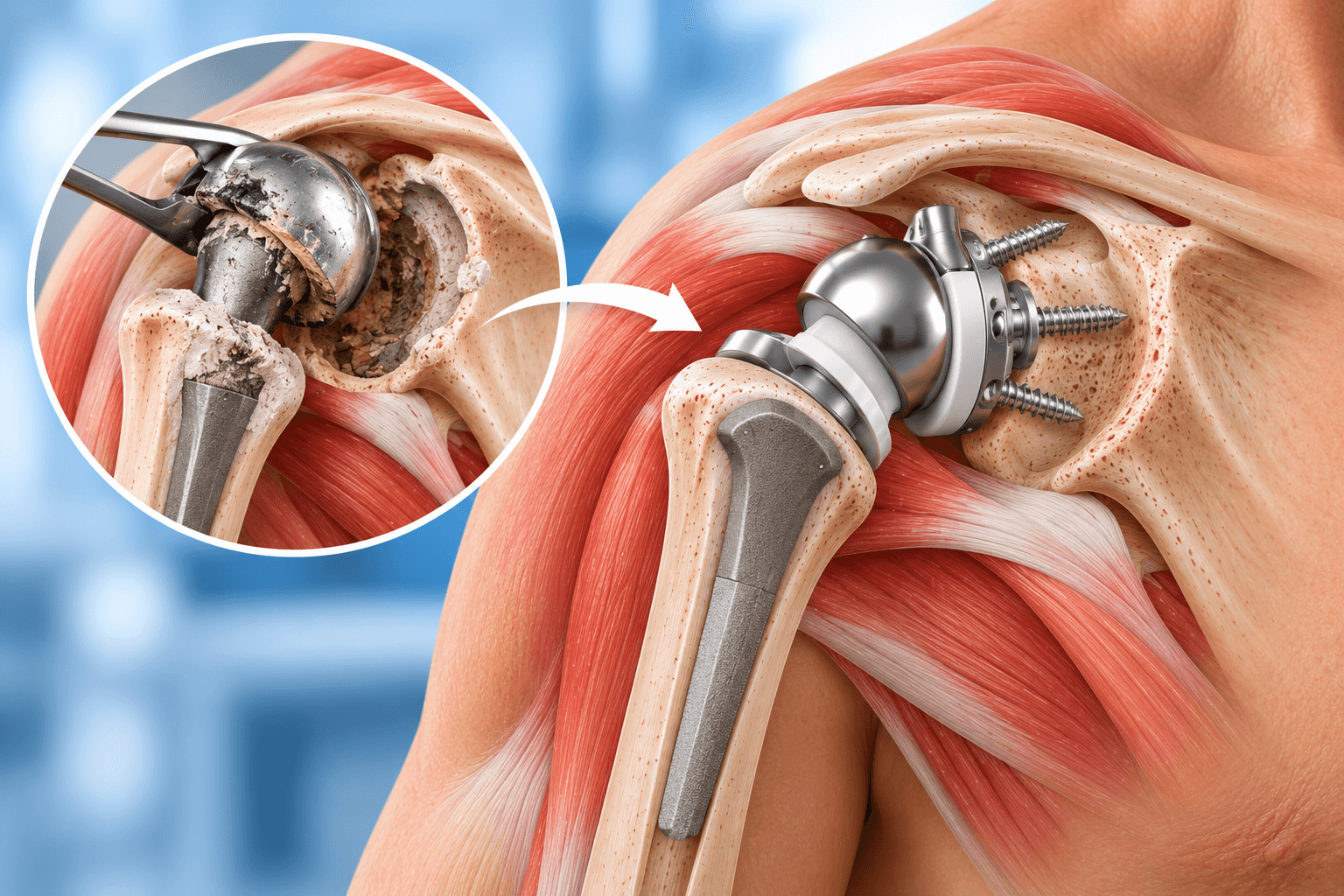
- 1Revision Shoulder Replacement is surgery to repair or replace a previous shoulder prosthesis that has failed, become painful or developed a major complication.
- 2Common reasons include component loosening, instability or dislocation, rotator cuff failure, infection, glenoid erosion, periprosthetic fracture, implant wear and severe bone loss.
- 3A failed anatomic shoulder replacement is frequently converted to a reverse shoulder replacement when rotator cuff failure, instability or complex glenoid problems are present.
- 4A failed hemiarthroplasty can also be converted to an anatomic or reverse replacement depending on rotator cuff function and glenoid condition.
- 5A failed reverse shoulder replacement can be revised, but rerevision is particularly complex because glenoid bone, humeral bone and soft tissues may already be deficient.
- 6Shoulder replacement infection can be subtle. Cutibacterium acnes is an especially important organism in shoulder periprosthetic joint infection and may cause pain and loosening without dramatic fever or redness.
- 7Infection reconstruction may involve one-stage revision, where implants are removed and replaced during one operation, or two-stage revision, where an antibiotic spacer is used before later reimplantation.
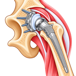
- 8Revision surgery often requires more advanced implants, including long humeral stems, augmented glenoid baseplates, bone grafts, revision glenospheres and occasionally patient-specific components.
- 9Revision surgery usually takes longer and has a higher risk of complications than primary shoulder replacement because scar tissue, bone loss, previous implants and soft-tissue damage make reconstruction more difficult.
- 10Patients can still obtain substantial pain relief and functional improvement after successful revision, but outcomes are generally less predictable than after uncomplicated primary shoulder replacement.
- 11Instability, fracture, infection and recurrent implant problems are particularly important risks after revision reverse shoulder arthroplasty.
- 12Rehabilitation must be individualized. A straightforward component exchange can recover differently from a revision requiring structural bone graft or treatment of infection.
- 13A painful shoulder replacement should not automatically be revised. The surgeon should first determine whether the pain comes from the implant, infection, rotator cuff failure, fracture, stiffness, nerve disease or another diagnosis.
Overview
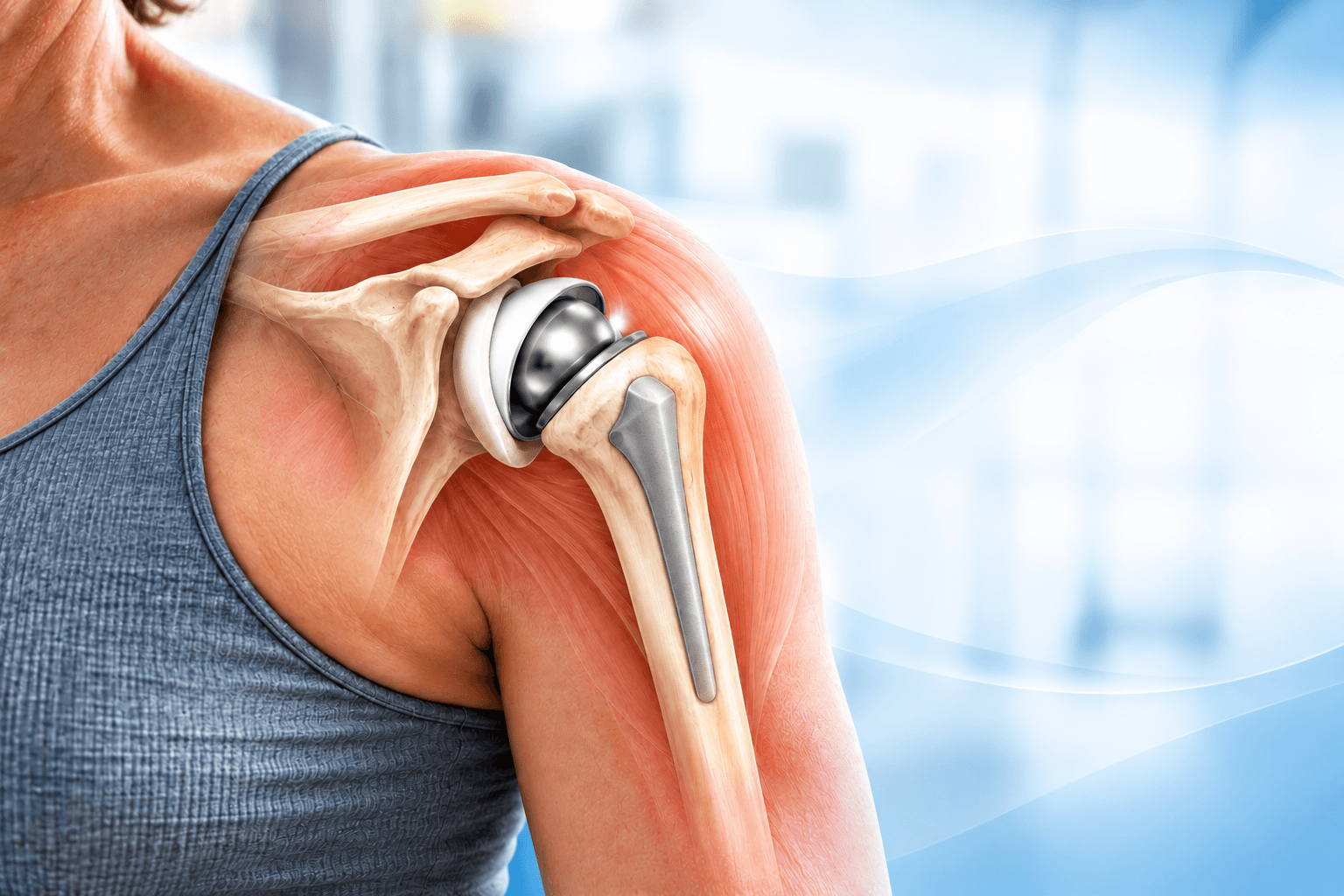
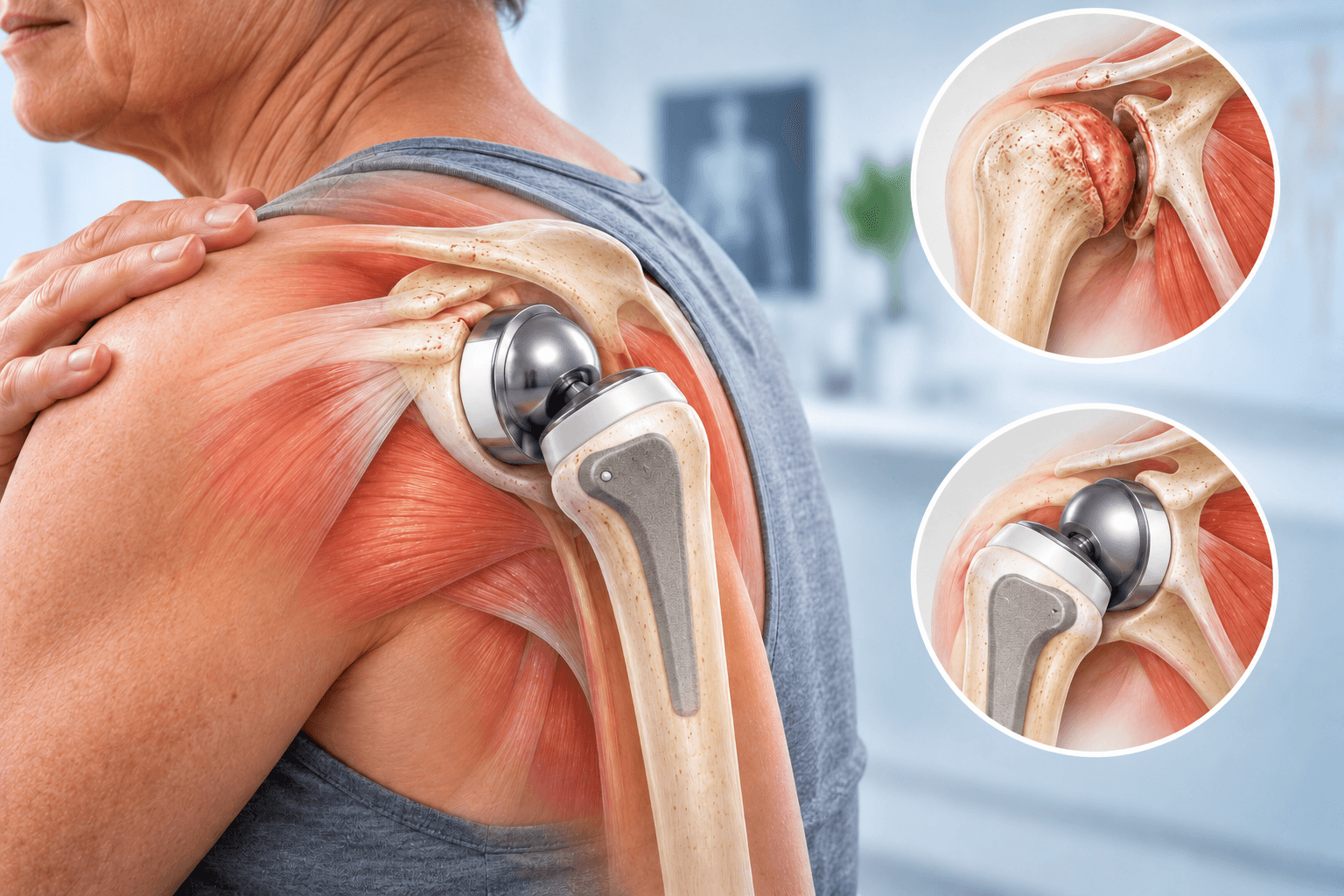
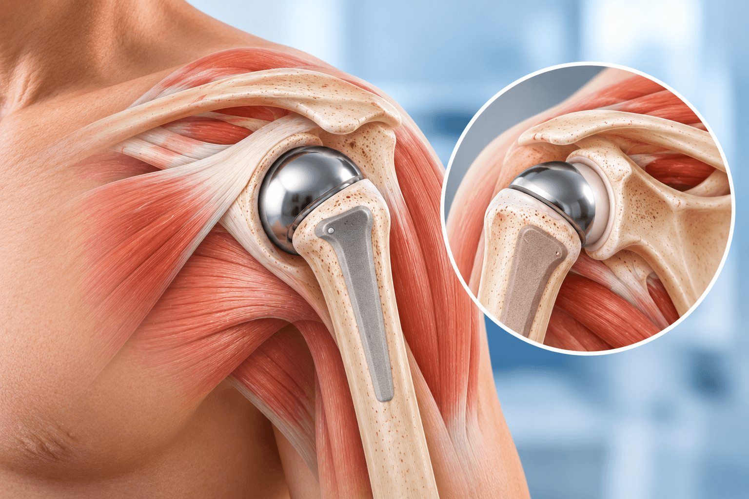
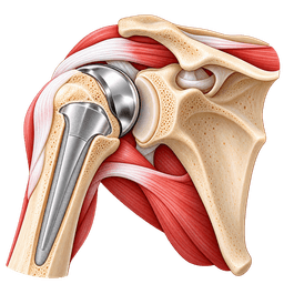
Revision Shoulder Replacement is an operation performed when a previous shoulder replacement needs to be repaired, removed, exchanged or reconstructed. The procedure can involve one implant component or the entire shoulder prosthesis.
A primary shoulder replacement is performed in a shoulder that has not previously undergone arthroplasty. Revision surgery enters a shoulder that already contains implants, scar tissue and altered anatomy. This distinction is important because removing a prosthesis can damage bone, previous surgery can compromise the rotator cuff and deltoid, and the surgeon may have much less native tissue available for reconstruction.
Some revisions are relatively contained. For example, an unstable reverse shoulder can occasionally be treated by exchanging a polyethylene insert or adjusting modular components while leaving well-fixed implants intact. Other revisions require removal of a deeply fixed humeral stem, reconstruction of major glenoid bone loss and implantation of a completely new prosthetic system.
The phrase Revision Shoulder Replacement therefore refers to a broad family of reconstructive procedures rather than one identical operation.
Why Would a Shoulder Replacement Need Revision?
Shoulder replacements can fail for mechanical, biological or infectious reasons.
Mechanical failure can include loosening, instability, dislocation, implant wear, fracture around the prosthesis or breakage of a component. Biological failure can include progressive rotator cuff deterioration, bone loss and poor integration of an implant. Infection can occur early after surgery or present years later.
The surgeon's first task is to identify the dominant cause of failure because revision strategies differ dramatically. Treating an infected implant as though it were simply loose can lead to persistent infection, while treating instability without correcting the underlying mechanical imbalance can lead to repeated dislocation.
Is Pain Alone Enough to Diagnose a Failed Shoulder Replacement?
No.
Pain deserves investigation, but it does not prove that the implant itself requires revision.
Shoulder pain after arthroplasty can arise from infection, rotator cuff disease, acromial stress fracture, scapular spine fracture, component loosening, instability, stiffness, nerve compression, cervical spine disease or muscular problems.
The timing and character of pain provide useful clues. Pain with every movement can suggest mechanical problems, whereas persistent unexplained pain and stiffness can raise concern for infection. Sudden pain after trauma can suggest fracture or dislocation.
Revision should therefore follow a diagnosis rather than simply treating an X-ray or a painful shoulder.
Types of Previous Shoulder Replacement That Can Be Revised
Revision surgery can be performed after:
Hemiarthroplasty
Humeral-head resurfacing
Anatomic total shoulder replacement
Reverse shoulder replacement
Previous revision shoulder replacement
Fracture hemiarthroplasty
Shoulder antibiotic spacer or temporary reconstruction
Each starting point creates different challenges.
A patient with a failed anatomic total shoulder replacement may still have good humeral bone but substantial glenoid damage. A patient with a failed reverse replacement can have baseplate loosening and severe scapular bone loss. A failed fracture hemiarthroplasty can be complicated by tuberosity malunion, cuff deficiency and abnormal humeral anatomy.
Revision of an Anatomic Total Shoulder Replacement
Anatomic total shoulder replacement relies on a functioning rotator cuff and appropriate prosthetic alignment.
Common reasons for revision include rotator cuff failure, glenoid component loosening, instability, infection and periprosthetic fracture.
Rotator cuff failure is particularly important. Once the cuff can no longer center the prosthetic humeral head, an anatomic replacement can migrate upward and become painful or unstable.
Conversion to reverse shoulder replacement has therefore become one of the most important modern solutions for failed anatomic arthroplasty.
The reverse design allows the deltoid to become the primary driver of arm elevation and is less dependent on a normal rotator cuff.
Revision of a Hemiarthroplasty
A hemiarthroplasty replaces the humeral head while preserving the natural glenoid.
Long-term failure can occur when the native glenoid progressively erodes.
Patients can develop deep joint pain and stiffness despite a well-fixed humeral implant.
Rotator cuff failure can also compromise shoulder mechanics.
If the cuff remains healthy and glenoid bone is adequate, conversion to an anatomic total replacement may occasionally be considered. When cuff deficiency, complex deformity or instability is present, conversion to reverse shoulder arthroplasty is frequently more predictable.
Revision of a Reverse Shoulder Replacement
Revision of a failed reverse shoulder is one of the most technically challenging forms of shoulder arthroplasty.
The reverse implant already represents a reconstruction designed to compensate for deficient cuff function. When it fails, the surgeon may face severe problems involving bone, soft tissue and fixation simultaneously.
Reasons include instability, baseplate loosening, infection, periprosthetic fracture, polyethylene wear, glenosphere or component problems and extensive bone loss.
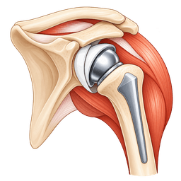
The surgeon can often revise a reverse replacement to another reverse construct, but maintaining secure glenoid fixation and restoring appropriate deltoid tension can be difficult.
Can a Reverse Shoulder Replacement Be Revised?
Yes.
A reverse shoulder replacement can be revised by exchanging one or more components or reconstructing the entire prosthesis.
The exact operation depends on the failure mechanism. Recurrent instability may require changes in liner thickness, glenosphere size, component position or lateralization. A loose baseplate may require revision fixation, bone graft or an augmented component. Infection can require complete implant exchange.
Rerevision is possible even after previous revision surgery, but risks increase as bone and soft tissues become progressively compromised.
Why Does Revision Surgery Become Harder After Each Previous Operation?
Every operation can alter anatomy.
Scar tissue forms around nerves, vessels and muscles. Bone can be removed during implant extraction. Previous screw holes reduce available fixation. Repeated surgical approaches can weaken the subscapularis and other soft tissues.
Infection can further destroy bone and soft tissue.
The surgeon therefore has fewer reconstructive options with each additional operation.
This does not mean successful rerevision is impossible, but expectations and complication risks need to be discussed realistically.
Shoulder Implant Loosening
Loosening means a component has lost stable fixation to bone.
A glenoid component can loosen in an anatomic replacement. A reverse baseplate can loosen from the scapula. A humeral stem can also become loose.
Patients can develop progressive pain, often during movement.
Radiographs can show migration, radiolucent lines or changes in implant position.
CT can provide more detailed evaluation, particularly around the glenoid.
Painful mechanical loosening often requires revision because a loose implant rarely becomes permanently stable again on its own.
Glenoid Component Loosening
Glenoid loosening is an important cause of failure after anatomic total shoulder arthroplasty.
The polyethylene socket can progressively lose fixation.
Bone around the component can erode.
During revision, the surgeon removes the failed component and assesses the remaining glenoid.
If adequate bone remains, a new component can sometimes be implanted. However, significant cuff failure or bone loss often makes conversion to reverse replacement more appropriate.
Reverse Baseplate Loosening
The reverse baseplate supports the glenosphere and must remain rigidly fixed to the scapula.
Loosening can occur because of inadequate initial fixation, bone loss, infection or long-term mechanical stress.
Revision requires identification of the reason the baseplate failed.
Simply placing another baseplate into poor bone without reconstructing the defect can lead to repeated failure.
Augmented baseplates, bone graft, longer central fixation, specialized screws or custom components can be required.
Humeral Stem Loosening
The humeral component can loosen whether it is cemented or cementless.
Patients often report pain with arm use or rotation.
A loose stem can usually be removed more easily than a well-fixed stem, although bone loss may already be present.
Revision can require a longer stem that bypasses deficient bone and obtains fixation farther down the humerus.
If infection caused the loosening, antimicrobial treatment becomes part of the revision strategy.
Rotator Cuff Failure After Anatomic Shoulder Replacement
The rotator cuff can deteriorate years after an apparently successful anatomic replacement.
The humeral head then loses its normal muscular balance.
It can migrate upward, placing abnormal stress on the glenoid and causing pain, weakness and instability.
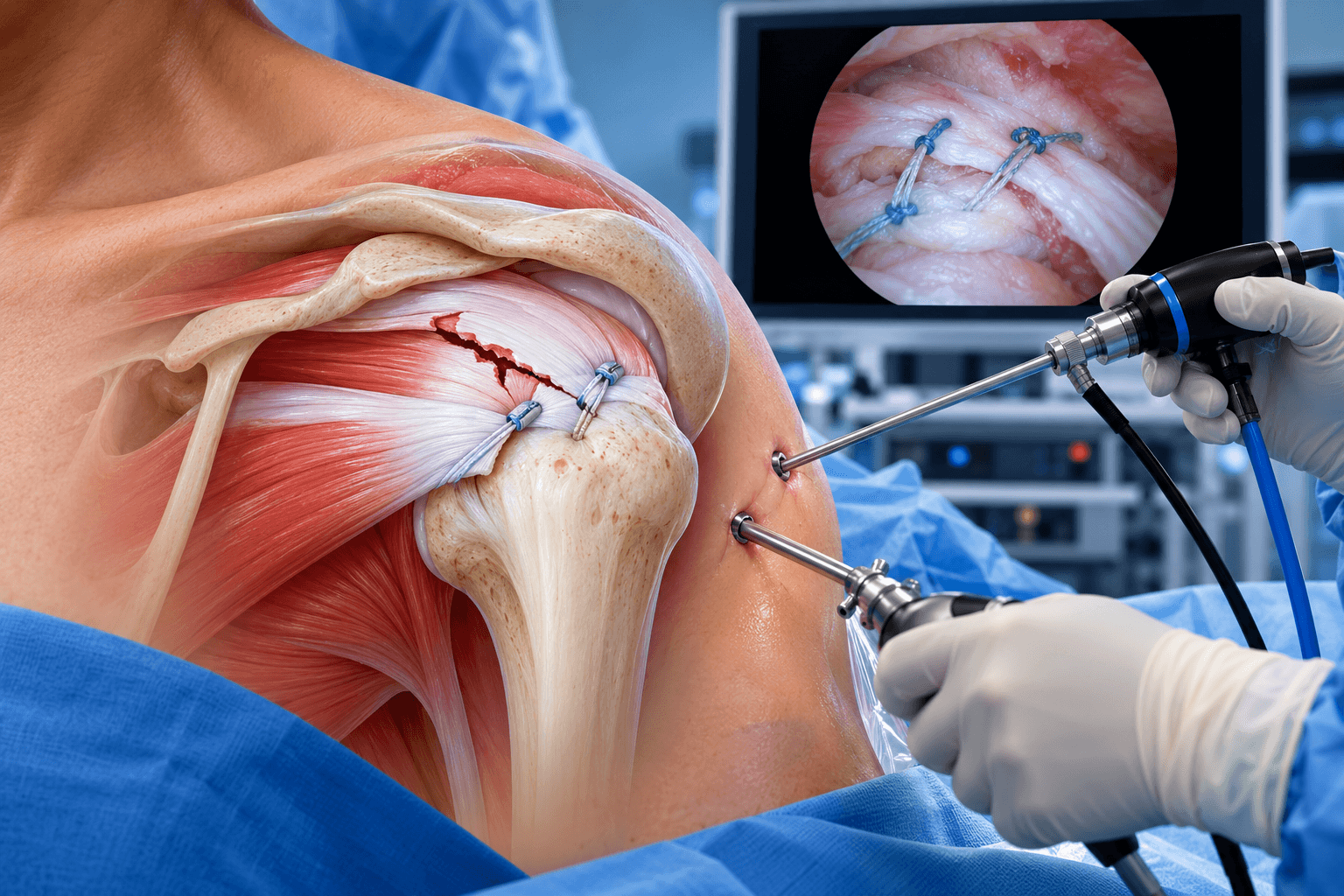
Repairing the cuff alone can be possible in selected acute tears, but chronic degenerative failure around a shoulder prosthesis is often difficult to repair reliably.
Conversion to reverse shoulder replacement provides a way to restore elevation without requiring a fully functional cuff.
Instability After Shoulder Replacement
Instability means the artificial joint moves abnormally or dislocates.
After an anatomic replacement, instability can result from cuff or subscapularis failure, component malposition, soft-tissue imbalance or bone deficiency.
After reverse replacement, instability can reflect inadequate deltoid tension, inappropriate component position, impingement, soft-tissue deficiency or implant design.
The surgeon should identify the mechanical cause before choosing revision components.
Simply inserting a thicker liner can occasionally solve a tension problem but will not correct severe malposition or infection.
Recurrent Dislocation of a Reverse Shoulder Replacement
Repeated reverse shoulder dislocation is a major revision indication.
The surgeon evaluates whether the humerus has become too short, whether the glenosphere is appropriately positioned and whether soft tissues can provide stability.
Component orientation is assessed with X-rays and often CT.
Revision options include increasing humeral tension, changing the liner, altering tray position, using a larger or more lateralized glenosphere or revising malpositioned components.
Every strategy changes shoulder forces, so excessive tension must also be avoided.
Infection After Shoulder Replacement
Periprosthetic joint infection is one of the most serious causes of revision.
Shoulder infection can be particularly difficult to diagnose because it may not present with obvious fever, redness or drainage.
Patients can have unexplained pain, stiffness or gradual loosening.
Cutibacterium acnes is especially important in shoulder arthroplasty infection. It is a low-virulence skin organism that can grow slowly in culture and can produce subtle clinical symptoms.
A negative routine blood test therefore does not completely exclude infection.
Cutibacterium Acnes and Shoulder Replacement
Cutibacterium acnes normally lives in skin follicles, particularly around the shoulder and upper trunk.
It can contaminate or infect implants.
Because it grows slowly and can be difficult to distinguish from contamination, diagnosis requires careful interpretation of multiple tissue cultures and the overall clinical picture.
Laboratories often need adequate culture incubation.
The surgeon should therefore investigate infection systematically before revising a painful unexplained shoulder implant.
Early vs Chronic Infection
An acute infection occurring soon after surgery can sometimes be treated differently from a chronic infection that has existed for months or years.
In selected acute infections where the implants remain stable, debridement with antibiotics and retention of components can sometimes be considered.
Chronic infection more often requires implant removal.
The appropriate treatment depends on organism, implant stability, soft-tissue condition, duration of symptoms and patient health.
One-Stage Revision for Shoulder Infection
In a one-stage exchange, the surgeon removes the infected prosthesis, performs extensive debridement and implants a new prosthesis during the same operation.
This avoids a second major reconstruction.
Modern systematic reviews suggest that carefully selected one-stage shoulder infection revisions can achieve good infection-control outcomes.
However, patient selection matters.
The organism, available antibiotics, bone loss and soft tissues all influence whether one-stage exchange is appropriate.
Two-Stage Revision for Shoulder Infection
Two-stage revision separates infection treatment and definitive reconstruction.
During the first operation, implants and infected tissue are removed.
An antibiotic-loaded spacer is often inserted.
The patient receives antimicrobial treatment and is observed for evidence of infection control.
A second operation later removes the spacer and implants the definitive shoulder replacement.
This strategy remains particularly useful when infection is difficult to characterize, bone or soft tissues need staged management or the treating team believes immediate reimplantation carries excessive risk.
Is One-Stage or Two-Stage Revision Better?
There is no universal answer.
Recent systematic reviews increasingly show that one-stage revision can achieve infection control comparable to, and in some datasets better than, two-stage treatment.
However, available studies are heterogeneous and often affected by selection bias.
Patients selected for one-stage surgery can have different organisms and bone conditions from those selected for two-stage surgery.
Treatment should therefore be individualized rather than declaring one strategy universally superior.
Antibiotic Spacer
An antibiotic spacer is a temporary implant made from antibiotic-loaded bone cement.
It occupies the joint space after infected components are removed.
The spacer can deliver high local antibiotic concentrations while maintaining some shoulder length and soft-tissue tension.
Some spacers articulate enough to permit limited function.
In certain medically fragile patients, a spacer can be retained long term, although this is generally considered a salvage strategy rather than a normal definitive shoulder replacement.
Periprosthetic Fracture
A periprosthetic fracture is a break in the bone around a shoulder implant.
It can involve the humerus, glenoid, acromion or scapular spine depending on prosthesis type and trauma.
Humeral fractures around a stem are particularly relevant to revision arthroplasty.
Treatment depends on whether the implant remains stable.
A stable stem with a fracture around it may be treated with fixation, while a loose stem often needs revision to a longer component that bypasses the fracture.
Intraoperative Fracture During Revision
Removing a well-fixed humeral stem can create a fracture.
The surgeon may need to perform a controlled humeral osteotomy to remove the implant.
This is not necessarily a surgical error; it can be a deliberate technique when cement or porous fixation makes extraction otherwise destructive.
The humerus is then repaired with cables, sutures, plates or a longer revision stem.
Preoperative planning includes having fracture-fixation equipment available.
Glenoid Bone Loss
Loss of glenoid bone is one of the central challenges of Revision Shoulder Replacement.
Previous component loosening can create cavitary or segmental defects.
Screws from a reverse baseplate can leave additional holes.
Infection can further reduce bone stock.
Successful reconstruction requires enough stable scapular bone to support a new baseplate.
Bone graft, metal augments and custom components can expand what is reconstructable.
Humeral Bone Loss
Humeral bone can be lost during loosening, fracture, infection or previous implant removal.
A revision stem can obtain fixation farther down the humeral canal.
Severe proximal deficiency can require allograft-prosthetic composites or specialized tumor-style components in extraordinary cases.
Preserving existing well-fixed components when compatible with the revision plan can therefore be valuable.
Failed Hemiarthroplasty With Glenoid Erosion
A shoulder hemiarthroplasty can remain firmly fixed while the native glenoid wears progressively.
This creates deep pain during movement.
If the cuff remains healthy and glenoid bone is adequate, the surgeon may consider adding a glenoid component and converting to an anatomic total shoulder.
However, chronic cuff deficiency and deformity are common in older failed hemiarthroplasties.
Conversion to reverse shoulder replacement often becomes the more reliable revision solution.
Fracture Hemiarthroplasty Failure
Older fracture hemiarthroplasties can fail because the greater tuberosity does not heal or heals in the wrong position.
The rotator cuff then loses its effective attachment.
The humeral implant can be technically stable while the shoulder remains weak and painful.
Reverse shoulder replacement can bypass much of the dependence on tuberosity and cuff function.
Revision may still be complicated by scar tissue and humeral deformity.
Painful Shoulder Replacement With Normal X-Rays
Normal standard radiographs do not exclude a clinically important problem.
Low-grade infection, early loosening, rotator cuff failure or nerve problems can exist without dramatic X-ray changes.
CT, ultrasound or aspiration can provide additional information.
Nuclear imaging is occasionally considered in difficult cases but does not automatically distinguish infection from mechanical loosening.
A specialist should interpret all tests within the clinical context.
Metal Allergy and Shoulder Revision
True clinically significant implant hypersensitivity is uncommon and difficult to prove.
Pain after arthroplasty should first be evaluated for more common causes such as infection, loosening, instability and cuff failure.
Patch or blood testing alone does not always establish that an implant is causing symptoms.
When a well-supported allergy concern exists, alternative implant materials can sometimes be considered during revision.
Shoulder Replacement Wear
Polyethylene components can gradually wear.
Microscopic particles can contribute to inflammatory bone loss in some prosthetic systems.
Wear is particularly relevant when component position creates abnormal contact.
A worn liner can occasionally be exchanged while stable metal components remain.
However, the surgeon needs to determine whether wear has already produced substantial bone damage.
Implant Breakage
Modern shoulder components rarely fracture, but mechanical failure can occur.
Screws, trays, glenoid components or other modular junctions can fail.
Revision requires removal of broken material and investigation of why the failure occurred.
If bone loss or implant malposition caused excessive stress, simply replacing the broken part without correcting the underlying problem is unlikely to be durable.
Scapular Notching After Reverse Replacement
Scapular notching is bone erosion along the inferior scapular neck caused by contact with the humeral component.
Mild notching often remains an imaging finding and does not require revision.
Severe progressive notching can contribute to bone loss and mechanical concerns.
Revision is generally considered only when notching forms part of a clinically significant failure rather than because it appears on an X-ray.
Acromial and Scapular Spine Fractures
Reverse shoulder replacement changes deltoid loading.
The acromion or scapular spine can develop stress fractures.
Many are initially treated without surgery.
Nonunion or severe displacement can compromise deltoid function and cause persistent pain.
Revision of the shoulder prosthesis itself is not always required, although implant tension and mechanical factors must be evaluated.
Revision After Failed Rotator Cuff Repair and Shoulder Arthroplasty
Some patients undergo multiple previous cuff repairs before eventually receiving shoulder replacement.
Scar tissue, muscle fatty degeneration and altered tendon anatomy can affect later arthroplasty outcomes.
When an anatomic replacement fails in this setting, conversion to reverse reconstruction can often improve function.
The surgeon should still counsel that repeated previous surgery can reduce the predictability of the final result.
Who it's for
- Painful loosening of a glenoid component after anatomic total shoulder replacement
- Loosening of a reverse shoulder baseplate
- Painful humeral stem loosening
- Rotator cuff failure after anatomic total shoulder arthroplasty
- Subscapularis failure associated with instability
- Recurrent shoulder replacement dislocation or instability
- Periprosthetic shoulder infection
- Chronic Cutibacterium acnes infection associated with painful arthroplasty
- Periprosthetic humeral fracture with an unstable prosthesis
- Certain fractures around a stable prosthesis requiring fixation with or without component revision
- Failed shoulder hemiarthroplasty with painful glenoid erosion
- Failed fracture hemiarthroplasty with tuberosity failure or cuff deficiency
- Failed shoulder resurfacing
- Mechanical failure or breakage of prosthetic components
- Polyethylene wear associated with symptomatic implant failure
- Severe glenoid bone loss around a failed component
- Severe humeral bone loss around a failed stem
- Recurrent instability after reverse shoulder replacement
- Failed reverse shoulder replacement requiring rerevision
- Component malposition causing pain, impingement or instability
- Painful arthroplasty where a clear surgically correctable cause has been identified
- Selected cases of prosthetic stiffness where mechanical obstruction or implant-related pathology cannot be managed nonoperatively
Good candidates
A good candidate has a clearly identified problem that revision surgery can realistically correct.
This distinction is important because revision does not reliably solve unexplained pain when investigations show stable components and no infection, fracture or mechanical failure.
The surgeon weighs symptom severity against the complexity of reconstruction.
A patient with mild pain but an extremely difficult extraction of a stable prosthesis can face more risk than benefit.
Conversely, progressive loosening, repeated dislocation or established infection generally creates a stronger reason for surgery.
Patients With Failed Anatomic Shoulder Replacement
Patients with anatomic replacements often require revision because of rotator cuff failure, glenoid loosening or instability.
Conversion to reverse replacement is especially useful when cuff function has been lost.
The surgeon evaluates the humeral stem carefully.
If the stem is well positioned and compatible with a convertible system, it can sometimes remain in place.
This can significantly reduce operative trauma.
Patients With Failed Reverse Shoulder Replacement
These patients require especially specialized evaluation.
The surgeon assesses glenoid fixation, bone loss, humeral fixation, deltoid function, component position and infection status.
Revision can still provide substantial improvement, but complication and rerevision risks are higher than in primary reverse replacement.
Patients should receive realistic counseling before undergoing another reconstruction.
Patients With Infection
Revision should be managed by a team experienced with periprosthetic shoulder infection.
Infectious-disease input can be useful.
Culture strategy, antimicrobial selection and implant plan should be coordinated.
A patient with infection should understand that the treatment priority is eradication of infection and preservation of useful function rather than simply replacing the implant as quickly as possible.
Patients With Major Bone Loss
Bone loss does not automatically make revision impossible.
Modern augmented baseplates, structural grafts, long stems and patient-specific reconstruction have expanded treatment options.
However, severe deficiency increases surgical complexity.
The surgeon should have access to multiple implant systems and backup strategies.
Complex reconstruction is best managed in a center familiar with shoulder revision rather than a facility that performs only occasional primary arthroplasty.
Older Patients
Advanced age alone does not prevent revision.
Painful instability or infection can significantly impair independence.
The decision should consider frailty, medical risk, bone quality and rehabilitation potential.
Sometimes a simpler salvage operation is more appropriate than an elaborate reconstruction when medical risk is high.
The surgical objective should match the patient's overall health and functional priorities.
Younger Patients
Younger revision patients face a different challenge because they may need the reconstruction to function for decades.
Preserving bone becomes particularly important.
Convertible implants and strategies that avoid unnecessary component removal can be valuable.
The patient should understand that each revision can make a future operation more difficult.
Long-term activity recommendations therefore matter.
Before surgery
Determining Why the Previous Replacement Failed
The most important part of revision surgery occurs before entering the operating room.
The surgeon needs to identify why the previous implant failed.
A complete evaluation includes history, examination, radiographs and frequently CT imaging.
Infection must be considered even when there are no obvious external signs.
Previous operative reports and implant records are extremely valuable because they reveal component sizes, fixation methods and whether existing parts are compatible with revision systems.
Understanding the Pain Pattern
Pain from loosening often occurs with movement or loading.
Infection can produce constant aching, stiffness or unexplained deterioration.
Instability can cause a feeling that the shoulder slips or clunks.
Fracture can cause sudden pain after trauma.
Cervical nerve disease can create arm pain that mimics a shoulder problem.
A careful history helps prevent unnecessary revision for pain that is not actually coming from the prosthesis.
Physical Examination
The surgeon evaluates the scar, swelling, deltoid, rotator cuff and neurological function.
Active and passive motion are compared.
Instability is assessed carefully rather than aggressively.
The surgeon examines the neck and peripheral nerves because neurological symptoms can coexist with arthroplasty problems.
Skin quality is also important because multiple previous incisions can complicate revision exposure.
Standard X-Rays
Radiographs are the initial imaging study.
They show component position, loosening, migration, fractures and bone loss.
Previous images are particularly valuable because progression over time can be more informative than one X-ray.
The surgeon looks for changes in glenoid position, humeral subsidence and evidence of scapular notching or erosion.
CT Scan
CT is especially useful for revision planning.
It defines three-dimensional glenoid bone loss, screw location and component orientation.
It can show whether enough scapular bone remains for a new baseplate.
Three-dimensional reconstruction can help plan augments or custom implants.
CT can also assess humeral bone around the stem.
MRI
Metal creates artifact on MRI, but modern metal-artifact reduction techniques can sometimes provide useful information.
MRI may help evaluate surrounding soft tissues and cuff integrity.
Ultrasound is often an alternative for evaluating rotator cuff tissue around a prosthesis.
The best imaging test depends on the question the surgeon needs to answer.
Ultrasound
Ultrasound can evaluate rotator cuff tendons dynamically without major metal artifact.
It can demonstrate tendon failure around an anatomic shoulder replacement.
It is also relatively accessible.
Operator expertise matters.
Ultrasound does not provide the same detailed assessment of glenoid bone that CT provides.
Infection Blood Tests
C-reactive protein and erythrocyte sedimentation rate can contribute to infection evaluation.
Normal results do not reliably exclude low-grade shoulder infection.
This is especially important with Cutibacterium acnes.
The surgeon therefore combines blood results with aspiration, imaging, clinical findings and intraoperative cultures rather than relying on one laboratory number.
Joint Aspiration
Aspiration can provide synovial fluid for cell count and culture.
A positive culture can be highly useful.
A negative aspiration does not completely exclude infection because shoulder organisms can be difficult to recover.
The surgeon determines whether aspiration is necessary according to the clinical level of suspicion.
Antibiotics taken before sampling can also reduce culture yield.
Tissue Cultures
When infection is suspected during revision, multiple deep tissue samples are obtained.
Sampling several sites improves diagnostic confidence.
The laboratory may need prolonged incubation for slow-growing organisms.
Antibiotics are coordinated carefully so they do not unnecessarily compromise culture accuracy when the patient is medically stable.
The exact protocol follows the treating institution's infection strategy.
Reviewing the Previous Implant
Knowing the manufacturer and exact implant model can simplify revision dramatically.
Some modern systems are convertible.
A well-fixed humeral stem can sometimes accept new modular components and be converted from an anatomic to reverse configuration without stem removal.
If the system is unknown or incompatible, a more extensive revision can become necessary.
Patients should therefore retain implant documentation whenever possible.
Previous Operative Reports
The previous surgical report reveals whether the subscapularis was repaired, whether bone graft was used and how components were fixed.
It can identify previous complications.
For revision surgery abroad, obtaining the operative report before travel can save significant diagnostic uncertainty.
Even photographs or implant stickers from the original procedure can be useful.
Medical Optimization
Revision surgery often lasts longer and can involve greater blood loss than primary replacement.
Heart, lung and kidney conditions should therefore be optimized.
Anemia is identified and treated where appropriate.
Diabetes control is important.
The anaesthesia team assesses whether additional monitoring or postoperative observation is necessary.
Smoking and Nicotine
Smoking impairs wound and bone healing.
Revision cases already have compromised tissues, making nicotine cessation particularly valuable.
Bone graft incorporation and fracture healing are biological processes that can be negatively affected by smoking.
Patients should ideally stop before surgery and remain nicotine-free during recovery.
Nutrition
Revision patients can have prolonged pain and decreased activity before surgery.
Poor nutrition can impair wound healing and immune function.
Adequate protein and correction of major nutritional deficiencies are important.
Complex infected revisions sometimes benefit from formal nutritional assessment.
Anticoagulants
Blood-thinning medication requires a coordinated plan.
Revision surgery can involve more bleeding than primary arthroplasty.
At the same time, stopping anticoagulation can create dangerous clotting risk.
Patients should never change these medications independently.
Planning for Blood Loss
Major extraction and reconstruction can cause substantial blood loss.
Preoperative hemoglobin is therefore important.
Some centers use tranexamic acid when appropriate.
Blood products can be available for particularly complex cases.
The need varies widely and should not be presented as routine for every shoulder revision.
Planning for Bone Graft
If CT demonstrates major glenoid deficiency, bone graft may be required.
The surgeon determines whether autograft from the patient, structural allograft or another strategy is likely.
Revision centers should have the necessary graft available before beginning the operation.
A case should not depend on improvisation after the defect is discovered.
Planning for Custom Implants
Extremely severe glenoid loss can require custom reconstruction.
CT data are used to manufacture a component designed for the remaining scapular anatomy.
Production takes time.
Custom implants therefore require considerable preoperative planning.
They are not necessary for most revision cases but can make reconstruction possible in situations where standard baseplates cannot achieve stable fixation.
Planning for Infection Revision
If infection is likely, the patient should know whether the surgeon plans one-stage or two-stage treatment.
Antibiotic spacer availability and culture protocols should be established.
The patient should understand that definitive implant selection can change if unexpected infection is discovered during surgery.
This is especially important for international patients who might otherwise expect one fixed package and one operation.
Consent for Alternative Reconstruction
Revision surgery contains uncertainty.
A surgeon may plan to retain a stem but discover that it is loose.
A planned anatomic revision may become reverse reconstruction after finding severe cuff failure.
Bone loss may be greater than CT suggested.
Patients should therefore understand the possible alternative procedures before anaesthesia rather than learning afterward that the plan changed.
Planning Rehabilitation
The rehabilitation protocol depends on what is reconstructed.
Component exchange without major soft-tissue disruption can progress relatively quickly.
A humeral osteotomy, structural glenoid graft or periprosthetic fracture requires more protection.
Patients should expect the final postoperative protocol to be determined partly by intraoperative findings.
How the operation is performed
Revision Shoulder Replacement usually begins by exposing the previous prosthesis, identifying which components are stable or failed, obtaining cultures when indicated and then removing or retaining components according to the reconstruction plan. The surgeon repairs bone defects, corrects implant position and builds a new shoulder construct designed to address the specific cause of failure.
Unlike primary replacement, revision frequently requires several possible plans.
The surgeon can begin intending to retain one component but change strategy if fixation is inadequate.
Backup implants, extraction tools and bone-grafting materials should therefore be available.
Anaesthesia
General anaesthesia is commonly combined with a regional block.
Complex operations may require arterial monitoring or other anaesthetic measures depending on medical condition and expected duration.
Pain management is planned before surgery.
Long procedures and previous scar tissue can increase postoperative swelling and discomfort.
Surgical Approach
The surgeon commonly reuses a previous deltopectoral incision when safe.
Scar tissue is dissected carefully.
The cephalic vein, deltoid and neurovascular structures can be more difficult to identify after previous operations.
Revision surgery therefore requires patience.
Rapid aggressive exposure risks nerve, vessel or muscle injury.
Obtaining Cultures
When infection is suspected, cultures are obtained before unnecessary contamination occurs.
Multiple deep tissue samples can be collected from different locations around the implant.
Synovial fluid and membrane tissue may also be sampled.
The surgeon avoids relying on a single superficial specimen.
Results are interpreted with the clinical picture because slow-growing organisms can complicate diagnosis.
Evaluating the Existing Components
The surgeon checks whether the humeral and glenoid components are actually loose.
A component that appears suspicious on imaging can occasionally remain firmly fixed.
Conversely, apparent radiographic stability can hide mechanical failure.
Retaining a well-fixed compatible component can preserve bone and reduce operative trauma.
A loose or infected component generally requires removal.
Removing an Anatomic Glenoid Component
A loose polyethylene glenoid component is carefully removed.
Cement and fibrous tissue are cleared while preserving as much native scapular bone as possible.
The resulting defect is assessed.
If the cuff remains functional and glenoid reconstruction is possible, an anatomic revision can sometimes be considered.
More commonly, severe cuff or bone problems favor conversion to a reverse baseplate.
Removing a Reverse Baseplate
A reverse baseplate can be difficult to remove because screws and porous surfaces may be strongly fixed.
The surgeon removes peripheral screws and disconnects the glenosphere.
Extraction must minimize additional damage to the remaining glenoid.
A failed baseplate often leaves a central cavity and screw holes.
These defects influence the design of the new reconstruction.
Removing a Humeral Head
A modular humeral head can be detached relatively easily.
The taper and underlying stem are inspected.
If a well-fixed stem is compatible with a conversion system, it can sometimes remain.
This is advantageous because stem extraction is often one of the most invasive parts of shoulder revision.
Removing a Well-Fixed Humeral Stem
A well-fixed stem can be difficult to remove without sacrificing bone.
The surgeon first clears tissue around the proximal implant.
Special extraction devices are used.
If necessary, the humerus can be opened through a controlled osteotomy.
This creates access to cement or porous surfaces and allows the stem to be removed with less uncontrolled bone loss.
The osteotomy is repaired afterward.
Humeral Osteotomy
An osteotomy deliberately opens part of the humerus.
It is similar in concept to opening a door around the implant.
After the old stem is removed, the bone is brought back into position.
Cables, sutures or a plate can provide fixation.
The new revision stem generally extends beyond the weakened region to obtain stable fixation.
Removing Cement
Older stems can be surrounded by bone cement.
The cement must sometimes be removed to allow implantation of a new stem.
This requires specialized instruments and careful technique because aggressive cement removal can perforate or fracture the humerus.
In selected circumstances, portions of a stable cement mantle may be retained according to the planned reconstruction.
Revising the Humeral Side
A long revision stem can bypass proximal bone loss.
The surgeon seeks secure fixation in healthy diaphyseal bone.
The component can be cemented or cementless depending on anatomy.
Modular revision systems allow adjustment of height, version and lateralization.
Restoring appropriate humeral length is particularly important for reverse stability and deltoid tension.
Managing Glenoid Bone Loss
The remaining glenoid is assessed after failed components are removed.
Small contained defects may be filled with graft.
Larger segmental deficiencies can require structural bone graft or a metal augment.
The aim is not simply to fill space.
The reconstruction must transmit forces into strong native scapular bone.
A new baseplate must achieve initial mechanical stability before biological fixation can occur.
Structural Bone Grafting
Structural graft can rebuild missing glenoid bone.
Allograft or autograft may be used according to the defect.
The baseplate can compress the graft against native bone.
Successful reconstruction requires graft incorporation over time.
Rehabilitation can therefore be more protective than after a routine reverse replacement.
Augmented Baseplates
Metal augments provide built-in wedges that fill asymmetric glenoid defects.
They can preserve native bone because less corrective reaming is required.
Posterior, superior and other augment configurations are available.
Three-dimensional CT planning helps match the augment to the defect.
Augments are particularly useful when enough native bone remains for secure central and peripheral fixation.
Custom Glenoid Components
Custom components are reserved for exceptionally difficult bone loss.
A CT-based three-dimensional model is used to design an implant that anchors into remaining scapular structures.
The objective is to obtain fixation where standard components cannot.
Custom revision is highly specialized.
It requires careful manufacturing and should be undertaken in experienced revision centers.
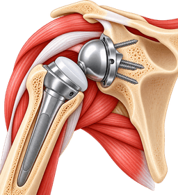
Conversion From Anatomic to Reverse Shoulder Replacement
This is one of the most common modern revision pathways.
The failed anatomic glenoid component is removed.
The glenoid is reconstructed and a reverse baseplate and glenosphere are implanted.
The humeral side is converted to a socket.
If the original stem is convertible and well positioned, it may be retained.
Otherwise, the humeral stem is revised.
This conversion is particularly effective when rotator cuff failure caused the original arthroplasty to fail.
Conversion From Hemiarthroplasty to Reverse Shoulder Replacement
The surgeon removes or converts the humeral prosthesis and reconstructs the glenoid with a reverse baseplate.
Scar tissue and tuberosity deformity can make exposure challenging.
A well-fixed humeral stem can sometimes be retained if the system supports conversion.
Otherwise, extraction and replacement with a revision stem are required.
Reverse mechanics reduce dependence on the compromised rotator cuff.
Revision Reverse to Reverse
A failed reverse replacement can often be revised to another reverse construct.
The surgeon changes components according to the problem.
Instability can require a different glenosphere, liner or humeral length.
Baseplate failure can require glenoid reconstruction.
Humeral loosening can require a longer stem.
Severe combined deficiency can require reconstruction on both sides.
Revision for Instability
The surgeon first evaluates implant position.
If components are well positioned but the shoulder lacks sufficient tension, increasing polyethylene thickness or changing humeral components can help.
A larger or more lateralized glenosphere can alter stability and impingement.
If a component is malpositioned, it may need complete revision.
Persistent instability should also trigger consideration of infection because occult infection can contribute to recurrent mechanical failure.
Revision for Infection — Debridement and Implant Retention
In selected acute infections, the surgeon can perform extensive irrigation and debridement while retaining well-fixed implants.
Modular components are often exchanged.
This strategy is not appropriate for every shoulder infection.
Chronic infection, loose implants or compromised tissue generally requires more extensive removal.
Antimicrobial therapy is coordinated with the infection team.
One-Stage Exchange Technique
All components judged infected are removed.
The surgeon performs aggressive debridement of infected tissue and cement.
The field is irrigated and new implants are inserted during the same operation.
Antibiotic strategies are individualized.
The potential advantage is avoiding an interval spacer and second major reconstruction.
Appropriate patient and organism selection remain important.
Two-Stage Exchange Technique
The first stage removes the prosthesis and infected tissue.
An antibiotic-loaded spacer is implanted.
The patient receives treatment and is monitored.
Later, once the infection-control plan has been completed and the shoulder is considered suitable for reconstruction, the spacer is removed and a definitive implant is inserted.
The interval can vary and should not be reduced to one standard number of weeks.
Revision After Antibiotic Spacer
The second-stage procedure can itself be technically difficult.
Bone loss can progress during the infection process.
The surgeon removes the cement spacer and evaluates glenoid and humeral bone.
Cultures can be repeated.
A reverse replacement is commonly used because the cuff and soft tissues can be substantially compromised after infection and multiple operations.
Revision for Periprosthetic Humeral Fracture
If the existing stem is loose, it is removed.
The fracture is reduced and stabilized.
A longer revision stem generally extends beyond the fracture to obtain fixation in intact bone.
Cables, plates and bone graft can be added.
If the existing stem is well fixed, the fracture can sometimes be treated with fixation alone rather than replacing the prosthesis.
Revision for Polyethylene Wear
When metal components remain secure and correctly positioned, modular polyethylene can sometimes be exchanged.
The joint is inspected for metal damage and bone loss.
If wear resulted from impingement or malposition, the underlying mechanical cause must be corrected.
A simple liner exchange is not durable when abnormal contact remains.
Managing Scar Tissue
Revision shoulders often contain dense adhesions.
Scar tissue can tether the humerus, deltoid and neurovascular structures.
Careful release restores exposure and can improve motion.
Excessive release can destabilize the shoulder.
The surgeon balances mobility with preservation of viable soft tissues.
Protecting the Axillary Nerve
The axillary nerve is particularly important in reverse shoulder reconstruction because it powers the deltoid.
Previous surgery and scar tissue can make the nerve more vulnerable.
The surgeon understands its expected location and avoids excessive traction.
In severe deformity or complex previous surgery, nerve identification can become a major part of the operation.
Restoring Deltoid Tension
Reverse shoulder stability depends on appropriate deltoid tension.
Too little length can cause instability.
Too much length can increase nerve stress and acromial loading.
Revision surgery must therefore restore the arm to an appropriate length rather than simply making it as tight as possible.
Trial components are essential for finding this balance.
Trial Reduction
Temporary components are inserted.
The surgeon checks range of motion, stability and impingement.
Different liner thicknesses and glenosphere options can be tested.
The arm should be stable without excessive tension.
Only then are definitive modular components selected.
Closure
Soft tissues are repaired according to what remains viable.
The subscapularis can be repaired in selected reconstructions.
Drains are used selectively.
The incision is closed in layers.
A sling is applied.
Complex infection or revision wounds receive particularly careful postoperative surveillance.
How Long Does Revision Shoulder Replacement Surgery Take?
Revision Shoulder Replacement commonly takes approximately two to four hours, although difficult rerevisions can require substantially longer.
A straightforward modular exchange can be shorter than this.
Removal of a well-fixed cemented stem, reconstruction of major glenoid bone loss or treatment of infection can extend the operation beyond four hours.
The patient should therefore receive a case-specific estimate after the surgeon has reviewed CT imaging and previous operative records.
Hospital stay
Recovery Room
After revision surgery, the patient is monitored closely as anaesthesia wears off.
Pain, circulation, neurological status and wound condition are checked.
The arm can remain numb from an interscalene block.
Complex revision patients can need closer observation than routine primary shoulder replacement because surgery has often been longer.
Typical Hospital Stay
Many revision patients remain in hospital for approximately one to three nights.
A limited revision can occasionally follow an outpatient or one-night pathway.
Major reconstruction, infection surgery, fracture treatment or substantial blood loss can require longer admission.
The goal is safe discharge rather than meeting a predetermined package duration.
Pain Control
Revision surgery can be more painful than primary replacement because of extensive scar dissection, implant extraction and bone reconstruction.
Regional anaesthesia, acetaminophen or paracetamol, anti-inflammatory medication where appropriate and short-term stronger analgesia can be combined.
Pain should gradually improve.
Severe escalating pain accompanied by swelling, neurological change or fever requires assessment.
Postoperative X-Rays
Radiographs are commonly obtained soon after surgery.
They confirm implant position and document fracture fixation or bone graft.
These images also establish a baseline for later comparison.
In complex reconstructions, additional views can be obtained at follow-up as graft and fixation mature.
Sling
A sling is typically used.
Duration depends on the reconstruction.
A limited component exchange can allow relatively early movement.
A humeral osteotomy, fracture repair or structural graft can require longer protection.
The final operative findings therefore determine sling duration more accurately than the preoperative diagnosis alone.
Blood Tests
Blood count and metabolic tests can be checked after extensive surgery.
The team monitors for anemia and other postoperative abnormalities.
In infection cases, antimicrobial therapy can require additional laboratory monitoring.
Antibiotics
Routine prophylactic antibiotics are used around revision surgery according to hospital protocols.
Established infection requires organism-directed therapy.
The duration and route depend on microbiology, surgical strategy and infectious-disease recommendations.
Patients should not assume that every revision requires several weeks of intravenous antibiotics.
Physical and Occupational Therapy
Therapists help patients dress, manage the sling and move safely.
The shoulder protocol is reviewed before discharge.
Patients learn not to push body weight through the operated arm when prohibited.
A written programme is especially important when rehabilitation will continue in another country.
Recovery
Recovery after Revision Shoulder Replacement commonly takes four to seven months for major function, while improvement can continue for six to twelve months or longer.
This is generally slower and less predictable than recovery after uncomplicated primary shoulder replacement.
The reason is not merely that the incision has been used before.
Bone grafts, fracture fixation, osteotomy healing, infection treatment and scar tissue can all influence recovery.
A patient whose revision consists only of a modular exchange can progress much faster than a patient whose humerus and glenoid both required reconstruction.
Revision Shoulder Replacement Protocol
There is no universal protocol.
The therapist needs to know exactly what was revised.
If the humeral stem was removed through an osteotomy, aggressive loading can jeopardize bone healing.
If a structural glenoid graft was used, the surgeon may limit activity while integration occurs.
If only modular reverse components were exchanged and fixation remains strong, motion can progress earlier.
The operative report therefore becomes part of the rehabilitation prescription.
First 24–72 Hours
The patient focuses on pain control, safe walking and protecting the surgical reconstruction.
Finger, wrist and usually elbow movement begins.
The shoulder remains in the sling except for permitted exercises and hygiene.
The patient should not test the strength of the revision simply because the nerve block makes the shoulder painless.
First Two Weeks
The incision begins healing.
Bruising and swelling can be more pronounced than after primary surgery.
The first follow-up checks the wound and radiographs.
If cultures were taken, final microbiology can sometimes become available only after a longer incubation period.
The surgeon may modify antibiotic management if unexpected organisms are identified.
Weeks 2–6
Sling use usually continues to some degree.
Passive or assisted shoulder motion progresses according to the reconstruction.
Patients should avoid lifting, pushing and pulling.
A fracture or osteotomy can require particularly cautious activity.
The objective is controlled mobility without jeopardizing fixation.
Weeks 6–12
Many patients transition toward active movement.
The sling is gradually reduced.
The deltoid and remaining cuff muscles begin working more directly.
Strengthening can start in selected cases.
Major bone reconstructions can remain more protected.
X-rays help determine whether bone healing is progressing.
Months 3–6
Strength and endurance improve.
Patients regain increasing independence with household tasks.
Desk work has usually resumed much earlier, while manual work remains limited.
The shoulder can continue to feel weak and fatigued.
Revision patients should expect a more gradual return than friends who underwent routine primary arthroplasty.
Months 6–12
Maximum improvement often continues throughout this period.
Muscle conditioning and confidence improve.
Complex infection and fracture revisions can take the entire year.
Some permanent loss of range or strength can remain even when pain relief is excellent.
The objective is frequently a stable, useful and comfortable shoulder rather than recreation of normal anatomy.
Recovery After Conversion to Reverse Shoulder Replacement
When an anatomic or hemiarthroplasty is converted to reverse replacement, the deltoid becomes more important.
Forward elevation often improves as the patient adapts to the new mechanics.
Rotation can remain limited when the cuff is extensively damaged.
Rehabilitation therefore emphasizes deltoid and scapular control.
Recovery After Revision of a Failed Reverse Replacement
Rerevision can require a slower protocol.
Bone and soft tissues have already undergone at least two major operations.
Instability risk can be higher.
The patient should avoid comparing their recovery with primary reverse-replacement timelines.
The surgeon can intentionally delay strengthening to protect fixation.
Recovery After Bone Grafting
Bone graft needs time to incorporate.
The shoulder can feel relatively comfortable while the graft remains biologically immature.
The surgeon can therefore restrict active loading even when pain is low.
Serial imaging helps assess integration.
Failure to follow restrictions can compromise baseplate fixation before the graft becomes structurally reliable.
Recovery After Humeral Osteotomy
An osteotomy behaves partly like a controlled fracture.
It needs bone healing.
The patient can therefore have additional lifting restrictions.
Cables or plates provide stability but do not eliminate the biological healing requirement.
Strenuous loading is delayed until radiographs demonstrate satisfactory progress.
Recovery After Infection Revision
The timeline is strongly affected by whether treatment is one-stage or two-stage.
After a one-stage exchange, rehabilitation can proceed around the definitive reconstruction while antibiotic treatment continues.
Two-stage treatment creates a much longer overall journey because the first surgery is followed by a spacer period and then another reconstruction.
Patients should distinguish recovery from each individual operation from the total infection-treatment timeline.
Pain After Revision Shoulder Replacement
Pain is expected during the early weeks.
Scar dissection and implant extraction can cause significant soreness.
Improvement should be progressive.
Persistent or increasing pain can indicate stiffness, infection, instability, fracture or another complication.
Pain that suddenly increases after a period of good progress deserves reassessment.
Sleeping
Many patients initially sleep partially upright.
A recliner or wedge pillows can improve comfort.
The arm is supported in the sling or on pillows according to instructions.
Sleeping directly on the revision shoulder is usually delayed until the incision and underlying tissues tolerate pressure.
Driving
Driving waits until sling use has ended or substantially decreased, sedating medication has stopped and the patient can safely control the car.
Revision patients can require longer than primary arthroplasty patients.
The ability to move the arm comfortably in the clinic does not automatically mean emergency steering is safe.
Desk Work
Selected patients can return to desk duties within several weeks.
A complex revision can delay this.
The arm should be supported.
Remote work can provide flexibility.
Commuting and driving limitations are often more restrictive than keyboard activity itself.
Manual Work
Heavy occupational activity can require six months or longer.
Some patients cannot safely return to repetitive heavy overhead work after major revision reconstruction.
The surgeon should discuss long-term job demands before surgery.
A successful revision can still require permanent work modification to protect limited bone stock and implants.
Lifting
Early lifting is restricted.
Over time, light and moderate daily loads are progressively reintroduced.
Very heavy repetitive lifting can be discouraged permanently, particularly after major glenoid reconstruction or rerevision.
There is no universal weight limit that applies to every revision shoulder.
Physiotherapy
Therapy initially focuses on safe motion.
Strength comes later.
Scarred shoulders can be prone to stiffness, but aggressive stretching must be balanced against fixation and soft-tissue healing.
The therapist should not use a generic primary shoulder-replacement protocol unless the surgeon specifically approves it.
Gym Exercise
Lower-body training can return relatively early when the operated arm remains protected.
Upper-body resistance progresses slowly.
Heavy presses, pull-ups and exercises involving body-weight support through the arm are late-stage activities and may not be recommended after complex revision.
Swimming
Swimming can be possible after adequate healing and strength.
Repetitive overhead activity places substantial demand on the revision shoulder.
The patient should begin with short sessions.
Complex bone-graft or rerevision patients require surgeon clearance before returning to swimming.
Golf
Many patients can eventually return to recreational golf.
Putting and short game activities come first.
Full swings require more rotation and power.
The final decision depends on stability, range of motion and the reason for revision.
Intimacy
Sexual activity can resume when the shoulder can remain protected without supporting body weight.
The arm should not be forced behind the body during early recovery.
A sling can be retained initially.
Comfort and surgical restrictions guide progression.
Flying After Revision Shoulder Replacement
International travel requires careful planning.
The patient should be medically stable, the wound should be satisfactory and early complications should have been excluded.
Luggage should be handled by someone else.
Patients receiving intravenous antibiotics or undergoing staged infection treatment require additional logistical planning.
When to Contact the Surgeon
Increasing wound drainage, fever, progressive redness, sudden severe pain, new deformity, a dislocation sensation or neurological change should be reported promptly.
Persistent unexplained deterioration is also important.
Shoulder infection can be subtle, so patients should not wait for high fever before contacting the team when the overall postoperative course appears abnormal.
Recovery timeline
- Protect the revision, manage pain and establish a safe postoperative routine1Protect the revision, manage pain and establish a safe postoperative routine
Days 0–14
The patient wears the sling, performs approved hand, wrist and elbow motion and begins only the shoulder exercises authorized by the surgeon. Wound surveillance is important. Culture results can still be pending in infection evaluations, and antibiotic plans can change if unexpected organisms are identified.
- Restore controlled mobility while preserving implant, bone and soft-tissue healing.2Restore controlled mobility while preserving implant, bone and soft-tissue healing.
Weeks 2–6
Passive and assisted range of motion gradually progresses. A relatively contained revision can move faster than a case involving osteotomy, fracture fixation or structural graft. Heavy lifting and pushing remain restricted.
- Transition toward active shoulder control.3Transition toward active shoulder control.
Weeks 6–12
The sling is reduced according to the surgeon's plan. Active movement increases, and selected patients begin low-resistance strengthening. Radiographs can be used to assess grafts, fractures and osteotomies before activity is advanced.
- Rebuild practical strength and daily function4Rebuild practical strength and daily function
Months 3–6
The patient progresses through functional strengthening, light work and recreational activity. Deltoid and scapular conditioning are particularly important after conversion to reverse arthroplasty. Heavy overhead loading remains limited.
- Achieve mature revision function and establish sustainable long-term activity.5Achieve mature revision function and establish sustainable long-term activity.
Months 6–12+
Strength and confidence continue improving. Final range of motion and lifting recommendations become clearer. Complex rerevision, infection and major bone-loss reconstruction can continue improving beyond one year.
Outcomes and success rates
How Successful Is Revision Shoulder Replacement?
Revision shoulder arthroplasty can provide meaningful pain relief and improved function, but it is less predictable than primary shoulder replacement.
Outcome depends on why the previous implant failed.
A patient revised for isolated component loosening with preserved bone and good soft tissues generally has a different prognosis from someone undergoing a third operation after infection and major bone loss.
The surgeon should therefore avoid quoting one universal success rate.
Revision of Failed Anatomic Replacement to Reverse Replacement
Conversion to reverse shoulder arthroplasty is one of the best-established revision strategies.
Modern systematic reviews show substantial improvements in pain, forward elevation and patient-reported shoulder function.
The procedure is particularly valuable when rotator cuff failure caused the original anatomic replacement to fail.
However, complication rates remain higher than after primary reverse shoulder replacement.
Revision of Failed Hemiarthroplasty to Reverse Replacement
Patients with painful glenoid erosion, tuberosity failure or cuff deficiency can improve after conversion to reverse arthroplasty.
Forward elevation and pain commonly improve.
Revision remains technically more difficult than primary reverse replacement because bone deformity and scar tissue are frequent.
Existing stems can occasionally be retained when compatible.
Revision of Failed Reverse Shoulder Replacement
A second reverse reconstruction can improve pain and shoulder scores.
The complication and rerevision burden is substantial, particularly when the initial failure involved instability, baseplate failure or infection.
This does not mean revision is ineffective.
It means the patient needs realistic counseling that successful reconstruction may require more than one operation and that final motion can remain limited.
Revision for Infection
Successful infection treatment is the first priority.
One-stage and two-stage strategies can both achieve infection control in appropriately selected patients.
Functional outcome depends on bone loss, cuff and deltoid status and whether a definitive prosthesis can be implanted.
A shoulder that is infection-free but has limited motion can still represent a medically successful salvage result.
Revision for Instability
Results depend on identifying the mechanical cause.
When inadequate tension or component configuration is corrected, stability can improve substantially.
Recurrent instability after multiple revisions is more difficult.
Poor soft tissue, bone loss and infection can contribute.
The surgeon should therefore investigate all of these factors rather than treating the dislocation as an isolated event.
Revision for Loosening
Removing a painful loose component and restoring stable fixation can provide major pain relief.
Outcome depends on remaining bone.
Contained defects are easier than severe segmental loss.
A stable revision baseplate or stem requires high-quality fixation to native bone, graft or appropriate augmentation.
Revision for Periprosthetic Fracture
Healing depends on fracture pattern, bone quality and implant stability.
A well-fixed stem can allow fracture fixation without complete replacement.
Loose components usually require revision.
Older patients with osteoporosis can require longer rehabilitation.
When fracture union and prosthetic stability are achieved, useful function can be restored.
How Long Does a Revision Shoulder Replacement Last?
There is no single lifespan for revision implants.
A well-reconstructed revision can function for many years.
Longevity depends on diagnosis, bone stock, infection history, patient activity and the number of previous operations.
The survival of a first revision is generally more favorable than the survival of repeated rerevisions in severely compromised bone.
A younger patient should understand that another operation can eventually be necessary.
Pain Relief
Pain improvement is often the most important benefit.
Mechanical loosening and painful instability can improve considerably once stable reconstruction is restored.
Infection-related pain can also improve after eradication.
Some muscular or nerve-related discomfort can remain because revision cannot completely reverse years of tissue damage.
Range of Motion
Forward elevation commonly improves after conversion to reverse shoulder replacement.
Rotation is less predictable.
Patients with severe cuff damage or multiple previous surgeries can remain limited in internal and external rotation.
The objective should be a useful functional arc rather than matching the opposite normal shoulder.
Patient Satisfaction
Satisfaction is strongly influenced by expectations.
A patient expecting a comfortable shoulder that allows eating, dressing and reaching a shelf may be very satisfied.
A patient expecting a multiply revised shoulder to perform unrestricted heavy overhead work may consider the same objective result disappointing.
Preoperative counseling therefore has a direct effect on perceived success.
Implants and technology
Revision Reverse Shoulder Prostheses
Reverse shoulder systems are central to modern revision surgery because they can compensate for deficient rotator cuff function.
Revision systems provide multiple glenosphere sizes, lateralization options and humeral components.
The surgeon can adjust shoulder length and tension.
Modularity is particularly useful when previous anatomy has been altered.
Convertible Humeral Systems
Some primary implants are designed to be converted later.
A well-fixed humeral stem can remain while the upper components are changed from anatomic to reverse configuration.
This avoids stem removal, preserves bone and can shorten surgery.
Conversion is only possible when the stem is compatible, well positioned and free of infection.
Long Revision Humeral Stems
Long stems bypass weak proximal bone and obtain fixation farther down the humerus.
They are commonly used after stem extraction, fracture or severe proximal bone loss.
The surgeon must choose sufficient length without unnecessarily occupying the entire humeral shaft.
Future revision still needs to be considered.
Modular Humeral Stems
Modular stems allow independent control of fixation, height and proximal reconstruction.
This flexibility can be valuable in complex revisions.
Every modular junction also introduces additional interfaces, so implant systems are designed carefully to balance flexibility and mechanical durability.
Augmented Glenoid Baseplates
Augmented baseplates contain wedges or built-in geometry designed to fill bone defects.
They can reduce the amount of remaining native bone that must be removed.
CT planning helps select the correct augment.
These implants have become increasingly useful for revision and severe primary glenoid deformity.
Revision Glenospheres
Different glenosphere sizes and offsets help restore stability and range.
Larger or lateralized options can improve certain instability patterns.
However, greater lateralization changes forces on the glenoid and deltoid.
The surgeon therefore chooses geometry according to fixation, bone quality and soft tissues rather than simply selecting the largest component.
Bone Graft
Bone graft remains an important reconstructive tool.
Small defects can use morselized graft.
Large segmental deficiencies can require structural graft.
Graft can come from the patient or donor tissue depending on the reconstruction.
Healing needs time and can affect rehabilitation.
Custom Glenoid Reconstruction
Patient-specific implants can be created for extreme bone loss.
CT scans are converted into three-dimensional models.
The implant is designed to obtain fixation in remaining scapular bone.
Custom surgery is expensive and highly specialized but can provide an option when standard baseplates have no secure foundation.
Patient-Specific Guides
Custom guides can help reproduce a preoperative plan.
They are particularly useful when normal glenoid landmarks have been destroyed.
A guide can improve trajectory but does not compensate for poor bone quality.
The surgeon must still assess fixation during the operation.
Computer Navigation
Navigation can provide real-time guidance for baseplate placement and screw direction.
This can be valuable in revision anatomy.
Evidence continues to evolve regarding how much it changes long-term clinical outcomes.
The technology should support, rather than replace, revision expertise.
Intraoperative Imaging
Fluoroscopy or other imaging can be used selectively during complex reconstruction.
It can help evaluate stem position and fracture fixation.
Routine use is not necessary in every revision.
The technique depends on the specific challenge.
Antibiotic-Loaded Cement
Bone cement can contain antibiotics in infection surgery.
It is used in temporary spacers and sometimes definitive fixation according to clinical strategy.
The type and dose of antimicrobial should be selected carefully because high doses can affect cement mechanics.
Infection treatment is coordinated with microbiology and infectious-disease recommendations.
Antibiotic Spacers
Spacers maintain joint space after infected prosthesis removal.
Prefabricated or surgeon-created designs exist.
They can include a stemmed shape and humeral-head component.
They are usually temporary but can occasionally remain as a long-term salvage solution in selected patients who cannot undergo another major reconstruction.
Risks and how they are managed
All surgery carries risk. Partner hospitals follow enhanced-recovery and infection-prevention protocols, and your surgeon will discuss the risks specific to your case before consent.
- Recurrent infection: Infection can persist or return even after extensive debridement and antibiotic treatment, particularly in multiply operated shoulders.
- Unexpected positive cultures: Revision performed for presumed mechanical failure can reveal organisms in deep tissue cultures, requiring reassessment of the postoperative treatment plan.
- Instability or dislocation: Revision reverse arthroplasty has a meaningful instability risk because soft tissues and bone can already be compromised.
- Repeat revision: A revision implant can fail and require another operation. Rerevision becomes increasingly complex.
- Glenoid baseplate loosening: Poor remaining bone or infection can prevent durable fixation of a new reverse baseplate.
- Humeral stem loosening: The revision stem can fail to integrate or loosen over time.
- Periprosthetic humeral fracture: Bone around the stem can fracture during surgery or after later trauma.
- Intraoperative fracture: Removing a well-fixed stem or cement can crack the humerus, occasionally requiring cables, plates or a longer stem.
- Glenoid fracture: Severely deficient scapular bone can fracture during preparation or fixation.
- Acromial or scapular spine fracture: Reverse reconstruction places increased stress through the deltoid origin and can cause postoperative stress fractures.
- Bone graft failure: Structural graft can fail to unite, resorb or lose fixation.
- Custom implant failure: Complex reconstruction remains vulnerable to loosening, fracture or infection even when patient-specific technology is used.
- Nerve injury: Scar tissue increases difficulty identifying nerves. The axillary nerve and brachial plexus can be stretched or injured.
- Deltoid dysfunction: A reverse reconstruction depends heavily on the deltoid, so damage to the muscle or axillary nerve can severely reduce function.
- Blood-vessel injury: Revision dissection around scarred anatomy carries a small risk of vascular injury.
- Bleeding or hematoma: Revision surgery can involve more blood loss than primary arthroplasty.
- Wound-healing problems: Multiple previous incisions, infection, diabetes, nicotine use and poor nutrition can compromise healing.
- Persistent pain: Even technically successful revision cannot always eliminate pain caused by scar tissue, nerve disease or irreversible muscle damage.
- Stiffness: Repeated surgery can produce substantial scar formation and limited range of motion.
- Loss of rotation: Functional forward elevation can improve while internal or external rotation remains limited.
- Component malposition: Complex distorted anatomy makes accurate reconstruction more challenging.
- Polyethylene wear: Revision components can still wear over time.
- Mechanical implant failure: Modular junctions, screws or other components can rarely fail.
- Need for staged treatment: Unexpected infection or severe bone loss can prevent definitive reconstruction during one operation.
- Medical complications: Older or medically complex patients can develop cardiovascular, pulmonary, urinary or other perioperative complications.
- Blood clots: Venous thromboembolism is uncommon compared with lower-limb replacement but remains possible.
- Anaesthetic complications: General and regional anaesthesia carry respiratory, neurological and cardiovascular risks.
- Loss of bone stock: Every further revision can remove additional glenoid or humeral bone and make future reconstruction more difficult.
Alternatives
- Observation and activity modification: Appropriate when implants remain stable and symptoms are mild enough to tolerate.
- Physical therapy: Can improve muscular function when pain is related to weakness or stiffness rather than mechanical implant failure.
- Pain management: Medication and carefully selected injections can sometimes manage symptoms when revision risk outweighs benefit.
- Debridement without component removal: Can be considered in selected acute infections with stable implants.
- Isolated modular component exchange: Can treat selected instability or wear problems without removing well-fixed major components.
- Open reduction and internal fixation: Certain periprosthetic fractures around a stable implant can be fixed without revising the prosthesis.
- Rotator cuff repair: Selected acute cuff failures around an anatomic prosthesis can occasionally be repaired when tissue quality remains favorable.
- Resection arthroplasty: Removal of implants without reimplantation can be used as a salvage strategy in severe infection or extreme reconstruction failure.
- Permanent antibiotic spacer: Can provide a salvage solution for selected patients who cannot tolerate definitive reimplantation.
- Nonoperative palliative management: Appropriate for medically frail patients when reconstructive surgery carries unacceptable risk.
What Revision Shoulder Arthroplasty costs
The contracted Turkey partner package next to approved self-pay benchmarks. Benchmarks are 20th–80th percentile ranges of approved records, normalised to USD.
Turkey package
$14,000 – $22,000
United States self-pay
$55,000 – $99,000
Germany self-pay
$18,500 – $40,350
Typical self-pay range by country
Surgeons who perform Revision Shoulder Arthroplasty
All surgeonsHospitals offering this procedure
Antalya Yaşam Hospital
Private hospital in Muratpaşa, Antalya, with an orthopedics and traumatology department
BHT Clinic Istanbul Tema Hospital
Large private hospital in Atakent, Küçükçekmece, with an orthopedics and traumatology unit
Biruni University Hospital
University hospital in Küçükçekmece, Istanbul, with a published orthopedics and traumatology department
Çankaya Hospital for Orthopedic Care
Çankaya Hospital for Orthopedic Care is an orthopedic hospital in central Ankara with robotic-arm assisted surgery
Sources and references
Peer-reviewed guidance and institutional sources used to write and review this page.
- 01Broad evidence base covering thousands of revision shoulder arthroplasties, including common revision indications such as component loosening, instability, rotator cuff failure and infection.
Bone & Joint Open / PubMed, 2021
https://pubmed.ncbi.nlm.nih.gov/34382837/
- 02Current systematic evidence for converting failed anatomic shoulder replacements to reverse arthroplasty. Rotator cuff failure, loosening and instability were major revision indications, with clinically meaningful improvements in pain and function despite a higher complication burden than primary surgery.
Journal of Shoulder and Elbow Surgery / PubMed, 2025
https://pubmed.ncbi.nlm.nih.gov/39694226/
- 03Systematic evidence showing substantial improvements in pain and range of motion after conversion of failed anatomic total shoulder replacement or hemiarthroplasty to reverse reconstruction, while documenting the clinically important complication and reoperation burden of revision surgery.
ournal of Shoulder and Elbow Surgery / PubMed, 2024
https://pubmed.ncbi.nlm.nih.gov/38754544/
- 04Contemporary evidence specifically addressing revision of failed reverse shoulder replacements. Instability, baseplate problems and infection were major revision causes, and the review highlights the substantial complication and rerevision burden associated with reverse rerevision. PubMed
ournal of Shoulder and Elbow Surgery / PubMed, 2025
https://pubmed.ncbi.nlm.nih.gov/40158875/
- 05Systematic review specifically examining conversion of failed hemiarthroplasty to reverse shoulder arthroplasty and documenting implant loosening, fracture, infection and further revision as important complications. PubMed
Shoulder & Elbow / PubMed, 2022
https://pubmed.ncbi.nlm.nih.gov/36199509/
- 06Evidence regarding the continuing but more selective role of anatomic revision for failed shoulder arthroplasty, particularly where glenoid arthrosis or component failure exists and cuff function remains suitable. PubMed
PubMed-indexed orthopedic literature, 2024
https://pubmed.ncbi.nlm.nih.gov/39545004/
- 07Major consensus resource addressing culture techniques, laboratory markers and diagnostic evaluation of periprosthetic shoulder infection.
: Journal of Shoulder and Elbow Surgery / PubMed, 2019
https://pubmed.ncbi.nlm.nih.gov/31196514/