

Key takeaways
- 1Flat feet describes a foot whose inner arch is low or collapsed, and in adults a sudden or progressive change usually points to adult acquired flatfoot rather than a lifelong variation.
- 2The most common cause of a painful, progressive flat foot in adults is posterior tibial tendon dysfunction, where the main arch-supporting tendon becomes inflamed, stretched or torn.
- 3Many people have flat feet that never cause pain, and a low arch alone does not need treatment if the foot is flexible and comfortable.
- 4Early flat feet usually improve with supportive footwear, custom or off-the-shelf insoles, calf stretching, strengthening and sometimes a short period in a boot or brace.
- 5Doctors grade adult acquired flatfoot in 4 stages, and the stage decides whether bracing, tendon surgery, bone realignment or fusion is the right choice.
- 6Flatfoot reconstruction combines tendon, bone and soft-tissue procedures to rebuild the arch, and it is considered when 3 to 6 months of good non-surgical care has not worked.
- 7Treatment for flat feet in turkey can suit planned, non-urgent reconstruction once your scans and records have been reviewed and your fitness for travel is confirmed.
Overview
What is flat feet?
Flat feet is a foot shape in which the arch along the inner side of the foot is low, or has flattened, so more of the sole rests on the floor. In adults, new or worsening flat feet with pain is a medical problem worth assessing. This page covers causes, stages, treatment and care, including treatment in turkey.
What are flat feet?
The arch is the curved span between the ball of the foot and the heel. When you stand, a healthy arch rises a little off the ground. With flat feet, that span sinks. Doctors call the condition pes planus, and when it develops later in life they call it adult acquired flatfoot.
It helps to separate two ideas. Some people are born with low arches and have flexible, pain-free feet for life. Others, usually after 40, slowly lose a previously normal arch because a tendon or ligament gives way. The second group is the main focus here.
Who gets flat feet?
Low arches are common, and most children are born with flat feet that develop an arch by about 6 to 10 years of age. A minority keep a low arch into adult life without any trouble. Adult acquired flatfoot is seen more often in women, in people over 40, and in those with obesity, diabetes, high blood pressure or inflammatory arthritis.
Past injury also matters. A fracture or sprain of the ankle or midfoot, or long years of work on hard floors, can place extra demand on the arch-supporting structures and bring on flat feet later.
How serious are flat feet?
A flexible flat foot that causes no pain is generally harmless. A painful flat foot that is getting flatter is different, because the tendon holding the arch may be failing. Left alone, the foot can stiffen in a collapsed shape and the ankle can start to wear. Early assessment keeps more treatment choices open.
Most adults with painful flat feet never need surgery. Even so, knowing which stage you are at is the single most useful piece of information for planning care.
How this page is organised
The sections below describe foot anatomy, symptoms and causes, then the 4 stages, tests and look-alike problems. We then cover insoles, exercises and self-care before moving to surgery, the decision to operate and the pathway for treatment abroad. Complications, prevention, outlook and common questions come last. For wider context, see our foot and ankle overview.
Anatomy
What happens in the body with flat feet
The arch of the foot is held up by bones shaped like a vault, by strong ligaments and, above all, by a tendon that runs behind the inner ankle bone. In flat feet this support system stretches or fails, so the vault sags and the heel drifts outward. Knowing the parts makes the symptoms easier to follow.
How is a healthy arch built?
Each foot has 26 bones. The ones that matter most for the arch are the heel bone (calcaneus), the ankle bone (talus), the boat-shaped navicular on the inner side and the 3 cuneiform bones in front of it. Together they form a curved vault rather than a flat plate.
The spring ligament is a thick band under the inner midfoot that cradles the head of the talus like a hammock. The plantar fascia, a broad band along the sole, ties the heel to the toes and tightens the arch when the big toe lifts as you push off.
What does the posterior tibial tendon do?
The posterior tibial tendon starts in a deep calf muscle, passes behind the inner ankle bone (the medial malleolus) and fans out under the navicular. It acts like a rope that pulls the arch up and turns the heel slightly inward every time you push off. It is the main dynamic stabiliser of the arch.
Because it is loaded with every step, this tendon is under heavy stress. It also has a relatively poor blood supply just behind the ankle bone, which helps explain why it is the usual site of trouble.
What changes in adult acquired flatfoot?
When the posterior tibial tendon becomes inflamed or torn, it can no longer hold the arch. The spring ligament then stretches, the talus slips inward and downward, and the heel tilts outward in a position called hindfoot valgus. The front of the foot rotates outward, which is why the toes seem to point away from the body line.
Over time the outer ankle bone can press against the heel bone, a problem known as sinus tarsi or subfibular impingement, and the inner side of the ankle joint can be overloaded. In long-standing cases the joints stiffen and the deformity becomes fixed.
Symptoms & causes
Flat feet symptoms and causes
Common symptoms
- Pain and swelling along the inner ankle and arch, often just behind or below the inner ankle bone, that is worse after standing or walking for long periods.
- A visibly lower arch on one or both feet, often noticed by a partner or in photographs before it is felt, and getting flatter over months.
- The heel tilting outward when seen from behind, with the ankle appearing to roll inward as you stand.
- Difficulty rising up on the toes of the affected side, or inability to do a single-leg heel raise, which suggests posterior tibial tendon weakness.
- The "too many toes" sign, where more toes can be seen on the outer side of the foot when viewed from behind compared with the healthy side.
- Aching, tiredness or a heavy feeling in the foot and calf by the end of the day, easing with rest and with supportive shoes.
- Pain on the outer side of the ankle in later stages, caused by the heel bone pressing against the fibula (outer ankle bone).
- Shoes wearing down on the inner edge of the sole, or footwear that no longer feels comfortable or stable.
- Stiffness in the midfoot and hindfoot when the deformity becomes fixed, so the arch cannot be recreated by hand or by standing on tiptoe.
- Pain on the inner side of the ankle joint, sometimes with a sense of instability on uneven ground, when the ankle itself has started to tilt.
Causes and risk factors
- Posterior tibial tendon dysfunction: the leading cause of adult acquired flatfoot, where the tendon is inflamed, stretched, partly torn or ruptured, often after years of low-grade overload.
- Obesity and rising age: extra body weight raises the load on the arch with every step, and tendon quality declines after about 40 years of age.
- Diabetes and high blood pressure: both reduce tendon blood supply and healing, and they are consistently linked with posterior tibial tendon problems.
- Inflammatory arthritis, especially rheumatoid arthritis: inflammation of the tendon sheath and joints weakens the arch supports, and it can occur on both sides.
- Injury: an ankle or midfoot fracture, a severe sprain, or a direct blow to the inner ankle can damage the tendon or ligaments, and arch collapse may follow later.
- Inherited foot shape: a naturally flexible, low-arched foot or loose ligaments (generalised laxity) can predispose to collapse in adult life.
- Tight calf muscles (gastrocnemius or Achilles tightness): they push the foot into a flatter position and increase strain on the arch during walking.
- Steroid injections near the tendon or long-term steroid use: these can weaken tendon tissue and, in some cases, contribute to rupture.
- Accessory navicular bone: an extra bone on the inner side of the midfoot can disturb the tendon's attachment and lead to pain and flattening.
Types
Types and stages of flat feet
Doctors divide adult acquired flatfoot into 4 stages according to how far the deformity has progressed and whether the foot can still be corrected by hand. The stage is the most important factor in choosing between non-surgical care and the type of operation. It does not always advance in a straight line, and many people stay at stage 1 or 2 for years.
What are the 4 stages of adult acquired flatfoot?
The widely used Johnson and Strom system, later expanded, describes the following stages. Imaging and a standing examination place you within it.
| Stage | What is happening | Foot flexibility | Usual direction of care |
|---|---|---|---|
| Stage 1 | Tendon is inflamed or has small splits; the arch height is near normal | Flexible; single-leg heel raise possible but painful | Rest, insoles, physiotherapy, short boot |
| Stage 2 | Tendon is stretched or torn; the arch has dropped and the heel tilts outward | Flexible; can still be corrected by hand | Bracing first; reconstruction if it fails |
| Stage 3 | Deformity has become fixed; hindfoot joints are stiff | Rigid; cannot be corrected by hand | Bracing or joint fusion (arthrodesis) |
| Stage 4 | The ankle joint itself tilts and wears down | Rigid hindfoot with ankle involvement | Fusion or ankle replacement with realignment |
Why does flexibility matter?
A flexible flat foot can be pushed back into a normal shape by the examiner, which means the joints are still healthy and the arch can be rebuilt without fusing them. A rigid flat foot cannot, so a surgeon usually needs to fuse certain joints to hold the foot in a better position. This is the reason many surgeons describe flexible and rigid flat feet as separate problems.
Are there other types of flat feet?
Yes. Fallen arches, the everyday name for this shape, come in several forms. Flexible flat feet that begin in childhood are the most common and usually cause no pain. Rigid flat feet in children may come from a tarsal coalition, in which two foot bones are joined by bone or fibrous tissue. Neurological conditions, such as cerebral palsy or Charcot arthropathy in diabetes, can also produce a collapsed arch.
These types need a different approach, and a correct diagnosis matters. If you are not sure which type applies to you, a review of your scans can often clarify it before any decision is made.
Diagnosis
How is flat feet diagnosed?
Flat feet in adults are diagnosed by a physical examination while you stand and walk, supported by weight-bearing X-rays and sometimes ultrasound or MRI. The key aim is to decide whether the posterior tibial tendon is working, whether the foot is still flexible, and whether the ankle joint is healthy. Good information at this point guides the whole plan.
What does the clinical history cover?
Your clinician will ask when the arch changed, whether one or both feet are involved, and where the pain sits. They will ask about injuries, work on hard surfaces, sports, footwear and any earlier injections. They will also check for diabetes, rheumatoid arthritis, high blood pressure, smoking and body weight, because these affect both the cause and the healing.
What happens during the examination?
You will be viewed standing from behind and from the side. The examiner looks for hindfoot tilt, a flattened arch, the "too many toes" sign and swelling behind the inner ankle bone. Tenderness is checked along the course of the tendon.
Strength is tested with the single-leg heel raise. In a healthy foot, the heel lifts and the heel turns inward. When the tendon fails, the heel stays tilted or cannot lift at all. The examiner also checks whether the heel corrects by hand, and how tight the calf is.
Which imaging is used?
Weight-bearing X-rays of the foot and ankle are the first test, because a flat foot looks different when loaded. They show arch height, the alignment of the talus and navicular, and signs of arthritis. An ankle view shows whether the ankle joint is tilting.
MRI shows the tendon directly, including splits, thickening, and the spring ligament. Ultrasound can also show tendon inflammation and fluid. CT is useful when joints are stiff or a coalition is suspected. Not everyone needs every scan.
What should you prepare for a remote review?
For an online review, gather recent weight-bearing X-ray images in digital form (not only the report), any MRI or ultrasound, a list of medicines, and a summary of treatment tried so far. A few clear photographs of both feet from behind and from the side, taken while standing, add very useful information. Our guide to medical record review explains how this works.
Tests you may have
- Weight-bearing foot and ankle X-rays: show arch height, talus and navicular alignment, hindfoot tilt, and early arthritis when the foot is loaded.
- Standing hindfoot alignment view: measures how far the heel has drifted outward and helps plan the size of bone correction.
- MRI of the ankle and foot: shows the posterior tibial tendon, the spring ligament and any tendon splits, tears or inflammation.
- Ultrasound of the tendon: a quick, radiation-free way to see tendon thickening, fluid in the sheath and, with movement, whether the tendon glides.
- CT scan: gives fine bone detail when joints look stiff, when a tarsal coalition is suspected, or when fusion planning needs more precision.
- Single-leg heel raise test: a bedside test of tendon strength, and one of the most reliable ways to separate stage 1 from later stages.
- Gait analysis or pressure mapping: records how load passes through the foot and can guide insole design in selected cases.
- Blood tests for diabetes or inflammatory arthritis: used when the history suggests a medical cause behind a progressive flat foot.
Look-alikes
Conditions that can feel like flat feet
Pain on the inner side of the foot and a dropping arch can come from several different problems, and sorting them out is important because treatment differs. The table below compares the main look-alikes. A careful examination and the right scan usually make the answer clear.
Which conditions can look like flat feet?
| Look-alike condition | How it differs | How doctors tell |
|---|---|---|
| Flexible flat feet since childhood | Present for life, usually pain-free, arch returns on tiptoe | History of lifelong low arch, normal tendon strength, normal MRI |
| Plantar fasciitis | Pain at the heel underside, worst with the first steps in the morning | Tenderness at the heel origin, arch may be normal or low; see plantar fasciitis |
| Tarsal coalition | Rigid flat foot starting in the teens, with limited hindfoot motion | CT or MRI showing a bone or fibrous bridge between 2 foot bones |
| Ankle sprain or chronic ankle instability | Outer-ankle pain and giving way, arch usually preserved | Examination of the ligaments and stress imaging |
| Midfoot arthritis | Pain over the top of the midfoot with bony lumps | X-rays showing narrowed joints and spurs |
| Charcot arthropathy | Warm, swollen foot in a person with neuropathy, often with little pain | X-ray or MRI, nerve testing, usually in diabetes |
| Tibialis posterior tendon rupture after injury | Sudden onset after a twist or fall | MRI showing a complete tear, often grouped with the tendon dysfunction spectrum |
Why is the difference important?
Treating a flat foot as if it were plantar fasciitis, or the reverse, wastes months and can allow the tendon to deteriorate. The same applies to a hidden tarsal coalition, which does not respond to arch supports in the same way. A sensible rule is to ask for a second look if a treatment has not helped within 6 to 12 weeks.
Flat feet can also overlap with forefoot problems. Many people with a collapsed arch develop a bunion because the big toe drifts as the foot rolls inward. The same applies to nerve pain between the toes, such as Morton's neuroma.
Non-surgical
Non-surgical treatment for flat feet
Most adults with painful flat feet are treated without surgery first, and early stages often settle with supportive footwear, bracing and a tailored exercise plan. The aim is to rest the overloaded tendon, hold the arch up while it recovers and build the calf and foot muscles that share the work. Treatment is usually tried for at least 3 to 6 months.
What is the first step?
Activity is adjusted rather than stopped. High-impact work such as running, jumping and long hours on hard floors is reduced for a period, while low-impact options such as swimming and cycling continue. Ice and pain-relieving medicines can calm flare-ups. A simple walk to build confidence usually remains possible.
Weight management is helpful because every extra kilogram adds load to the arch with each step. Even a modest loss often eases the inner ankle pain.
Which footwear and insoles help?
Shoes with a firm heel counter, a stiff midsole and a roomy toe box give the foot a stable base. Soft, flexible shoes and flat sandals tend to make the problem worse. Supportive insoles, either ready-made or custom, raise the arch and share load across the sole.
Custom orthoses are usually moulded from the foot and may include a medial heel wedge and arch support. Studies suggest they reduce pain and slow progression in early stages, although they do not rebuild a torn tendon.
When is a boot or brace needed?
For a flare-up in stage 1 or 2, a walking boot for 4 to 8 weeks gives the tendon complete rest. After that, many people move to a lace-up ankle brace or a custom ankle-foot orthosis that wraps around the ankle and supports the hindfoot. Braces are the main non-surgical option when surgery is not suitable, such as for older or medically frail patients.
What does physiotherapy include?
A physiotherapist will check calf flexibility and strength, and teach a progressive programme. This often includes calf stretching, tibialis posterior strengthening with resistance bands, balance training and foot-control exercises. Programmes typically run for 6 to 12 weeks, then continue as home exercise.
Are medicines and injections useful?
Simple analgesics and anti-inflammatory medicines (NSAIDs) may reduce pain and swelling, taken as advised by a doctor. Corticosteroid injections around the posterior tibial tendon are generally avoided, because they may weaken the tendon and lead to rupture. If an injection is suggested, ask about the risks and alternatives.
What does the evidence say?
Evidence for early-stage care is encouraging but not strong, with small studies showing that bracing combined with exercise can relieve pain in a large share of people. Many people avoid surgery for years. What the evidence does not show is that insoles alone can reverse an established collapse, so regular review is useful.
Self-care
Exercises and self-care for flat feet
Good daily habits protect the tendon and make the foot more comfortable, whatever treatment you follow. Check with your doctor or physiotherapist before starting any new exercise, particularly if you have diabetes, circulation problems or a recent flare-up, because the right exercise at the wrong stage can aggravate the tendon.
Which flat feet exercises are commonly used?
These flat feet exercises are typical examples of what a physiotherapist may teach. They should be introduced gradually and stopped if they cause sharp pain.
- Calf stretch against a wall: hold for 30 seconds, 3 times on each side, with the heel down and the toes pointing straight ahead.
- Short-foot exercise: sit or stand and draw the ball of the foot toward the heel without curling the toes, hold for 5 seconds and repeat 10 times.
- Resisted foot turn-in: with a resistance band around the forefoot, turn the foot inward slowly, 3 sets of 10 repetitions.
- Double-leg heel raise: rise onto both toes and lower slowly over 3 seconds, progressing to a single leg only once cleared by a physiotherapist.
- Towel scrunches and marble pick-ups: small-muscle exercises that train the intrinsic foot muscles over 2 to 3 minutes a day.
How should you plan the day?
Wear supportive shoes from the moment you get out of bed, even indoors, because bare feet on hard floors increase arch load. Alternate between sitting and standing at work. If your job needs long periods on your feet, anti-fatigue mats and a pair of sturdy shoes make a visible difference.
Keep a simple diary of pain, walking distance and swelling. This helps your care team judge whether the plan is working, and it gives a clear picture if you later ask for a remote opinion.
What should you avoid?
Avoid walking barefoot for long periods, high heels, worn-out shoes and flat flip-flops. Avoid sudden jumps in walking or running distance, and avoid pushing through sharp pain on the inner ankle. If the arch looks to be dropping quickly or the heel is tilting more, stop the activity and seek review.
If you have diabetes, check your feet daily for blisters, red areas or sores, because insoles and braces can rub where sensation is reduced.
Can flat feet be fixed at home?
Self-care can reduce pain and slow progression, but exercises alone do not rebuild a ruptured tendon or reverse a rigid deformity. Think of home care as the foundation of every plan, not a replacement for assessment when symptoms persist beyond 3 months.
Check with your doctor or physiotherapist before starting new exercises, and stop any movement that causes sharp pain.
Treatment
Flat feet treatment options
When non-surgical care does not control symptoms, flat feet surgery rebuilds the arch by repairing or replacing the failing tendon and correcting the bones and ligaments. The right operation depends on the stage, the flexibility of the foot and your general health. No single procedure suits everyone, and surgeons usually combine several steps.
What is flatfoot reconstruction?
Flatfoot reconstruction is the standard surgery for painful, flexible flat feet that have not improved with bracing. It is described in more detail on our flatfoot reconstruction procedure page. You can also read how flatfoot reconstruction in turkey is organised.
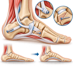
The operation is built from several parts, chosen to suit your foot. Most patients need 2 to 4 of them.
- Tendon transfer: a healthy tendon, often the one that bends the toes (flexor digitorum longus), is moved to take over from the damaged posterior tibial tendon.
- Heel bone osteotomy: the heel bone is cut and shifted inward so the heel sits back under the leg, which reduces strain on the repair.
- Lateral column lengthening: the outer side of the foot is lengthened with a bone graft or wedge to rotate the forefoot back into line.
- Spring ligament repair or reinforcement: the stretched ligament under the inner midfoot is tightened or reinforced.
- Calf lengthening: a tight gastrocnemius or Achilles tendon is released to reduce pressure on the arch.
When is joint fusion needed?
For a stiff, rigid foot (stage 3), the joints are no longer healthy enough to be rebuilt and are instead fused. A triple arthrodesis, or a more limited fusion, holds the hindfoot in a corrected position. Fusion reduces pain and gives a stable foot, at the cost of some flexibility in the joints that are joined.
What if the ankle is involved?
In stage 4, the ankle itself has started to tilt. Options include fusion of the ankle, sometimes together with the hindfoot, or realignment combined with total ankle replacement, a procedure also offered as total ankle replacement in turkey. Ankle fusion is also discussed on the ankle fusion page.
Are there minimally invasive choices?
Some surgeons use ankle arthroscopy to look inside the joint or clear inflamed tissue, and certain smaller procedures use small incisions. These are suitable for selected, less severe cases. A small implant placed in the hindfoot (a subtalar spacer) is occasionally used in flexible deformity, though it is not suited to every foot.
How do the options compare?
| Option | Best suited to | Main trade-off |
|---|---|---|
| Bracing and orthoses | Stage 1 to 2, or people not fit for surgery | Does not repair a torn tendon |
| Tendon transfer with heel osteotomy | Flexible stage 2 foot | Longer recovery, some residual stiffness |
| Joint fusion | Rigid stage 3 foot | Loss of motion in the fused joints |
| Ankle fusion or replacement with realignment | Stage 4 with ankle wear | Larger operation, more rehabilitation |
For an overview of the specialty, see foot and ankle surgery. For costs, read our flatfoot reconstruction cost guide.
When surgery is considered
Surgery for flat feet is worth discussing when pain and weakness still limit daily life after 3 to 6 months of good non-surgical care. It is a quality-of-life decision rather than an emergency, so you have time to gather information, ask questions and compare options. The decision is shared between you and your surgeon.
What are the usual signs that it is time to consider surgery?
- Pain on the inner ankle or arch that persists despite properly fitted insoles or a brace used for 3 to 6 months.
- Inability to walk the distances you need, work on your feet, or take part in the activities that matter to you.
- A foot that is getting visibly flatter or the heel tilting further outward, even with bracing.
- Outer ankle pain from the heel bone pressing against the fibula.
- Evidence on MRI of a significant tendon tear that is not likely to heal alone.
- Starting ankle joint tilt, which suggests that waiting may allow joint wear to progress.
When is surgery less suitable?
Surgery is approached with great care in people who smoke, who have poorly controlled diabetes, poor circulation, active infection or severe neuropathy. Healing is slower in these groups, and complications are more likely. Doctors often ask patients to stop smoking and improve blood sugar control for several weeks before elective foot surgery.
It may also be less suitable for very frail patients or those who cannot follow the weight-bearing restrictions after surgery. Bracing is a reasonable long-term choice in those cases.
What questions should you ask your surgeon?
- Which stage am I at, and what scans show that?
- Which parts of the operation do you recommend for my foot, and why?
- How many flatfoot reconstructions do you perform each year?
- What are the main risks for me, including nonunion and wound healing?
- How long will I be non-weight-bearing, and when can I drive and return to work?
Our guide to questions to ask before surgery abroad adds further prompts.
Procedures
Procedures that may treat flat feet
Costs
Flat feet treatment cost in Turkey
| Procedure | Turkey package | US self-pay | Saving |
|---|---|---|---|
| Flatfoot Reconstruction | $6,500 – $10,500 | $39,850 | ~79% |
Packages are contracted partner prices with validity dates; benchmarks are source-backed estimates. Open a cost guide for the full country comparison.
In Turkey
Treating flat feet in Turkey
Treatment for flat feet in turkey can be a sensible choice for planned, non-urgent reconstruction when your scans are available and a surgeon has reviewed your case remotely first. Foot and ankle reconstruction is elective, so there is time to prepare, compare options and travel when you are ready. This section explains the pathway and its limits.
When does treatment in turkey make sense?
Flat feet surgery in turkey is chosen for several reasons. People often consider flat feet treatment in turkey when waiting lists at home are long, when they want a surgeon who performs reconstruction regularly, or when they wish to combine treatment with a recovery period away from daily duties. It suits stable adults with stage 2 or 3 deformity whose general health is good. See why patients choose us on our page about why turkey.
What does the pathway look like?
- Free case review: you send weight-bearing X-rays, MRI if available and a short history through our free case review form.
- Remote opinion: a foot and ankle surgeon reviews your records and proposes a plan, including which parts of the operation are likely.
- Preparation: blood tests, medicine review and any needed medical clearance are arranged before you travel.
- Surgery and hospital stay: the operation is usually followed by 1 to 2 nights in hospital, then a period of rest nearby.
- Early follow-up in turkey: wound check, cast or boot change and X-rays are done before you fly home.
- Rehabilitation at home: your local physiotherapist continues the programme, supported by our team.
How can you judge safety and quality?
Ask whether the hospital holds international accreditation, whether the surgeon is a specialist in foot and ankle surgery and how many reconstructions they perform each year. Ask which implants are used, and who will manage complications after you leave. Our overview of orthopedic care in turkey explains how we vet partners, and the cities with a major hospital hub include Istanbul, Ankara, Izmir and Antalya.
How long should you plan to stay?
Most people plan roughly 10 to 14 days in turkey, which covers surgery, the first dressing change and early checks. You will not be able to put weight on the foot at first, so a companion is useful. Read our companion guide and the page on flying after surgery, because long flights and reduced movement raise the risk of blood clots.
When should you not travel?
Do not travel for surgery if you have an active foot infection, an unhealed ulcer, uncontrolled diabetes or a recent illness that needs medical attention at home. If your flat foot follows a recent injury with a sudden change, get local assessment first. Travel is for stable, planned cases only, and only when your treating team agrees.
Before you decide, compare the cost drivers on our flatfoot reconstruction cost guide and read about follow-up after returning home.
Complications
Complications of flat feet
Untreated adult acquired flatfoot can progress, and every operation carries risk, so both sides of the decision deserve an honest look. Most people with flat feet never face serious complications. When they do arise, early recognition makes them easier to manage.
What can happen if flat feet are left untreated?
A failing posterior tibial tendon can lead to a progressively flatter arch. Over months or years the deformity can become fixed, so what could have been treated with a tendon-sparing reconstruction may later need joint fusion. The outer ankle can become painful, and the inner ankle joint can wear down, producing arthritis.
Altered walking mechanics can also overload the knee, hip and lower back in some people, though the evidence on this link is mixed. Pain can reduce activity, which affects weight, fitness and mood.
What are the risks of non-surgical treatment?
Braces and insoles are low-risk, but they can cause skin pressure sores, particularly in people with reduced sensation. Steroid injections near the tendon may increase the risk of rupture. Long periods of immobilisation can weaken muscles and stiffen joints, so boots are used for limited periods.
What are the risks of surgery?
Risks of flatfoot reconstruction include wound healing problems, infection, nerve irritation, stiffness and swelling that can last for months. Bone cuts and fusions can fail to join (nonunion), and a minority of patients need further surgery. Hardware may become prominent and need removal. Blood clots are uncommon but serious, and surgeons use preventive measures.
Not every patient gets a perfectly normal-looking arch, and some residual pain or reduced push-off strength is possible. Smoking, diabetes and obesity raise the risk of complications. Your surgeon should talk about these chances in terms that apply to you.
How are complications reduced?
Careful patient selection, stopping smoking, optimising blood sugar, choosing the right procedure for the stage, protecting the foot during healing and attending follow-up appointments all lower risk. If you are travelling for care, ask in advance who will deal with a problem once you are home.
Urgent care
When to seek urgent care for flat feet
- Sudden severe pain, swelling or a "pop" in the inner ankle after a twist or fall: seek urgent local assessment, because a tendon rupture or fracture may be present.
- A hot, red, swollen foot with fever or feeling unwell: contact a doctor or emergency service the same day, as infection must be treated promptly.
- A new open sore, blister or black area on the foot, especially with diabetes: see a doctor within 24 hours because ulcers can deepen quickly.
- A foot that is cold, pale, blue or numb: seek emergency care, because blood supply may be reduced.
- Calf pain, swelling or breathlessness after surgery or a long flight: call emergency services, since these may point to a blood clot.
- After surgery, a wound that leaks, opens or smells, or pain that is rising rather than falling: contact your surgical team the same day.
Prevention
How to lower your risk of flat feet
Not every case of flat feet can be prevented, because inherited foot shape, age and some medical conditions play a part. Even so, protecting the posterior tibial tendon and the arch reduces the chance of a painful collapse, and it can slow progression if it has started. The measures below are practical and low-cost.
How can you protect the arch day to day?
Wear supportive, well-fitting shoes with a firm heel and a stiff sole, and replace them when the inner heel is worn down, usually after 6 to 12 months of regular use. Avoid prolonged barefoot walking on hard surfaces. Rotating shoes and using a cushioned insole can reduce repetitive load on the tendon.
What about weight and general health?
Keeping a healthy weight cuts the load on the arch with every step. Good control of blood sugar and blood pressure protects tendon blood supply. Stopping smoking improves tendon healing. If you have rheumatoid arthritis, regular review of your inflammatory disease also helps protect the foot.
Which habits help the foot and calf?
Regular calf stretching keeps the Achilles tendon from tugging the heel outward. Strengthening the foot and ankle muscles, balance work and gradual increases in walking or running volume all protect the tendon. A sensible rule is to raise weekly activity by no more than about 10% at a time.
Should you act early on symptoms?
Yes. Early inner ankle pain that lasts more than 2 to 3 weeks deserves attention. Early supportive care when the foot is still flexible gives the best chance of keeping the arch without surgery. A visit to a foot specialist when you first see your arch dropping is far easier than waiting for a fixed deformity.
What cannot be prevented?
Age-related tendon changes, family history and a naturally loose-jointed foot cannot be altered. What you can do is reduce the extra load and notice early signs. Many people with these risk factors keep a comfortable foot for life.
Treating other foot problems promptly, such as hallux rigidus or ankle osteoarthritis, also helps because pain in one part of the foot changes how you walk and loads the arch differently.
Outlook
Living with flat feet: outlook and recovery
The outlook for flat feet is generally good, and many adults stay active with bracing, insoles and exercise, while those who need surgery usually report less pain and better walking. The path depends on the stage, your general health and how closely you follow the rehabilitation plan. Realistic expectations help you plan well.
What is the natural course without surgery?
Mild flexible flat feet may stay stable for years. Painful posterior tibial tendon dysfunction can progress, though not always. Some people hold at stage 1 or 2 for a long time with a brace, while others slowly stiffen. Because it cannot be predicted for each person, periodic review makes sense.
What is recovery after flatfoot reconstruction like?
Recovery is gradual. A typical pattern is a cast or splint for about 2 weeks, then a boot with no weight on the foot for around 6 weeks. Weight-bearing in the boot usually starts between 6 and 8 weeks, and the move into supportive shoes happens around 10 to 12 weeks. Swelling can last for 6 months or longer.
Physiotherapy begins once bones have started to heal. Your surgeon will set the timetable, as it differs between a tendon transfer and a fusion.
When can you return to work and driving?
Desk work is often possible after around 6 to 8 weeks with the foot elevated. Driving depends on which foot was operated on and on being off strong pain medicines, often 8 to 12 weeks for the right foot. Jobs that need standing or climbing may need 4 to 6 months before full return.
What about sport and long-term results?
Walking, cycling and swimming are usually back within 4 to 6 months. Running and impact sport are often discussed from about 6 to 12 months, and some patients choose not to return to high-impact activity. Most people report good pain relief and function after reconstruction, though the foot may feel slightly stiffer.
Long-term, a corrected foot can last many years, but arthritis in neighbouring joints can still appear. Keeping to your exercises, supportive shoes and a healthy weight protects the result. Our rehabilitation guide describes what a recovery plan can look like.
Surgeons
Specialists who treat flat feet
FAQ
Flat feet: frequently asked questions
What are the main flat feet symptoms in adults?
Can flat feet be corrected without surgery?
Why are my feet getting flatter as an adult?
Do flat feet always need treatment?
Are insoles good for flat feet?
How long does recovery take after flat feet surgery?
Is flat feet surgery a major operation?
Can walking make flat feet worse?
Is flat feet treatment in turkey safe?
What should I send for a remote review of flat feet?
How long should I stay in turkey after flatfoot surgery?
Can flat feet cause knee or back pain?
Sources
Sources for this flat feet guide
Peer-reviewed guidance and institutional sources used to write and review this page.
- 01Adult Acquired Flatfoot
American Academy of Orthopaedic Surgeons (OrthoInfo), 2022
https://orthoinfo.aaos.org/en/diseases--conditions/adult-acquired-flatfoot/
- 02
- 03Posterior Tibial Tendon Dysfunction
American Academy of Orthopaedic Surgeons (OrthoInfo), 2023
https://orthoinfo.aaos.org/en/diseases--conditions/posterior-tibial-tendon-dysfunction/
- 04Flatfoot
Mayo Clinic, 2023
https://www.mayoclinic.org/diseases-conditions/flatfeet/symptoms-causes/syc-20372604
- 05
- 06
- 07Foot problems
National Institute of Arthritis and Musculoskeletal and Skin Diseases, 2023
https://www.niams.nih.gov/health-topics